
Rabbit Anti-HES1 antibody
bHLHb39; C-HAIRY1; c-hairy1A; Class B basic helix-loop-helix protein 39; FLJ20408; Hairy and enhancer of split 1 (Drosophila); Hairy and enhancer of split 1; Hairy Enhancer of Split 1; Hairy homolog (Drosophila); Hairy Homolog; Hairy like; Hairy, Drosophi
View History [Clear]
Details
Product Name HES1 Chinese Name 转录因子HES-1Recombinant rabbit monoclonal anti Alias bHLHb39; C-HAIRY1; c-hairy1A; Class B basic helix-loop-helix protein 39; FLJ20408; Hairy and enhancer of split 1 (Drosophila); Hairy and enhancer of split 1; Hairy Enhancer of Split 1; Hairy homolog (Drosophila); Hairy Homolog; Hairy like; Hairy, Drosophila, homolog of; Hairy-like protein; Hairy/enhancer of split, Drosophila, homolog of, 1; HAIRY1; HES-1; hes1; Hes1 hairy and enhancer of split 1 (Drosophila); HES1_HUMAN; HHL; HL; HRY; RHL; Transcription factor HES 1; Transcription factor HES-1. Research Area immunology Chromatin and nuclear signals Neurobiology transcriptional regulatory factor Epigenetics Immunogen Species Rabbit Clonality Monoclonal React Species (predicted: Human, Mouse, Rat, ) Applications WB=1:500-1000 IHC-P=1:100-500 ICC=1:100-500 IF=1:100-500 (Paraffin sections need antigen repair)
not yet tested in other applications.
optimal dilutions/concentrations should be determined by the end user.Theoretical molecular weight 30kDa Cellular localization The nucleus Form Liquid Concentration 1mg/ml immunogen KLH conjugated synthetic peptide derived from human HES1 Lsotype IgG Purification affinity purified by Protein A Buffer Solution 0.01M TBS(pH7.4) with 1% BSA, 0.03% Proclin300 and 50% Glycerol. Storage Shipped at 4℃. Store at -20 °C for one year. Avoid repeated freeze/thaw cycles. Attention This product as supplied is intended for research use only, not for use in human, therapeutic or diagnostic applications. PubMed PubMed Product Detail Transcriptional repressor of genes that require a bHLH protein for their transcription. May act as a negative regulator of myogenesis by inhibiting the functions of MYOD1 and ASH1. Binds DNA on N-box motifs: 5'-CACNAG-3' with high affinity and on E-box motifs: 5'-CANNTG-3' with low affinity.
Function:
Transcriptional repressor of genes that require a bHLH protein for their transcription. May act as a negative regulator of myogenesis by inhibiting the functions of MYOD1 and ASH1. Binds DNA on N-box motifs: 5'-CACNAG-3' with high affinity and on E-box motifs: 5'-CANNTG-3' with low affinity.
Subunit:
Transcription repression requires formation of a complex with a corepressor protein of the Groucho/TLE family. Interacts (via WPRW motif) with TLE1, and more weakly with TLE2. Interacts with HES6 (By similarity). Interacts with SIRT1. Interacts with an FA complex, composed of FANCA, FANCF, FANCG and FANCL, but not of FANCC, nor FANCE.
Subcellular Location:
Nucleus.
Similarity:
Contains 1 basic helix-loop-helix (bHLH) domain. Contains 1 Orange domain.
SWISS:
Q14469
Gene ID:
3280
Database links:Entrez Gene: 395128 Chicken
Entrez Gene: 3280 Human
Entrez Gene: 15205 Mouse
Omim: 139605 Human
SwissProt: O57337 Chicken
SwissProt: Q14469 Human
SwissProt: P35428 Mouse
Unigene: 250666 Human
Unigene: 390859 Mouse
Unigene: 19727 Rat
Product Picture 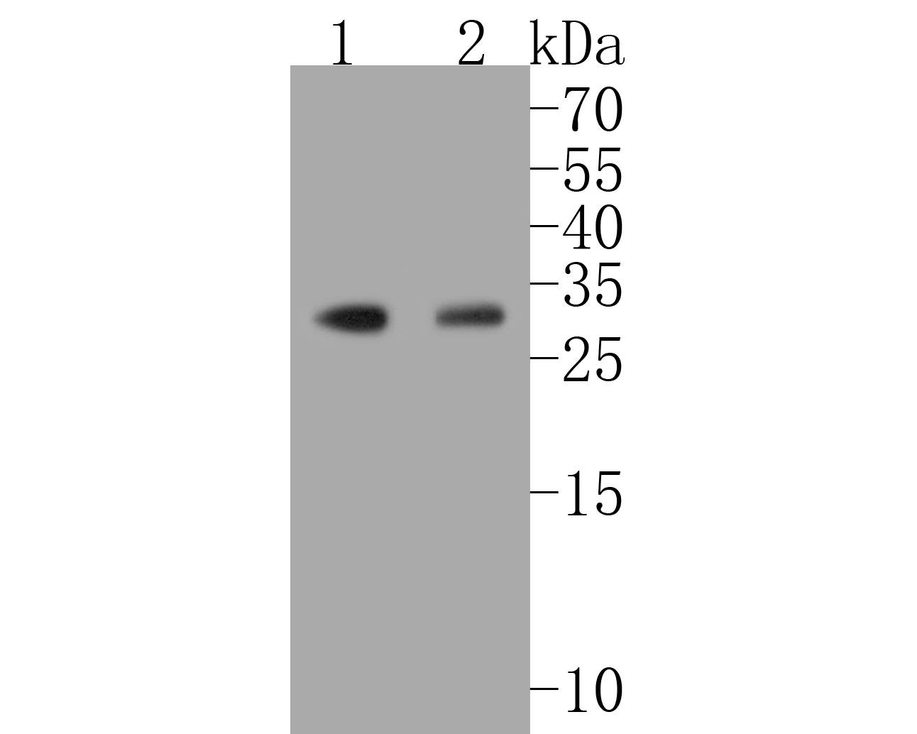 Western blot analysis of Hes1 on different lysates. Proteins were transferred to a PVDF membrane and blocked with 5% BSA in PBS for 1 hour at room temperature. The primary antibody (SLM-52568R, 1/500) was used in 5% BSA at room temperature for 2 hours. Goat Anti-Rabbit IgG - HRP Secondary Antibody (HA1001) at 1:5,000 dilution was used for 1 hour at room temperature.
Positive control:
Lane 1: SiHa cell lysate
Lane 2: SK-Br-3 cell lysate
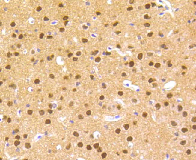 Immunohistochemical analysis of paraffin-embedded mouse brain tissue using anti-Hes1 antibody. The section was pre-treated using heat mediated antigen retrieval with sodium citrate buffer (pH 6.0) for 20 minutes. The tissues were blocked in 5% BSA for 30 minutes at room temperature, washed with ddH2O and PBS, and then probed with the primary antibody (SLM-52568R, 1/200) for 30 minutes at room temperature. The detection was performed using an HRP conjugated compact polymer system. DAB was used as the chromogen. Tissues were counterstained with hematoxylin and mounted with DPX.
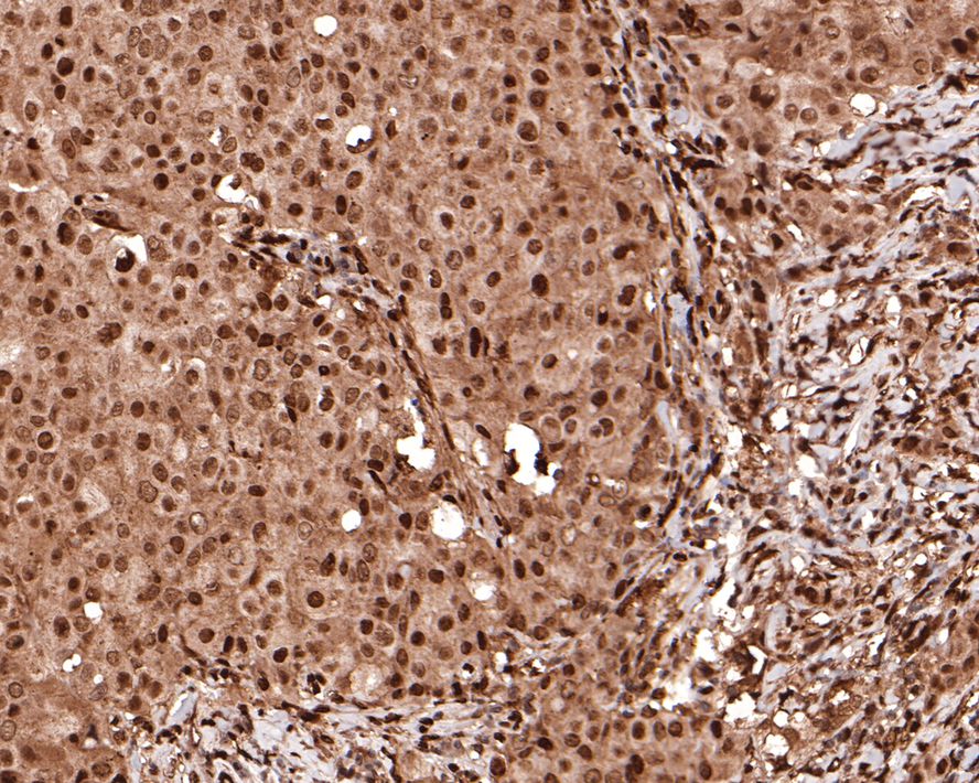 Immunohistochemical analysis of paraffin-embedded human breast carcinoma tissue with Rabbit anti-Hes1 antibody (SLM-52568R) at 1/400 dilution. The section was pre-treated using heat mediated antigen retrieval with sodium citrate buffer (pH 6.0) for 2 minutes. The tissues were blocked in 1% BSA for 20 minutes at room temperature, washed with ddH2O and PBS, and then probed with the primary antibody (SLM-52568R) at 1/400 dilution for 1 hour at room temperature. The detection was performed using an HRP conjugated compact polymer system. DAB was used as the chromogen. Tissues were counterstained with hematoxylin and mounted with DPX.
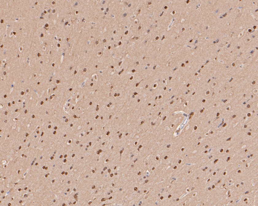 Immunohistochemical analysis of paraffin-embedded rat brain tissue using anti-Hes1 antibody. The section was pre-treated using heat mediated antigen retrieval with sodium citrate buffer (pH 6.0) for 20 minutes. The tissues were blocked in 5% BSA for 30 minutes at room temperature, washed with ddH2O and PBS, and then probed with the primary antibody (SLM-52568R, 1/200) for 30 minutes at room temperature. The detection was performed using an HRP conjugated compact polymer system. DAB was used as the chromogen. Tissues were counterstained with hematoxylin and mounted with DPX.
 ICC staining of Hes1 in 293 cells (green). Formalin fixed cells were permeabilized with 0.1% Triton X-100 in TBS for 10 minutes at room temperature and blocked with 1% Blocker BSA for 15 minutes at room temperature. Cells were probed with the primary antibody (SLM-52568R, 1/50) for 1 hour at room temperature, washed with PBS. Alexa Fluor®488 Goat anti-Rabbit IgG was used as the secondary antibody at 1/1,000 dilution. The nuclear counter stain is DAPI (blue).
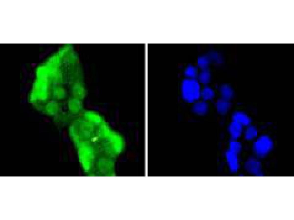 ICC staining of Hes1 in Hela cells (green). Formalin fixed cells were permeabilized with 0.1% Triton X-100 in TBS for 10 minutes at room temperature and blocked with 1% Blocker BSA for 15 minutes at room temperature. Cells were probed with the primary antibody (SLM-52568R, 1/50) for 1 hour at room temperature, washed with PBS. Alexa Fluor®488 Goat anti-Rabbit IgG was used as the secondary antibody at 1/1,000 dilution. The nuclear counter stain is DAPI (blue).
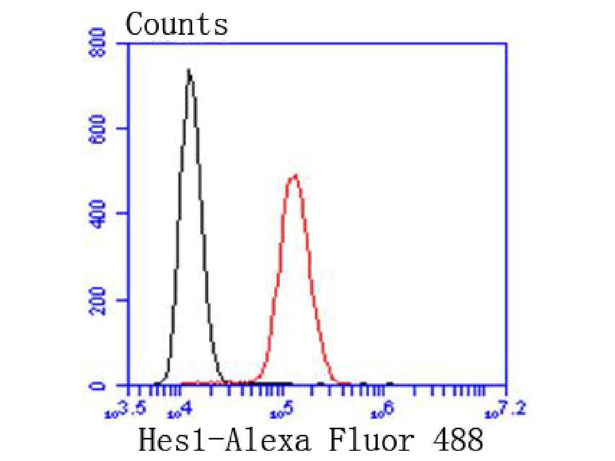 Flow cytometric analysis of Hes1 was done on SH-SY5Y cells. The cells were fixed, permeabilized and stained with the primary antibody (SLM-52568R, 1/50) (red). After incubation of the primary antibody at room temperature for an hour, the cells were stained with a Alexa Fluor 488-conjugated Goat anti-Rabbit IgG Secondary antibody at 1/1000 dilution for 30 minutes.Unlabelled sample was used as a control (cells without incubation with primary antibody; black).
Cartpieces
Totalgoods,subtotals:¥Checkout
References (0)
No References
Bought notes(bought amounts latest0)
No one bought this product
User Comment(Total0User Comment Num)
- No comment


 +86 571 56623320
 +86 18668110335

