
Rabbit Anti-TARDBP antibody
TAR DNA-binding protein 43; TAR DNA binding protein 43; ALS10; OTTHUMP00000002171; TAR DNA binding protein 43; TAR DNA binding protein; TDP 43; TDP-43; TDP43; tar DNA binding protein; ALS10; TADBP_HUMAN.
View History [Clear]
Details
Product Name TARDBP Chinese Name Tar DNA Binding protein43抗体 Alias TAR DNA-binding protein 43; TAR DNA binding protein 43; ALS10; OTTHUMP00000002171; TAR DNA binding protein 43; TAR DNA binding protein; TDP 43; TDP-43; TDP43; tar DNA binding protein; ALS10; TADBP_HUMAN. Research Area Neurobiology Signal transduction Bacteria and viruses Epigenetics Immunogen Species Rabbit Clonality Monoclonal React Species (predicted: Human, Mouse, Rat, Zebrafish, ) Applications WB=1:500-5000 IHC-P=1:100-500 ICC=1:100-500 IF=1:100-500 (Paraffin sections need antigen repair)
not yet tested in other applications.
optimal dilutions/concentrations should be determined by the end user.Theoretical molecular weight 45kDa Cellular localization The nucleus Form Liquid Concentration 1mg/ml immunogen KLH conjugated synthetic peptide derived from human TDP43 Lsotype IgG Purification affinity purified by Protein A Buffer Solution 0.01M TBS(pH7.4) with 1% BSA, 0.03% Proclin300 and 50% Glycerol. Storage Shipped at 4℃. Store at -20 °C for one year. Avoid repeated freeze/thaw cycles. Attention This product as supplied is intended for research use only, not for use in human, therapeutic or diagnostic applications. PubMed PubMed Product Detail HIV-1, the causative agent of acquired immunodeficiency syndrome (AIDS), contains an RNA genome that produces a chromosomally integrated DNA during the replicative cycle. Activation of HIV-1 gene expression by the transactivator Tat is dependent on an RNA regulatory element (TAR) located downstream of the transcription initiation site. The protein encoded by this gene is a transcriptional repressor that binds to chromosomally integrated TAR DNA and represses HIV-1 transcription. In addition, this protein regulates alternate splicing of the CFTR gene. A similar pseudogene is present on chromosome 20. [provided by RefSeq, Jul 2008]
Function:
DNA and RNA-binding protein which regulates transcription and splicing. Involved in the regulation of CFTR splicing. It promotes CFTR exon 9 skipping by binding to the UG repeated motifs in the polymorphic region near the 3'-splice site of this exon. The resulting aberrant splicing is associated with pathological features typical of cystic fibrosis. May also be involved in microRNA biogenesis, apoptosis and cell division. Can repress HIV-1 transcription by binding to the HIV-1 long terminal repeat. Stabilizes the low molecular weight neurofilament (NFL) mRNA through a direct interaction with the 3' UTR.
Subcellular Location:
Nucleus. In patients with frontotemporal lobar degeneration and amyotrophic lateral sclerosis, it is absent from the nucleus of affected neurons but it is the primary component of cytoplasmic ubiquitin-positive inclusion bodies.
Tissue Specificity:
Ubiquitously expressed. In particular, expression is high in pancreas, placenta, lung, genital tract and spleen.
Post-translational modifications:
Hyperphosphorylated in hippocampus, neocortex, and spinal cord from individuals affected with ALS and FTLDU.
Ubiquitinated in hippocampus, neocortex, and spinal cord from individuals affected with ALS and FTLDU.
Cleaved to generate C-terminal fragments in hippocampus, neocortex, and spinal cord from individuals affected with ALS and FTLDU.
DISEASE:
Defects in TARDBP are the cause of amyotrophic lateral sclerosis type 10 (ALS10) [MIM:612069]. ALS is a neurodegenerative disorder affecting upper and lower motor neurons and resulting in fatal paralysis. Sensory abnormalities are absent. Death usually occurs within 2 to 5 years. The etiology of ALS is likely to be multifactorial, involving both genetic and environmental factors. The disease is inherited in 5-10% of the cases.
Similarity:
Contains 2 RRM (RNA recognition motif) domains.
SWISS:
Q13148
Gene ID:
23435
Database links:Entrez Gene: 23435 Human
Entrez Gene: 230908 Mouse
Omim: 605078 Human
SwissProt: Q13148 Human
SwissProt: Q921F2 Mouse
Unigene: 300624 Human
Unigene: 635053 Human
Unigene: 22453 Mouse
Unigene: 2633 Rat
变异蛋白质TDP-43 在额颞叶退行性病变(FTLD-U)和萎缩性侧索硬化症(ALS)中表达较高。TDP-43在大脑中堆积能导致神经细胞衰竭,从而引发疾病肌萎缩性侧索硬化(ALS,也被称为Lou Gehrig氏病)
TDP-43这种痴呆是由大脑额叶的退化引起的,退化能延伸到颞叶。这是仅次于阿尔默海兹症的让65岁以下患者痴呆的第二种最常见的原因,通常影响40几岁和50几岁的人。TDP-43过去在神经退化疾病患者病理的错误折叠蛋白中缺失。识别出这个蛋白应该有助于痴呆以及运动神经元疾病的研究。Product Picture  Western blot analysis of TDP43 on different lysates. Proteins were transferred to a PVDF membrane and blocked with 5% BSA in PBS for 1 hour at room temperature. The primary antibody (SLM-52949R, 1/500) was used in 5% BSA at room temperature for 2 hours. Goat Anti-Rabbit IgG - HRP Secondary Antibody (HA1001) at 1:5,000 dilution was used for 1 hour at room temperature. Positive control: Lane 1: K562 cell lysate Lane 2: Hela cell lysate
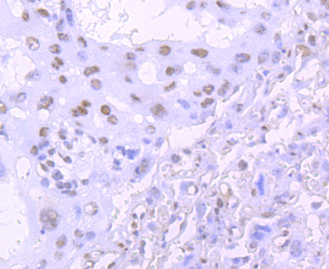 Immunohistochemical analysis of paraffin-embedded mouse placenta tissue using anti-TDP43 antibody. The section was pre-treated using heat mediated antigen retrieval with Tris-EDTA buffer (pH 8.0-8.4) for 20 minutes.The tissues were blocked in 5% BSA for 30 minutes at room temperature, washed with ddH2O and PBS, and then probed with the primary antibody (SLM-52949R, 1/50) for 30 minutes at room temperature. The detection was performed using an HRP conjugated compact polymer system. DAB was used as the chromogen. Tissues were counterstained with hematoxylin and mounted with DPX.
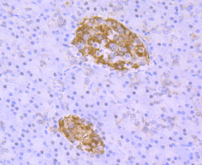 Immunohistochemical analysis of paraffin-embedded human pancreas tissue using anti-TDP43 antibody. The section was pre-treated using heat mediated antigen retrieval with Tris-EDTA buffer (pH 8.0-8.4) for 20 minutes.The tissues were blocked in 5% BSA for 30 minutes at room temperature, washed with ddH2O and PBS, and then probed with the primary antibody (SLM-52949R, 1/50) for 30 minutes at room temperature. The detection was performed using an HRP conjugated compact polymer system. DAB was used as the chromogen. Tissues were counterstained with hematoxylin and mounted with DPX.
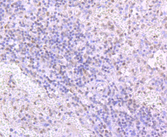 Immunohistochemical analysis of paraffin-embedded human spleen tissue using anti-TDP43 antibody. The section was pre-treated using heat mediated antigen retrieval with Tris-EDTA buffer (pH 8.0-8.4) for 20 minutes.The tissues were blocked in 5% BSA for 30 minutes at room temperature, washed with ddH2O and PBS, and then probed with the primary antibody (SLM-52949R, 1/50) for 30 minutes at room temperature. The detection was performed using an HRP conjugated compact polymer system. DAB was used as the chromogen. Tissues were counterstained with hematoxylin and mounted with DPX.
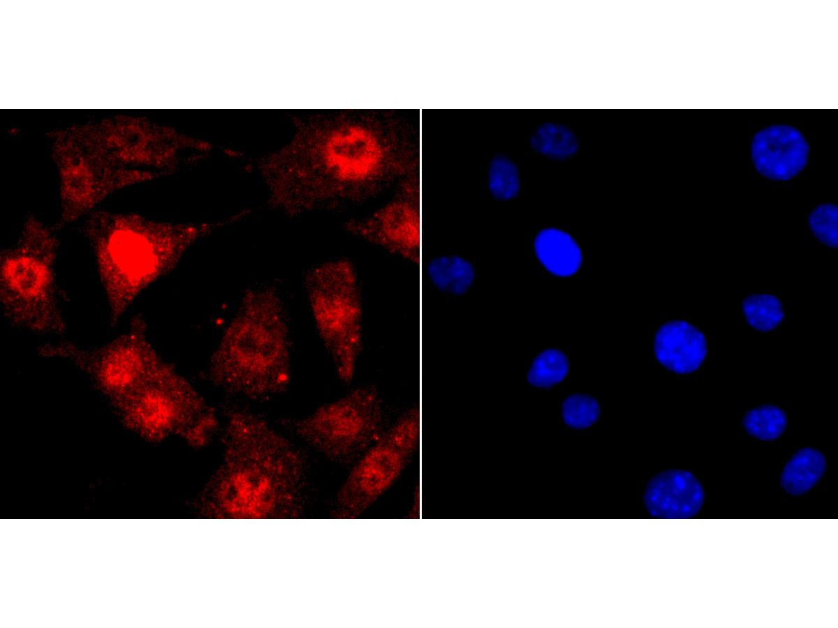 ICC staining of TDP43 in SH-SY5Y cells (red). Formalin fixed cells were permeabilized with 0.1% Triton X-100 in TBS for 10 minutes at room temperature and blocked with 1% Blocker BSA for 15 minutes at room temperature. Cells were probed with the primary antibody (SLM-52949R, 1/50) for 1 hour at room temperature, washed with PBS. Alexa Fluor®594 Goat anti-Rabbit IgG was used as the secondary antibody at 1/1,000 dilution. The nuclear counter stain is DAPI (blue)
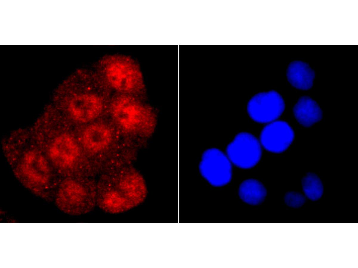 ICC staining of TDP43 in Hela cells (red). Formalin fixed cells were permeabilized with 0.1% Triton X-100 in TBS for 10 minutes at room temperature and blocked with 1% Blocker BSA for 15 minutes at room temperature. Cells were probed with the primary antibody (SLM-52949R, 1/50) for 1 hour at room temperature, washed with PBS. Alexa Fluor®594 Goat anti-Rabbit IgG was used as the secondary antibody at 1/1,000 dilution. The nuclear counter stain is DAPI (blue).
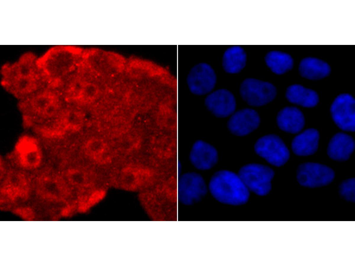 ICC staining of TDP43 in 293T cells (red). Formalin fixed cells were permeabilized with 0.1% Triton X-100 in TBS for 10 minutes at room temperature and blocked with 1% Blocker BSA for 15 minutes at room temperature. Cells were probed with the primary antibody (SLM-52949R, 1/50) for 1 hour at room temperature, washed with PBS. Alexa Fluor®594 Goat anti-Rabbit IgG was used as the secondary antibody at 1/1,000 dilution. The nuclear counter stain is DAPI (blue).
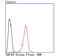 Flow cytometric analysis of TDP43 was done on Hela cells. The cells were fixed, permeabilized and stained with the primary antibody (SLM-52949R, 1/50) (red). After incubation of the primary antibody at room temperature for an hour, the cells were stained with a Alexa Fluor 488-conjugated Goat anti-Rabbit IgG Secondary antibody at 1/1000 dilution for 30 minutes.Unlabelled sample was used as a control (cells without incubation with primary antibody; black).
Cartpieces
Totalgoods,subtotals:¥Checkout
References (0)
No References
Bought notes(bought amounts latest0)
No one bought this product
User Comment(Total0User Comment Num)
- No comment


 +86 571 56623320
 +86 18668110335

