
Rabbit Anti-Ku80 antibody
86 kDa subunit of Ku antigen; ATP dependent DNA helicase 2 subunit 2; ATP dependent DNA helicase II 80 kDa subunit; ATP dependent DNA helicase II 86 Kd subunit; ATP dependent DNA helicase II; ATP-dependent DNA helicase 2 subunit 2; ATP-dependent DNA helic
View History [Clear]
Details
Product Name Ku80 Chinese Name DNA修复酶Ku-80Recombinant rabbit monoclonal anti Alias 86 kDa subunit of Ku antigen; ATP dependent DNA helicase 2 subunit 2; ATP dependent DNA helicase II 80 kDa subunit; ATP dependent DNA helicase II 86 Kd subunit; ATP dependent DNA helicase II; ATP-dependent DNA helicase 2 subunit 2; ATP-dependent DNA helicase II 80 kDa subunit; CTC box binding factor 85 kDa; CTC box-binding factor 85 kDa subunit; CTC85; CTCBF; DNA repair protein XRCC5; KARP 1; KARP1; Ku 80; Ku autoantigen 80kDa; Ku86; Ku86 autoantigen related protein 1; KUB 2; KUB2; Lupus Ku autoantigen protein p86; NFIV; Nuclear factor IV; Thyroid lupus autoantigen; Thyroid-lupus autoantigen; TLAA; X ray repair complementing defective repair in Chinese hamster cells 5 (double strand break rejoining); X-ray repair complementing defective repair in Chinese hamster cells 5 (double-strand-break rejoining); X-ray repair cross-complementing protein 5; Xray repair complementing defective repair in Chinese hamster cells 5; XRCC 5; XRCC5; XRCC5_HUMAN. Research Area Chromatin and nuclear signals Immunogen Species Rabbit Clonality Monoclonal React Species (predicted: Human, ) Applications WB=1:500-2000 IHC-P=1:100-500 ICC=1:50-200 IF=1:50-200 (Paraffin sections need antigen repair)
not yet tested in other applications.
optimal dilutions/concentrations should be determined by the end user.Theoretical molecular weight 83kDa Cellular localization The nucleus Form Liquid Concentration 1mg/ml immunogen KLH conjugated synthetic peptide derived from human Ku80 Lsotype IgG Purification affinity purified by Protein A Buffer Solution 0.01M TBS(pH7.4) with 1% BSA, 0.03% Proclin300 and 50% Glycerol. Storage Shipped at 4℃. Store at -20 °C for one year. Avoid repeated freeze/thaw cycles. Attention This product as supplied is intended for research use only, not for use in human, therapeutic or diagnostic applications. PubMed PubMed Product Detail The protein encoded by this gene is the 80-kilodalton subunit of the Ku heterodimer protein which is also known as ATP-dependant DNA helicase II or DNA repair protein XRCC5. Ku is the DNA-binding component of the DNA-dependent protein kinase, and it functions together with the DNA ligase IV-XRCC4 complex in the repair of DNA double-strand break by non-homologous end joining and the completion of V(D)J recombination events. This gene functionally complements Chinese hamster xrs-6, a mutant defective in DNA double-strand break repair and in ability to undergo V(D)J recombination. A rare microsatellite polymorphism in this gene is associated with cancer in patients of varying radiosensitivity. [provided by RefSeq, Jul 2008]
Function:
Single stranded DNA-dependent ATP-dependent helicase. Has a role in chromosome translocation. The DNA helicase II complex binds preferentially to fork-like ends of double-stranded DNA in a cell cycle-dependent manner. It works in the 3'-5' direction. Binding to DNA may be mediated by XRCC6. Involved in DNA non-homologous end joining (NHEJ) required for double-strand break repair and V(D)J recombination. The XRCC5/6 dimer acts as regulatory subunit of the DNA-dependent protein kinase complex DNA-PK by increasing the affinity of the catalytic subunit PRKDC to DNA by 100-fold. The XRCC5/6 dimer is probably involved in stabilizing broken DNA ends and bringing them together. The assembly of the DNA-PK complex to DNA ends is required for the NHEJ ligation step. In association with NAA15, the XRCC5/6 dimer binds to the osteocalcin promoter and activates osteocalcin expression. The XRCC5/6 dimer probably also acts as a 5'-deoxyribose-5-phosphate lyase (5'-dRP lyase), by catalyzing the beta-elimination of the 5' deoxyribose-5-phosphate at an abasic site near double-strand breaks. XRCC5 probably acts as the catalytic subunit of 5'-dRP activity, and allows to 'clean' the termini of abasic sites, a class of nucleotide damage commonly associated with strand breaks, before such broken ends can be joined. The XRCC5/6 dimer together with APEX1 acts as a negative regulator of transcription.
Subunit:
Heterodimer of a 70 kDa and a 80 kDa subunit.
Subcellular Location:
Nucleus. Chromosome.
Similarity:
Belongs to the ku80 family.
Contains 1 Ku domain.
SWISS:
P13010
Gene ID:
7520
Database links:Entrez Gene: 7520 Human
Entrez Gene: 22596 Mouse
Omim: 194364 Human
SwissProt: P13010 Human
SwissProt: P27641 Mouse
Unigene: 388739 Human
Unigene: 246952 Mouse
Ku80也是一种DNA修复蛋白,当细胞在受到辐射损伤而发生DNA双链断裂时,Ku80可迅速将其修复,从而提高细胞存活率。
Ku是一种多功能的蛋白,在许多重要的细胞生命过程中起着直接或间接的作用,如DNA双链断裂的修复,免疫球蛋白和T细胞受体V(D)J重排,免疫球蛋白构型转换,DNA复制,DNA转录的调节,同时在细胞周期的G2和M时相中起着特殊的作用。Product Picture 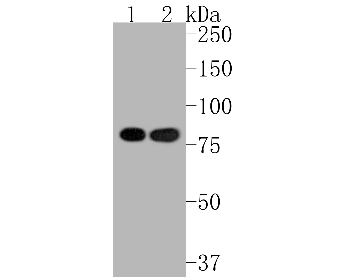 Western blot analysis of Ku80 on different lysates. Proteins were transferred to a PVDF membrane and blocked with 5% BSA in PBS for 1 hour at room temperature. The primary antibody (SLM-52512R, 1/500) was used in 5% BSA at room temperature for 2 hours. Goat Anti-Rabbit IgG - HRP Secondary Antibody (HA1001) at 1:5,000 dilution was used for 1 hour at room temperature.
Positive control:
Lane 1: MCF-7 cell lysate
Lane 2: A549 cell lysate
 Immunohistochemical analysis of paraffin-embedded human breast carcinoma tissue with Rabbit anti-Ku80 antibody (SLM-52512R) at 1/50 dilution. The section was pre-treated using heat mediated antigen retrieval with Tris-EDTA buffer (pH 8.0-8.4) for 20 minutes. The tissues were blocked in 1% BSA for 20 minutes at room temperature, washed with ddH2O and PBS, and then probed with the primary antibody (SLM-52512R) at 1/50 dilution for 1 hour at room temperature. The detection was performed using an HRP conjugated compact polymer system. DAB was used as the chromogen. Tissues were counterstained with hematoxylin and mounted with DPX.
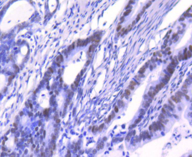 Immunohistochemical analysis of paraffin-embedded human colon cancer tissue with Rabbit anti-Ku80 antibody (SLM-52512R) at 1/50 dilution. The section was pre-treated using heat mediated antigen retrieval with Tris-EDTA buffer (pH 8.0-8.4) for 20 minutes. The tissues were blocked in 1% BSA for 20 minutes at room temperature, washed with ddH2O and PBS, and then probed with the primary antibody (SLM-52512R) at 1/50 dilution for 1 hour at room temperature. The detection was performed using an HRP conjugated compact polymer system. DAB was used as the chromogen. Tissues were counterstained with hematoxylin and mounted with DPX.
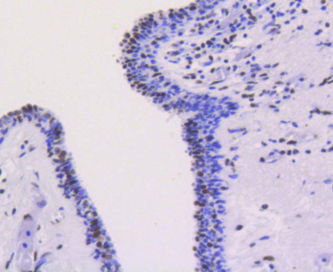
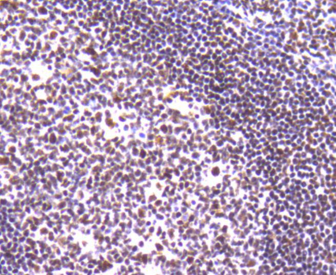 Immunohistochemical analysis of paraffin-embedded human tonsil tissue with Rabbit anti-Ku80 antibody (SLM-52512R) at 1/50 dilution. The section was pre-treated using heat mediated antigen retrieval with Tris-EDTA buffer (pH 8.0-8.4) for 20 minutes. The tissues were blocked in 1% BSA for 20 minutes at room temperature, washed with ddH2O and PBS, and then probed with the primary antibody (SLM-52512R) at 1/50 dilution for 1 hour at room temperature. The detection was performed using an HRP conjugated compact polymer system. DAB was used as the chromogen. Tissues were counterstained with hematoxylin and mounted with DPX.
 Immunohistochemical analysis of paraffin-embedded human kideny tissue using anti-Ku80 antibody. Counter stained with hematoxylin. Immunohistochemical analysis of paraffin-embedded human kideny tissue with Rabbit anti-Ku80 antibody (SLM-52512R) at 1/50 dilution. The section was pre-treated using heat mediated antigen retrieval with Tris-EDTA buffer (pH 8.0-8.4) for 20 minutes. The tissues were blocked in 1% BSA for 20 minutes at room temperature, washed with ddH2O and PBS, and then probed with the primary antibody (SLM-52512R) at 1/50 dilution for 1 hour at room temperature. The detection was performed using an HRP conjugated compact polymer system. DAB was used as the chromogen. Tissues were counterstained with hematoxylin and mounted with DPX.
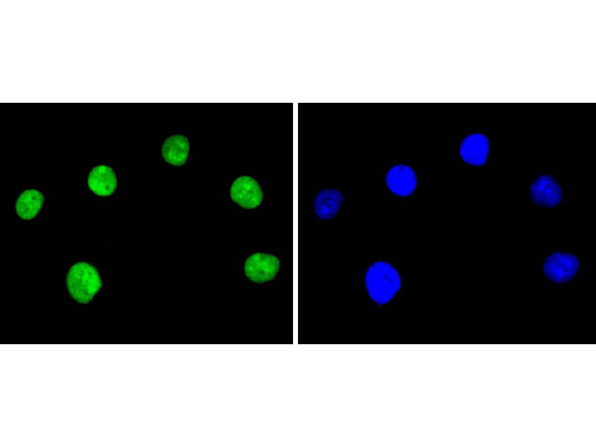 ICC staining of Ku80 in A549 cells (green). Formalin fixed cells were permeabilized with 0.1% Triton X-100 in TBS for 10 minutes at room temperature and blocked with 1% Blocker BSA for 15 minutes at room temperature. Cells were probed with the primary antibody (SLM-52512R, 1/50) for 1 hour at room temperature, washed with PBS. Alexa Fluor®488 Goat anti-Rabbit IgG was used as the secondary antibody at 1/1,000 dilution. The nuclear counter stain is DAPI (blue).
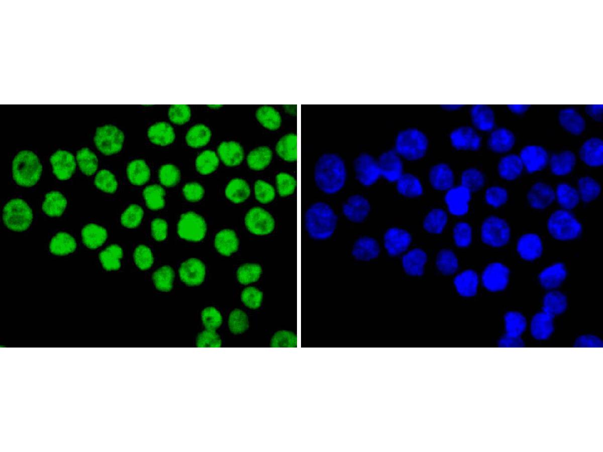 ICC staining of Ku80 in SW480 cells (green). Formalin fixed cells were permeabilized with 0.1% Triton X-100 in TBS for 10 minutes at room temperature and blocked with 1% Blocker BSA for 15 minutes at room temperature. Cells were probed with the primary antibody (SLM-52512R, 1/50) for 1 hour at room temperature, washed with PBS. Alexa Fluor®488 Goat anti-Rabbit IgG was used as the secondary antibody at 1/1,000 dilution. The nuclear counter stain is DAPI (blue).
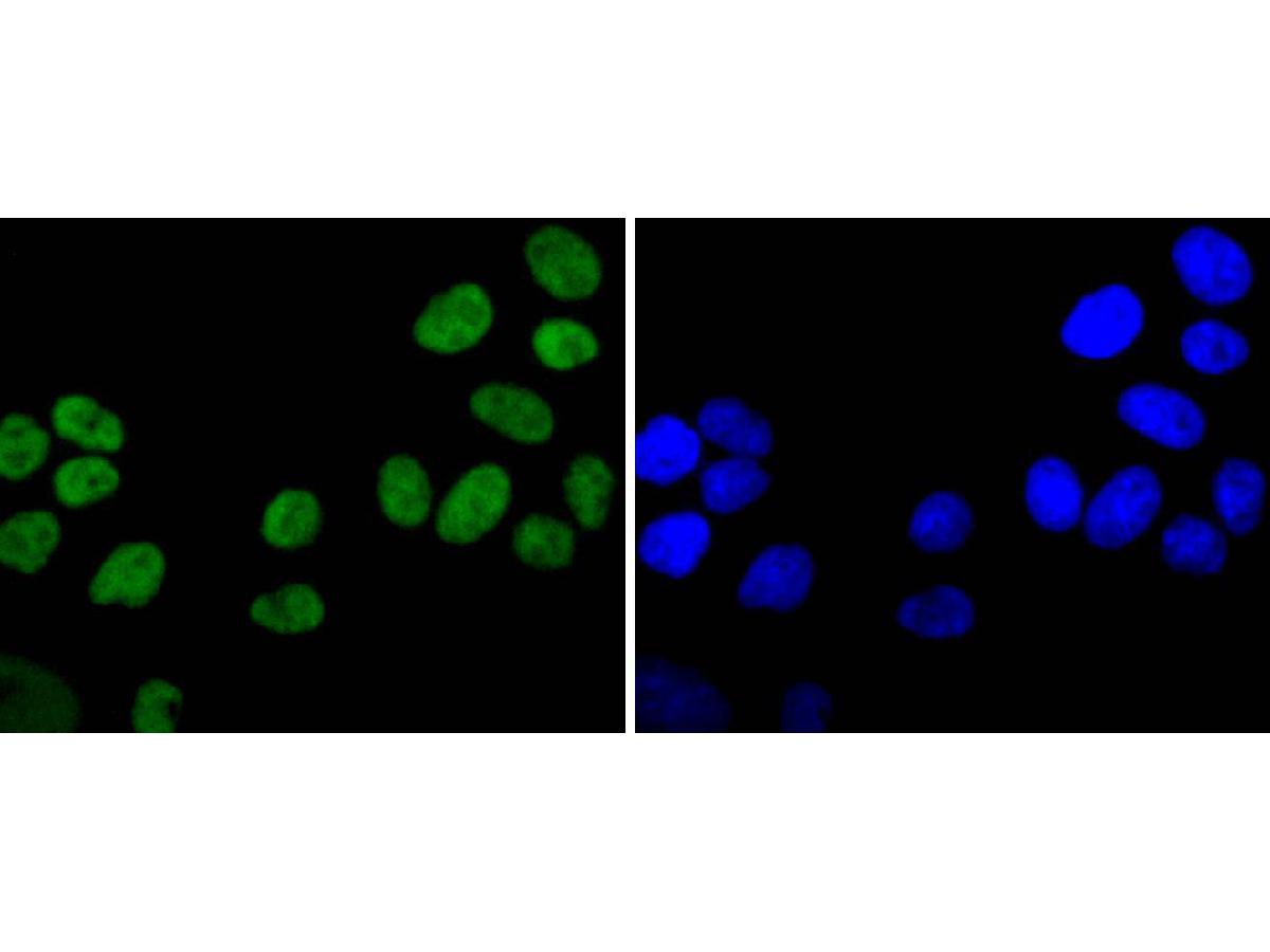 ICC staining of Ku80 in Hela cells (green). Formalin fixed cells were permeabilized with 0.1% Triton X-100 in TBS for 10 minutes at room temperature and blocked with 1% Blocker BSA for 15 minutes at room temperature. Cells were probed with the primary antibody (SLM-52512R, 1/50) for 1 hour at room temperature, washed with PBS. Alexa Fluor®488 Goat anti-Rabbit IgG was used as the secondary antibody at 1/1,000 dilution. The nuclear counter stain is DAPI (blue).
Cartpieces
Totalgoods,subtotals:¥Checkout
References (0)
No References
Bought notes(bought amounts latest0)
No one bought this product
User Comment(Total0User Comment Num)
- No comment


 +86 571 56623320
 +86 18668110335

