
Rabbit Anti-nNOS antibody
NOS1_HUMAN; Nitric oxide synthase, brain; NOS1; EC:1.14.13.39; Constitutive NOS; N NOS; N-NOS; NC-NOS; NOS type I; Neuronal NOS (N-NOS; nNOS); Peptidyl-cysteine S-nitrosylase NOS1; bNOS; neuronal Nitric Oxide Synthase;
View History [Clear]
Details
Product Name nNOS Chinese Name 一氧化氮合成酶-1(神经型)Recombinant rabbit monoclonal anti Alias NOS1_HUMAN; Nitric oxide synthase, brain; NOS1; EC:1.14.13.39; Constitutive NOS; N NOS; N-NOS; NC-NOS; NOS type I; Neuronal NOS (N-NOS; nNOS); Peptidyl-cysteine S-nitrosylase NOS1; bNOS; neuronal Nitric Oxide Synthase; Research Area Neurobiology Immunogen Species Rabbit Clonality Monoclonal React Species Mouse, Rat, (predicted: Human, ) Applications WB=1:500-1000 IHC-P=1:100-500 ICC=1:50-200 (Paraffin sections need antigen repair)
not yet tested in other applications.
optimal dilutions/concentrations should be determined by the end user.Theoretical molecular weight 130kDa Cellular localization The cell membrane Form Liquid Concentration 1mg/ml immunogen KLH conjugated synthetic peptide derived from human nNOS Lsotype IgG Purification affinity purified by Protein A Buffer Solution 0.01M TBS(pH7.4) with 1% BSA, 0.03% Proclin300 and 50% Glycerol. Storage Shipped at 4℃. Store at -20 °C for one year. Avoid repeated freeze/thaw cycles. Attention This product as supplied is intended for research use only, not for use in human, therapeutic or diagnostic applications. PubMed PubMed Product Detail The protein encoded by this gene belongs to the family of nitric oxide synthases, which synthesize nitric oxide from L-arginine. Nitric oxide is a reactive free radical, which acts as a biologic mediator in several processes, including neurotransmission, and antimicrobial and antitumoral activities. In the brain and peripheral nervous system, nitric oxide displays many properties of a neurotransmitter, and has been implicated in neurotoxicity associated with stroke and neurodegenerative diseases, neural regulation of smooth muscle, including peristalsis, and penile erection. This protein is ubiquitously expressed, with high level of expression in skeletal muscle. Multiple transcript variants that differ in the 5' UTR have been described for this gene but the full-length nature of these transcripts is not known. Additionally, alternatively spliced transcript variants encoding different isoforms (some testis-specific) have been found for this gene.[provided by RefSeq, Feb 2011].
Function:
Produces nitric oxide (NO) which is a messenger molecule with diverse functions throughout the body. In the brain and peripheral nervous system, NO displays many properties of a neurotransmitter. Probably has nitrosylase activity and mediates cysteine S-nitrosylation of cytoplasmic target proteins such SRR.
Subunit:
Homodimer. Interacts with DLG4; the interaction possibly being prevented by the association between NOS1 and CAPON. Forms a ternary complex with CAPON and RASD1. Forms a ternary complex with CAPON and SYN1. Interacts with ZDHHC23. Interacts with NOSIP; which may impair its synaptic location (By similarity). Interacts with HTR4. Interacts with VAC14 (By similarity). Interacts with SLC6A4.
Subcellular Location:
Cell membrane, sarcolemma; Peripheral membrane protein. Cell projection, dendritic spine. Note=In skeletal muscle, it is localized beneath the sarcolemma of fast-twitch muscle fiber by associating with the dystrophin glycoprotein complex. In neurons, enriched in dendritic spines (By similarity).
Tissue Specificity:
Isoform 1 is ubiquitously expressed: detected in skeletal muscle and brain, also in testis, lung and kidney, and at low levels in heart, adrenal gland and retina. Not detected in the platelets. Isoform 3 is expressed only in testis. Isoform 4 is detected in testis, skeletal muscle, lung, and kidney, at low levels in the brain, but not in the heart and adrenal gland.
Post-translational modifications:
Ubiquitinated; mediated by STUB1/CHIP in the presence of Hsp70 and Hsp40 (in vitro).
Similarity:
Belongs to the NOS family.
Contains 1 FAD-binding FR-type domain.
Contains 1 flavodoxin-like domain.
Contains 1 PDZ (DHR) domain.
SWISS:
P29475
Gene ID:
4842
Database links:Entrez Gene: 4842 Human
Entrez Gene: 18125 Mouse
SwissProt: P29475 Human
SwissProt: Q9Z0J4 Mouse
Product Picture 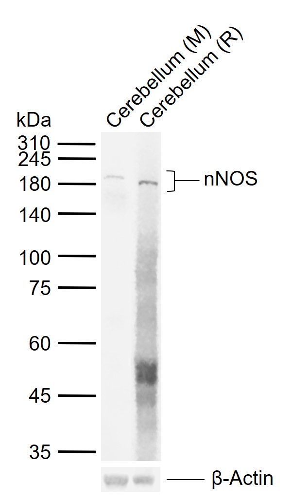 Sample:
Lane 1: Mouse Cerebellum tissue lysates
Lane 2: Rat Cerebellum tissue lysates
Primary: Anti-nNOS (SLM-52474R) at 1/1000 dilution
Secondary: IRDye800CW Goat Anti-Rabbit IgG at 1/20000 dilution
Predicted band size: 130 kDa
Observed band size: 180 kDa
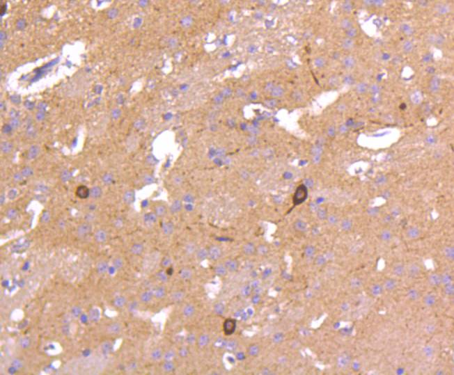 Immunohistochemical analysis of paraffin-embedded mouse brain tissue using anti-nNOS antibody. The section was pre-treated using heat mediated antigen retrieval with Tris-EDTA buffer (pH 8.0-8.4) for 20 minutes.The tissues were blocked in 5% BSA for 30 minutes at room temperature, washed with ddH2O and PBS, and then probed with the primary antibody (SLM-52474R, 1/50) for 30 minutes at room temperature. The detection was performed using an HRP conjugated compact polymer system. DAB was used as the chromogen. Tissues were counterstained with hematoxylin and mounted with DPX.
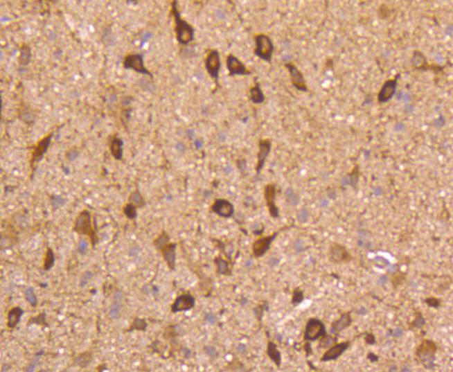 Immunohistochemical analysis of paraffin-embedded rat brain tissue using anti-nNOS antibody. The section was pre-treated using heat mediated antigen retrieval with Tris-EDTA buffer (pH 8.0-8.4) for 20 minutes.The tissues were blocked in 5% BSA for 30 minutes at room temperature, washed with ddH2O and PBS, and then probed with the primary antibody (SLM-52474R, 1/50) for 30 minutes at room temperature. The detection was performed using an HRP conjugated compact polymer system. DAB was used as the chromogen. Tissues were counterstained with hematoxylin and mounted with DPX.
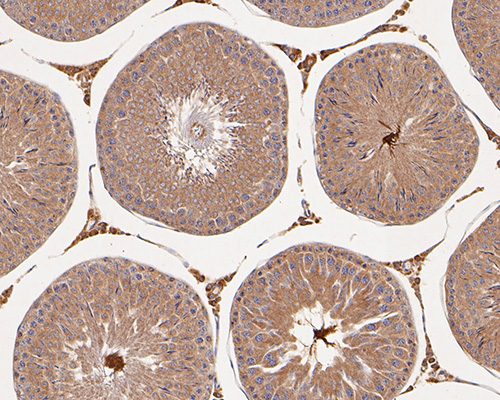 Immunohistochemical analysis of paraffin-embedded rat testis tissue using anti-nNOS antibody. The section was pre-treated using heat mediated antigen retrieval with Tris-EDTA buffer (pH 8.0-8.4) for 20 minutes.The tissues were blocked in 5% BSA for 30 minutes at room temperature, washed with ddH2O and PBS, and then probed with the primary antibody (SLM-52474R, 1/50) for 30 minutes at room temperature. The detection was performed using an HRP conjugated compact polymer system. DAB was used as the chromogen. Tissues were counterstained with hematoxylin and mounted with DPX.
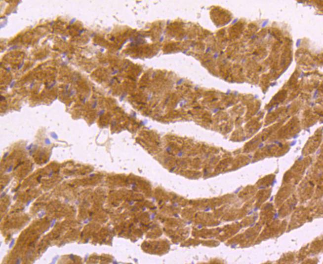 Immunohistochemical analysis of paraffin-embedded mouse heart tissue using anti-nNOS antibody. The section was pre-treated using heat mediated antigen retrieval with Tris-EDTA buffer (pH 8.0-8.4) for 20 minutes.The tissues were blocked in 5% BSA for 30 minutes at room temperature, washed with ddH2O and PBS, and then probed with the primary antibody (SLM-52474R, 1/50) for 30 minutes at room temperature. The detection was performed using an HRP conjugated compact polymer system. DAB was used as the chromogen. Tissues were counterstained with hematoxylin and mounted with DPX.
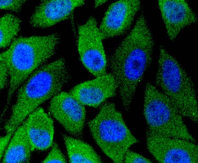 ICC staining of nNOS in PC-3M cells (green). Formalin fixed cells were permeabilized with 0.1% Triton X-100 in TBS for 10 minutes at room temperature and blocked with 1% Blocker BSA for 15 minutes at room temperature. Cells were probed with the primary antibody (SLM-52474R, 1/50) for 1 hour at room temperature, washed with PBS. Alexa Fluor®488 Goat anti-Rabbit IgG was used as the secondary antibody at 1/1,000 dilution. The nuclear counter stain is DAPI (blue).
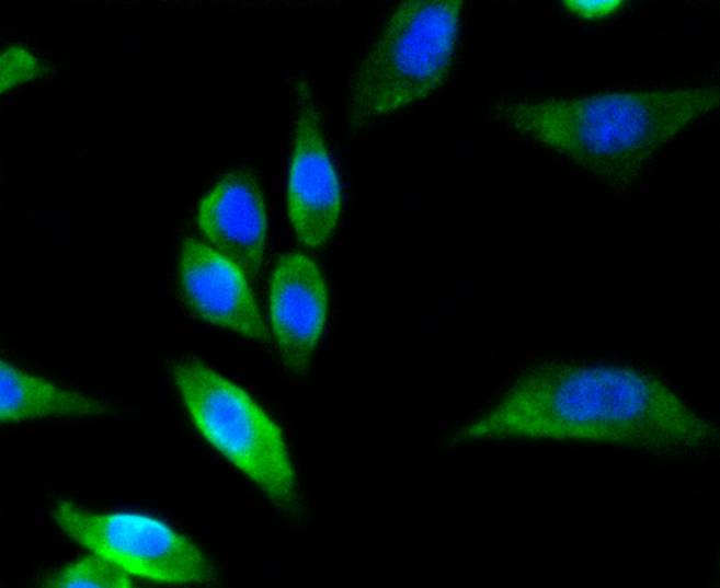 ICC staining of nNOS in SH-SY5Y cells (green). Formalin fixed cells were permeabilized with 0.1% Triton X-100 in TBS for 10 minutes at room temperature and blocked with 1% Blocker BSA for 15 minutes at room temperature. Cells were probed with the primary antibody (SLM-52474R, 1/50) for 1 hour at room temperature, washed with PBS. Alexa Fluor®488 Goat anti-Rabbit IgG was used as the secondary antibody at 1/1,000 dilution. The nuclear counter stain is DAPI (blue).
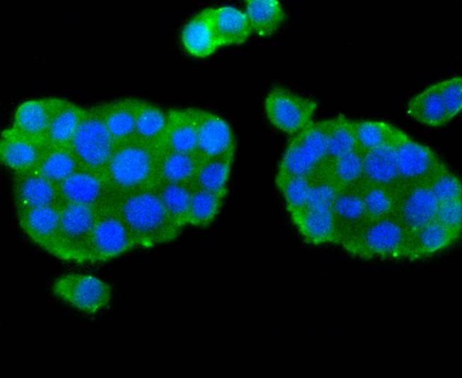 ICC staining of nNOS in PC-12 cells (green). Formalin fixed cells were permeabilized with 0.1% Triton X-100 in TBS for 10 minutes at room temperature and blocked with 1% Blocker BSA for 15 minutes at room temperature. Cells were probed with the primary antibody (SLM-52474R, 1/50) for 1 hour at room temperature, washed with PBS. Alexa Fluor®488 Goat anti-Rabbit IgG was used as the secondary antibody at 1/1,000 dilution. The nuclear counter stain is DAPI (blue).
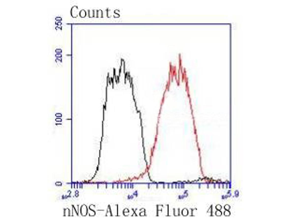 Flow cytometric analysis of nNOS was done on PC-12 cells. The cells were fixed, permeabilized and stained with the primary antibody (SLM-52474R, 1/100) (red). After incubation of the primary antibody at room temperature for an hour, the cells were stained with a Alexa Fluor 488-conjugated Goat anti-Rabbit IgG Secondary antibody at 1/1000 dilution for 30 minutes.Unlabelled sample was used as a control (cells without incubation with primary antibody; black).
Cartpieces
Totalgoods,subtotals:¥Checkout
References (0)
No References
Bought notes(bought amounts latest0)
No one bought this product
User Comment(Total0User Comment Num)
- No comment


 +86 571 56623320
 +86 18668110335

