
Rabbit Anti-beta Tubulin antibody
Beta 4 tubulin; Tubulin-beta; Tubulin beta; Beta 5 tubulin; BetaTubulin; Beta-Tubulin; dJ40E16.7; TUBB; TUBB2; TUBB2A; TUBB5; tubulin beta 2A; Tubulin beta chain; Tubulin beta-5 chain; TUBB4A; TUBB4; Tubulin 5 beta; Tubulin beta-4 chain; TBB4A_HUMAN; Tubu
View History [Clear]
Details
Product Name beta Tubulin Chinese Name 微管蛋白Recombinant rabbit monoclonal anti Alias Beta 4 tubulin; Tubulin-beta; Tubulin beta; Beta 5 tubulin; BetaTubulin; Beta-Tubulin; dJ40E16.7; TUBB; TUBB2; TUBB2A; TUBB5; tubulin beta 2A; Tubulin beta chain; Tubulin beta-5 chain; TUBB4A; TUBB4; Tubulin 5 beta; Tubulin beta-4 chain; TBB4A_HUMAN; Tubulin beta-4A chain. literatures Research Area Cell biology immunology Neurobiology Cytoskeleton Immunogen Species Rabbit Clonality Monoclonal Clone NO. 2F11 React Species (predicted: Human, Mouse, Rat, ) Applications WB=1:10000-20000 IHC-P=1:100-500 ICC=1:20-100 IF=1:20-100 (Paraffin sections need antigen repair)
not yet tested in other applications.
optimal dilutions/concentrations should be determined by the end user.Theoretical molecular weight 50kDa Cellular localization cytoplasmic Form Liquid Concentration 1mg/ml immunogen KLH conjugated synthetic peptide derived from human beta Tubulin Lsotype IgG Purification affinity purified by Protein A Buffer Solution 0.01M TBS(pH7.4) with 1% BSA, 0.03% Proclin300 and 50% Glycerol. Storage Shipped at 4℃. Store at -20 °C for one year. Avoid repeated freeze/thaw cycles. Attention This product as supplied is intended for research use only, not for use in human, therapeutic or diagnostic applications. PubMed PubMed Product Detail Microtubules are constituent parts of the mitotic apparatus, cilia, flagella, and elements of the cytoskeleton. They consist principally of 2 soluble proteins, alpha- and beta-tubulin, each of about 55,000 kDa. Antibodies against beta Tubulin are useful as loading controls for Western Blotting. However it should be noted that levels of beta Tubulin may not be stable in certain cells. For example, expression of tubulin in adipose tissue is very low (Spiegelman and Farmer, Cell, 1982, 29(1):53-60) and therefore beta Tubulin should not be used as loading control for these tissues.
SWISS:
Q13509
Gene ID:
203068
Product Picture 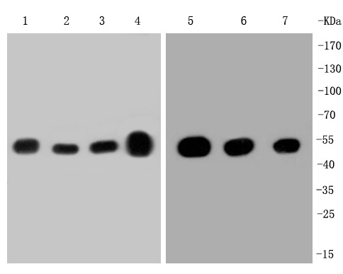 Western blot analysis of beta Tubulin on different lysates. Proteins were transferred to a PVDF membrane and blocked with 5% BSA in PBS for 1 hour at room temperature. The primary antibody (SLM-52290R, 1/500) was used in 5% BSA at room temperature for 2 hours. Goat Anti-Rabbit IgG - HRP Secondary Antibody (HA1001) at 1:200,000 dilution was used for 1 hour at room temperature. Positive control:
Lane 1: A431 cell lysate
Lane 2: MCF-7 cell lysate
Lane 3: SH-SY5Y cell lysate
Lane 4: mouse brain tissue lysate
Lane 5: Hela cell lysate
Lane 6: NIH/3T3 cell lysate
Lane 7: PC-12 cell lysate
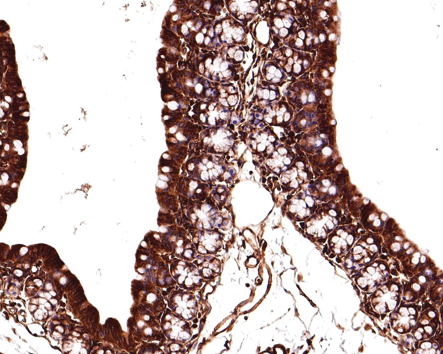 Immunohistochemical analysis of paraffin-embedded mouse large intestine tissue with Rabbit anti-beta Tubulin antibody (SLM-52290R) at 1/400 dilution. The section was pre-treated using heat mediated antigen retrieval with Tris-EDTA buffer (pH 9.0) for 20 minutes. The tissues were blocked in 1% BSA for 20 minutes at room temperature, washed with ddH2O and PBS, and then probed with the primary antibody (SLM-52290R) at 1/400 dilution for 1 hour at room temperature. The detection was performed using an HRP conjugated compact polymer system. DAB was used as the chromogen. Tissues were counterstained with hematoxylin and mounted with DPX.
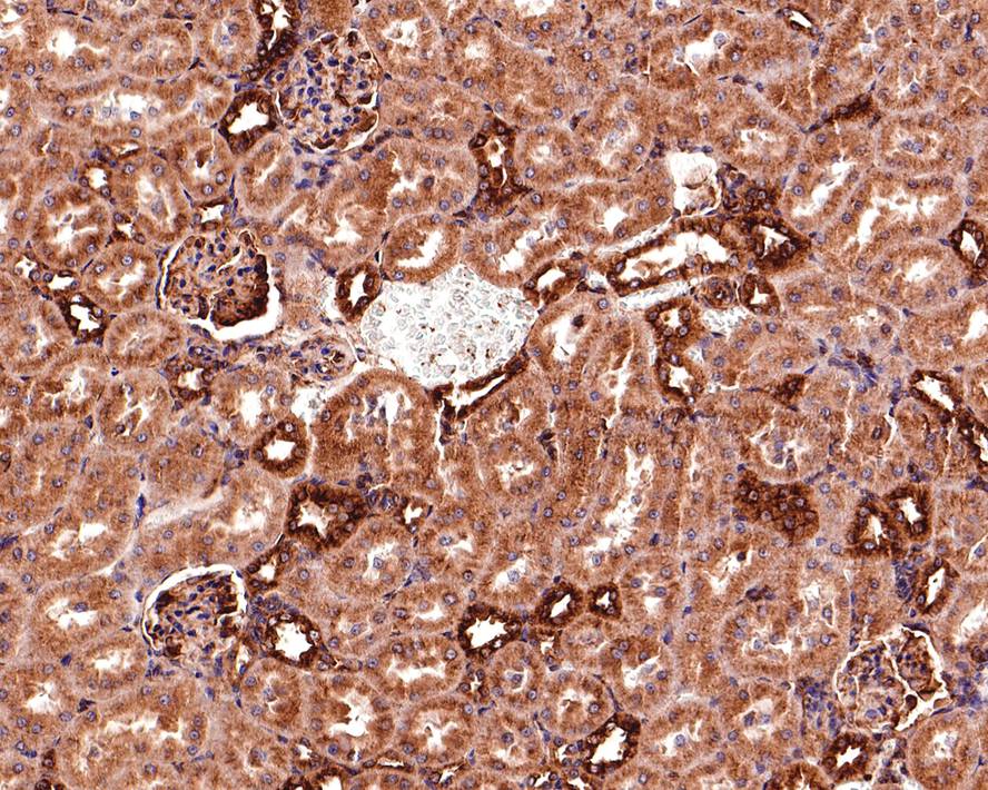 Immunohistochemical analysis of paraffin-embedded rat kidney tissue with Rabbit anti-beta Tubulin antibody (SLM-52290R) at 1/400 dilution. The section was pre-treated using heat mediated antigen retrieval with Tris-EDTA buffer (pH 9.0) for 20 minutes. The tissues were blocked in 1% BSA for 20 minutes at room temperature, washed with ddH2O and PBS, and then probed with the primary antibody (SLM-52290R) at 1/400 dilution for 1 hour at room temperature. The detection was performed using an HRP conjugated compact polymer system. DAB was used as the chromogen. Tissues were counterstained with hematoxylin and mounted with DPX.Immunohistochemical analysis of paraffin-embedded human colon carcinoma tissue with Rabbit anti-beta Tubulin antibody (SLM-52290R) at 1/400 dilution. The section was pre-treated using heat mediated antigen retrieval with Tris-EDTA buffer (pH 9.0) for 20 minutes. The tissues were blocked in 1% BSA for 20 minutes at room temperature, washed with ddH2O and PBS, and then probed with the primary antibody (SLM-52290R) at 1/400 dilution for 1 hour at room temperature. The detection was performed using an HRP conjugated compact polymer system. DAB was used as the chromogen. Tissues were counterstained with hematoxylin and mounted with DPX.
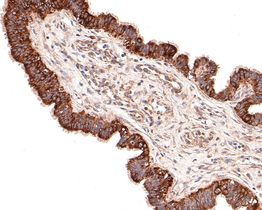 Immunohistochemical analysis of paraffin-embedded human fallopian tube tissue with Rabbit anti-beta Tubulin antibody (SLM-52290R) at 1/100 dilution. The section was pre-treated using heat mediated antigen retrieval with Tris-EDTA buffer (pH 9.0) for 20 minutes. The tissues were blocked in 1% BSA for 20 minutes at room temperature, washed with ddH2O and PBS, and then probed with the primary antibody (SLM-52290R) at 1/100 dilution for 1 hour at room temperature. The detection was performed using an HRP conjugated compact polymer system. DAB was used as the chromogen. Tissues were counterstained with hematoxylin and mounted with DPX.
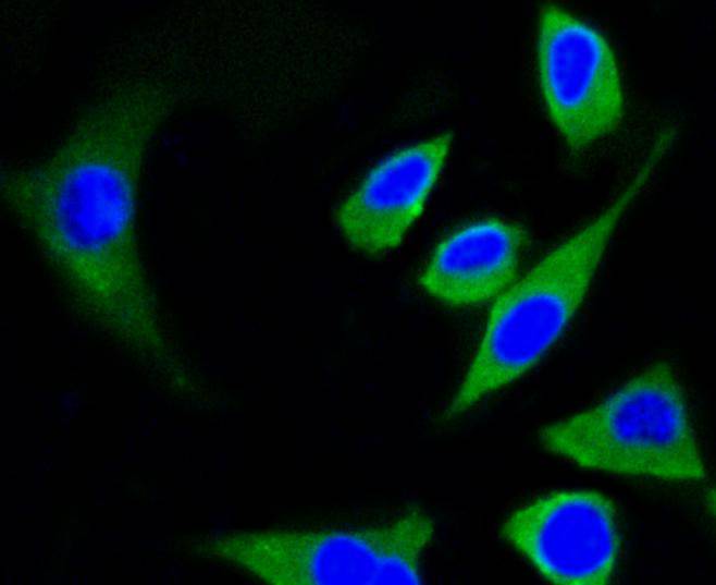 ICC staining of beta Tubulin in SH-SY5Y cells (green). Formalin fixed cells were permeabilized with 0.1% Triton X-100 in TBS for 10 minutes at room temperature and blocked with 10% negative goat serum for 15 minutes at room temperature. Cells were probed with the primary antibody (SLM-52290R, 1/50) for 1 hour at room temperature, washed with PBS. Alexa Fluor®488 conjugate-Goat anti-Rabbit IgG was used as the secondary antibody at 1/1,000 dilution. The nuclear counter stain is DAPI (blue).
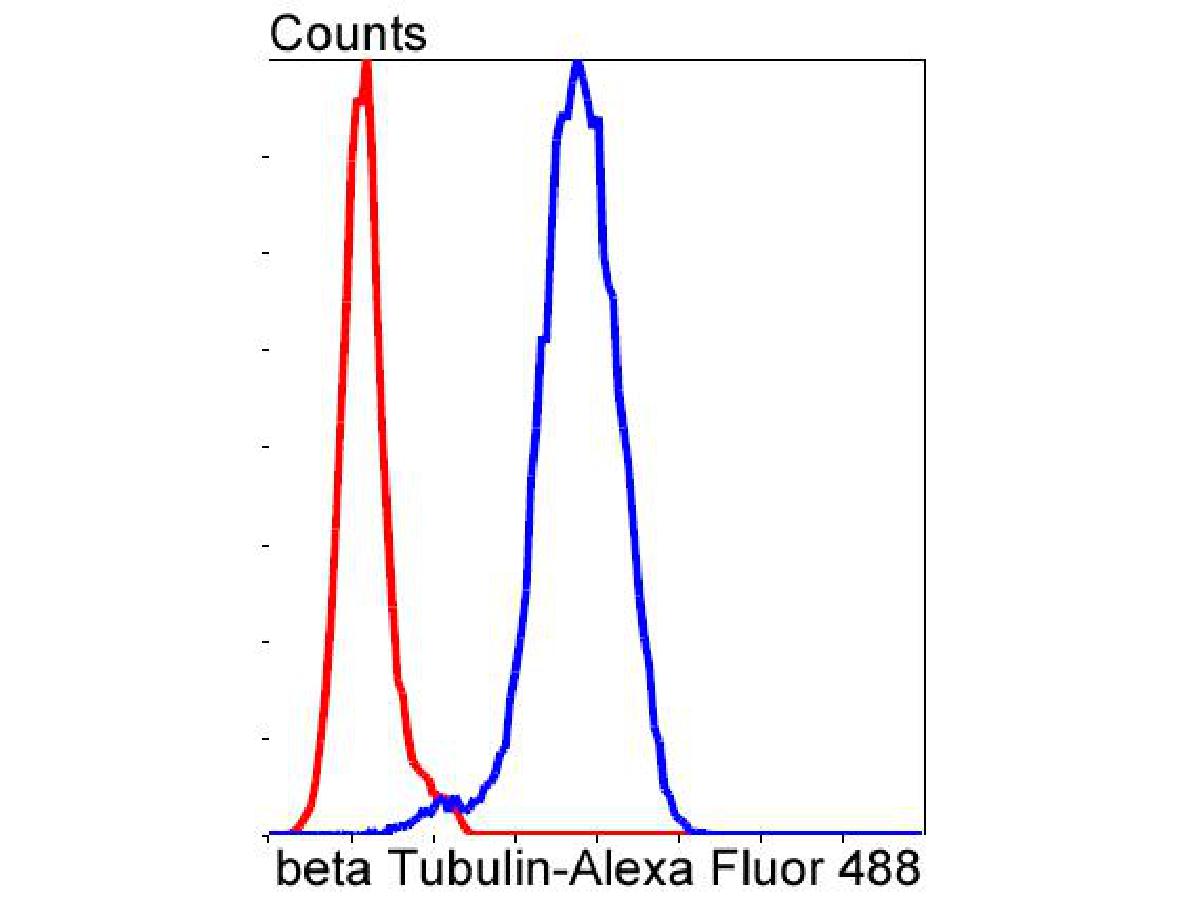 ICC staining of beta Tubulin in PC-12 cells (green). Formalin fixed cells were permeabilized with 0.1% Triton X-100 in TBS for 10 minutes at room temperature and blocked with 10% negative goat serum for 15 minutes at room temperature. Cells were probed with the primary antibody (SLM-52290R, 1/50) for 1 hour at room temperature, washed with PBS. Alexa Fluor®488 conjugate-Goat anti-Rabbit IgG was used as the secondary antibody at 1/1,000 dilution. The nuclear counter stain is DAPI (blue).
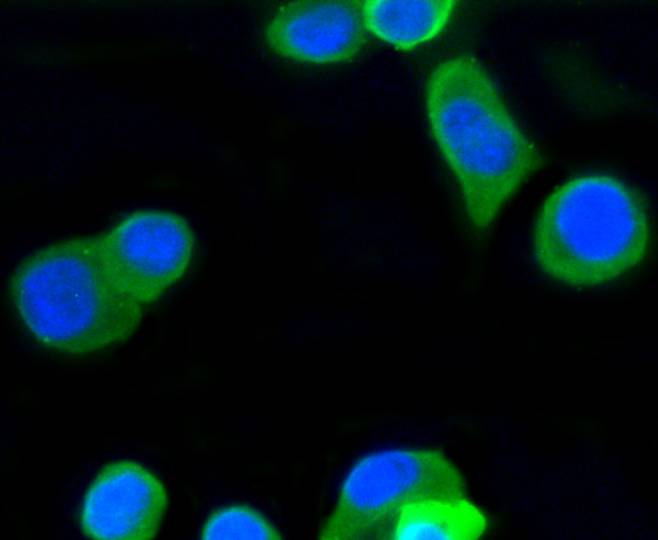 ICC staining of beta Tubulin in N2A cells (green). Formalin fixed cells were permeabilized with 0.1% Triton X-100 in TBS for 10 minutes at room temperature and blocked with 10% negative goat serum for 15 minutes at room temperature. Cells were probed with the primary antibody (SLM-52290R, 1/50) for 1 hour at room temperature, washed with PBS. Alexa Fluor®488 conjugate-Goat anti-Rabbit IgG was used as the secondary antibody at 1/1,000 dilution. The nuclear counter stain is DAPI (blue).
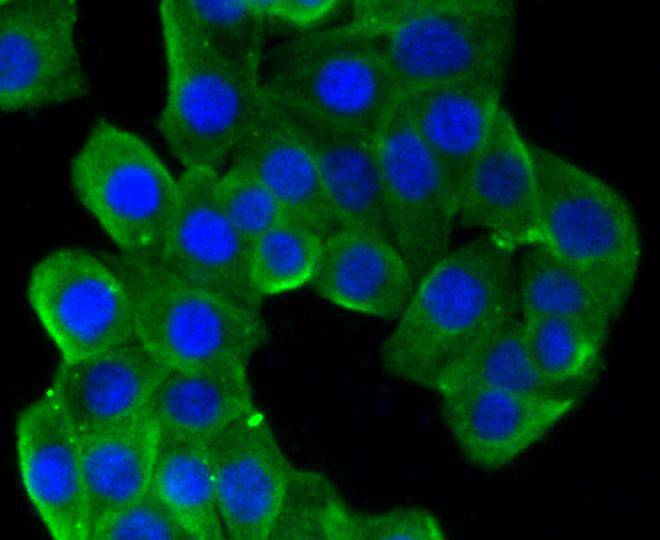 ICC staining of beta Tubulin in SH-SY5Y cells (green). Formalin fixed cells were permeabilized with 0.1% Triton X-100 in TBS for 10 minutes at room temperature and blocked with 10% negative goat serum for 15 minutes at room temperature. Cells were probed with the primary antibody (SLM-52290R, 1/50) for 1 hour at room temperature, washed with PBS. Alexa Fluor®488 conjugate-Goat anti-Rabbit IgG was used as the secondary antibody at 1/1,000 dilution. The nuclear counter stain is DAPI (blue).
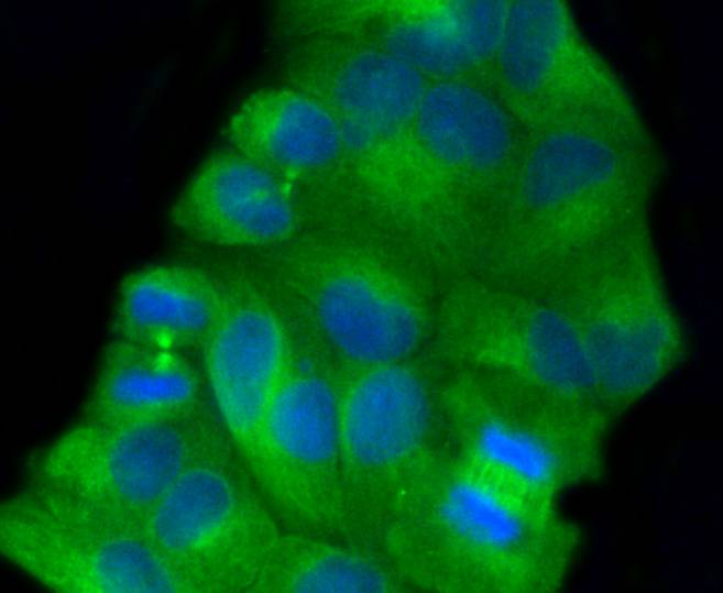 ICC staining of beta Tubulin in Hela cells (green). Formalin fixed cells were permeabilized with 0.1% Triton X-100 in TBS for 10 minutes at room temperature and blocked with 10% negative goat serum for 15 minutes at room temperature. Cells were probed with the primary antibody (SLM-52290R, 1/50) for 1 hour at room temperature, washed with PBS. Alexa Fluor®488 conjugate-Goat anti-Rabbit IgG was used as the secondary antibody at 1/1,000 dilution. The nuclear counter stain is DAPI (blue).Flow cytometric analysis of beta Tubulin was done on NIH/3T3 cells. The cells were fixed, permeabilized and stained with the primary antibody (SLM-52290R, 1/50) (blue). After incubation of the primary antibody at room temperature for an hour, the cells were stained with a Alexa Fluor®488 conjugate-Goat anti-Rabbit IgG Secondary antibody at 1/1,000 dilution for 30 minutes.Unlabelled sample was used as a control (cells without incubation with primary antibody; red).
Cartpieces
Totalgoods,subtotals:¥Checkout
References (0)
No References
Bought notes(bought amounts latest0)
No one bought this product
User Comment(Total0User Comment Num)
- No comment


 +86 571 56623320
 +86 18668110335

