
Rabbit Anti-Hsp27 antibody
Heat shock 27kDa protein; 28 kDa heat shock protein; CMT2F; DKFZp586P1322; Estrogen regulated 24 kDa protein; Estrogen-regulated 24 kDa protein; Heat shock 25kDa protein 1; Heat shock 25kDa protein 1; Heat shock 27 kDa protein; Heat shock 27kD protein 1;
View History [Clear]
Details
Product Name Hsp27 Chinese Name 热休克蛋白27Recombinant rabbit monoclonal anti Alias Heat shock 27kDa protein; 28 kDa heat shock protein; CMT2F; DKFZp586P1322; Estrogen regulated 24 kDa protein; Estrogen-regulated 24 kDa protein; Heat shock 25kDa protein 1; Heat shock 25kDa protein 1; Heat shock 27 kDa protein; Heat shock 27kD protein 1; Heat shock 27kDa protein 1; Heat shock 27kDa protein 1; Heat shock 28kDa protein 1; Heat shock 28kDa protein 1; Heat Shock Protein 27; Heat Shock Protein 27; Heat shock protein beta 1; Heat shock protein beta-1; Heat Shock Protein27; Heat Shock Protein27; HMN2B; HS.76067; Hsp 25; Hsp 25; Hsp 27; Hsp 27; Hsp 28; Hsp 28; Hsp B1; Hsp B1; Hsp25; Hsp25; HSP27; Hsp28; Hsp28; HspB1; HspB1; HSPB1_HUMAN; SRP27; Stress responsive protein 27; Stress-responsive protein 27. Research Area Tumour immunology Signal transduction Immunogen Species Rabbit Clonality Monoclonal React Species Human, (predicted: Rat, ) Applications WB=1:500-1000 IHC-P=1:100-500 Flow-Cyt=1:50 ICC=1:50-300 IF=1:100-500 (Paraffin sections need antigen repair)
not yet tested in other applications.
optimal dilutions/concentrations should be determined by the end user.Theoretical molecular weight 23kDa Cellular localization The nucleus cytoplasmic Form Liquid Concentration 1mg/ml immunogen Recombinant human Hsp27 Lsotype IgG Purification affinity purified by Protein A Buffer Solution 0.01M TBS(pH7.4) with 1% BSA, 0.03% Proclin300 and 50% Glycerol. Storage Shipped at 4℃. Store at -20 °C for one year. Avoid repeated freeze/thaw cycles. Attention This product as supplied is intended for research use only, not for use in human, therapeutic or diagnostic applications. PubMed PubMed Product Detail The protein encoded by this gene is induced by environmental stress and developmental changes. The encoded protein is involved in stress resistance and actin organization and translocates from the cytoplasm to the nucleus upon stress induction. Defects in this gene are a cause of Charcot-Marie-Tooth disease type 2F (CMT2F) and distal hereditary motor neuropathy (dHMN). [provided by RefSeq, Oct 2008]
Function:
Involved in stress resistance and actin organization.
Subunit:
Interacts with TGFB1I1. Associates with alpha- and beta-tubulin, microtubules and CRYAB. Interacts with HSPB8 and HSPBAP1.
Subcellular Location:
Cytoplasm. Nucleus. Cytoplasm, cytoskeleton, spindle. Note=Cytoplasmic in interphase cells. Colocalizes with mitotic spindles in mitotic cells. Translocates to the nucleus during heat shock and resides in sub-nuclear structures known as SC35 speckles or nuclear splicing speckles. Tissue Specificity : Detected in all tissues tested: skeletal muscle, heart, aorta, large intestine, small intestine, stomach, esophagus, bladder, adrenal gland, thyroid, pancreas, testis, adipose tissue, kidney, liver, spleen, cerebral cortex, blood serum and cerebrospinal fluid. Highest levels are found in the heart and in tissues composed of striated and smooth muscle.
Tissue Specificity:
Detected in all tissues tested: skeletal muscle, heart, aorta, large intestine, small intestine, stomach, esophagus, bladder, adrenal gland, thyroid, pancreas, testis, adipose tissue, kidney, liver, spleen, cerebral cortex, blood serum and cerebrospinal fluid. Highest levels are found in the heart and in tissues composed of striated and smooth muscle.
Post-translational modifications:
Phosphorylated in MCF-7 cells on exposure to protein kinase C activators and heat shock.
Phosphorylation by MAPKAPK2 and MAPKAPK3 in response to stress leads to dissociate HSP27/HSPB1 from large small heat-shock protein (sHsps) oligomers and impair its chaperone activity and ability to protect against oxidative stress effectively. Phosphorylation by MAPKAPK5 in response to PKA stimulation induces F-actin rearrangement.
DISEASE:
Defects in HSPB1 are the cause of Charcot-Marie-Tooth disease type 2F (CMT2F) [MIM:606595]. CMT2F is a form of Charcot-Marie-Tooth disease, the most common inherited disorder of the peripheral nervous system. Charcot-Marie-Tooth disease is classified in two main groups on the basis of electrophysiologic properties and histopathology: primary peripheral demyelinating neuropathy or CMT1, and primary peripheral axonal neuropathy or CMT2. Neuropathies of the CMT2 group are characterized by signs of axonal regeneration in the absence of obvious myelin alterations, normal or slightly reduced nerve conduction velocities, and progressive distal muscle weakness and atrophy. Nerve conduction velocities are normal or slightly reduced. CMT2F onset is between 15 and 25 years with muscle weakness and atrophy usually beginning in feet and legs (peroneal distribution). Upper limb involvement occurs later. CMT2F inheritance is autosomal dominant.
Defects in HSPB1 are a cause of distal hereditary motor neuronopathy type 2B (HMN2B) [MIM:608634]. Distal hereditary motor neuronopathies constitute a heterogeneous group of neuromuscular disorders caused by selective impairment of motor neurons in the anterior horn of the spinal cord, without sensory deficit in the posterior horn. The overall clinical picture consists of a classical distal muscular atrophy syndrome in the legs without clinical sensory loss. The disease starts with weakness and wasting of distal muscles of the anterior tibial and peroneal compartments of the legs. Later on, weakness and atrophy may expand to the proximal muscles of the lower limbs and/or to the distal upper limbs.
Similarity:
Belongs to the small heat shock protein (HSP20) family.
SWISS:
P04792
Gene ID:
3315
Database links:Entrez Gene: 3315 Human
Entrez Gene: 15507 Mouse
Omim: 602195 Human
SwissProt: P04792 Human
SwissProt: P14602 Mouse
Unigene: 3849 Dog
Unigene: 520973 Human
Unigene: 13849 Mouse
Unigene: 3841 Rat
信号传导(Signaling Intermediates)
适用组织:石蜡切片 Cellular localization:胞浆和部分胞核
HSPs是细胞受应激原刺激后诱导产生的一组应激蛋白,与Tumour发生、 增殖及分化有关。按其分子量不同可分为3种类型,每组的HSPs的分布及功能有所不同。 热休克蛋白27是人体中最常见而又最小的热休克蛋白。HSP27和其它HSPs可能与Tumour耐药和Tumour的分化程度以及病人的预后有关。Product Picture 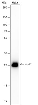 Blocking buffer: 5% NFDM/TBST
Primary Ab dilution: 1:1000
Primary Ab incubation condition: 2 hours at room temperature
Secondary Ab: Goat Anti-Rabbit IgG H&L (HRP)
Lysate: HeLa
Protein loading quantity: 20 μg
Exposure time: 60 s
Predicted MW: 23 kDa
Observed MW: 27 kDa
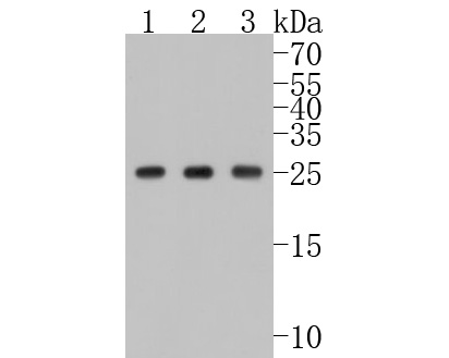 Western blot analysis of Hsp27 on different lysates. Proteins were transferred to a PVDF membrane and blocked with 5% BSA in PBS for 1 hour at room temperature. The primary antibody (SLM-52757R, 1/500) was used in 5% BSA at room temperature for 2 hours. Goat Anti-Rabbit IgG - HRP Secondary Antibody (HA1001) at 1:5,000 dilution was used for 1 hour at room temperature.
Positive control:
Lane 1: Hela cell lysate
Lane 2: A549 cell lysate
Lane 3: Jurkat cell lysate
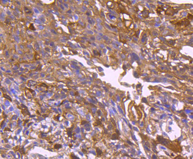 Immunohistochemical analysis of paraffin-embedded human colon carcinoma tissue using anti-Hsp27 antibody. The section was pre-treated using heat mediated antigen retrieval with Tris-EDTA buffer (pH 8.0-8.4) for 20 minutes.The tissues were blocked in 5% BSA for 30 minutes at room temperature, washed with ddH2O and PBS, and then probed with the primary antibody (SLM-52757R, 1/50) for 30 minutes at room temperature. The detection was performed using an HRP conjugated compact polymer system. DAB was used as the chromogen. Tissues were counterstained with hematoxylin and mounted with DPX.
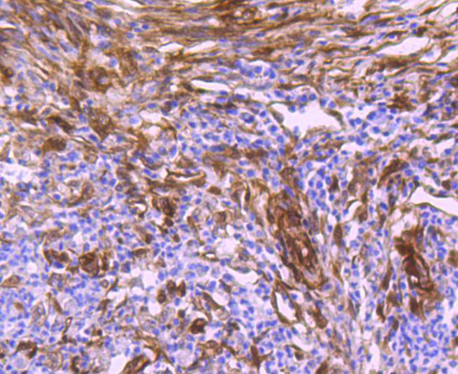 Immunohistochemical analysis of paraffin-embedded human colon carcinoma tissue using anti-Hsp27 antibody. The section was pre-treated using heat mediated antigen retrieval with Tris-EDTA buffer (pH 8.0-8.4) for 20 minutes.The tissues were blocked in 5% BSA for 30 minutes at room temperature, washed with ddH2O and PBS, and then probed with the primary antibody (SLM-52757R, 1/50) for 30 minutes at room temperature. The detection was performed using an HRP conjugated compact polymer system. DAB was used as the chromogen. Tissues were counterstained with hematoxylin and mounted with DPX.
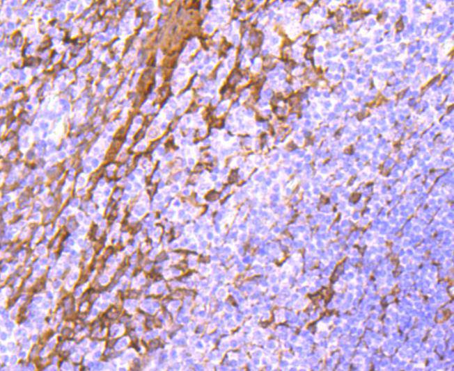 Immunohistochemical analysis of paraffin-embedded human tonsil tissue using anti-Hsp27 antibody. The section was pre-treated using heat mediated antigen retrieval with Tris-EDTA buffer (pH 8.0-8.4) for 20 minutes.The tissues were blocked in 5% BSA for 30 minutes at room temperature, washed with ddH2O and PBS, and then probed with the primary antibody (SLM-52757R, 1/50) for 30 minutes at room temperature. The detection was performed using an HRP conjugated compact polymer system. DAB was used as the chromogen. Tissues were counterstained with hematoxylin and mounted with DPX.
 Immunohistochemical analysis of paraffin-embedded human breast carcinoma tissue using anti-Hsp27 antibody. The section was pre-treated using heat mediated antigen retrieval with Tris-EDTA buffer (pH 8.0-8.4) for 20 minutes.The tissues were blocked in 5% BSA for 30 minutes at room temperature, washed with ddH2O and PBS, and then probed with the primary antibody (SLM-52757R, 1/50) for 30 minutes at room temperature. The detection was performed using an HRP conjugated compact polymer system. DAB was used as the chromogen. Tissues were counterstained with hematoxylin and mounted with DPX.
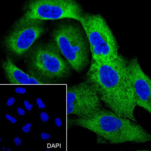 Cell line: A549
Fixative: 4% Paraformaldehyde
Permeabilization: 0.1% TritonX-100
Primary Ab dilution: 1:300
Primary Ab incubation condition: 4°C overnight
Secondary Ab: Goat Anti-Rabbit IgG
Nuclear counter stain: DAPI (Blue)
Comment: Color green is the positive signal for SLM-52757R
 ICC staining of Hsp27 in HepG2 cells (green). Formalin fixed cells were permeabilized with 0.1% Triton X-100 in TBS for 10 minutes at room temperature and blocked with 1% Blocker BSA for 15 minutes at room temperature. Cells were probed with the primary antibody (SLM-52757R, 1/50) for 1 hour at room temperature, washed with PBS. Alexa Fluor®488 Goat anti-Rabbit IgG was used as the secondary antibody at 1/1,000 dilution. The nuclear counter stain is DAPI (blue).
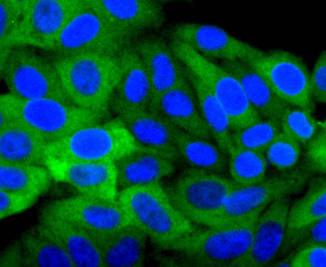
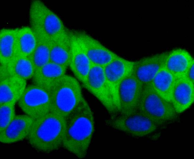 ICC staining of Hsp27 in Hela cells (green). Formalin fixed cells were permeabilized with 0.1% Triton X-100 in TBS for 10 minutes at room temperature and blocked with 1% Blocker BSA for 15 minutes at room temperature. Cells were probed with the primary antibody (SLM-52757R, 1/50) for 1 hour at room temperature, washed with PBS. Alexa Fluor®488 Goat anti-Rabbit IgG was used as the secondary antibody at 1/1,000 dilution. The nuclear counter stain is DAPI (blue).
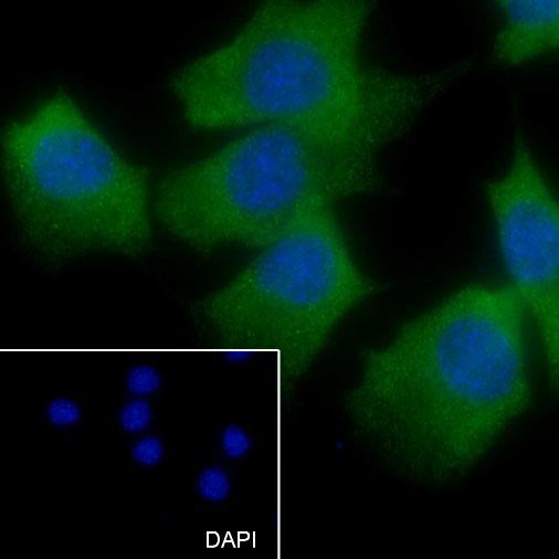 Cell line: C2C12
Fixative: 4% Paraformaldehyde
Permeabilization: 0.1% TritonX-100
Primary Ab dilution: 1:50
Primary Ab incubation condition: 4°C overnight
Secondary Ab: Goat Anti-Rabbit IgG
Nuclear counter stain: DAPI (Blue)
Comment: Color green is the positive signal for SLM-52757R
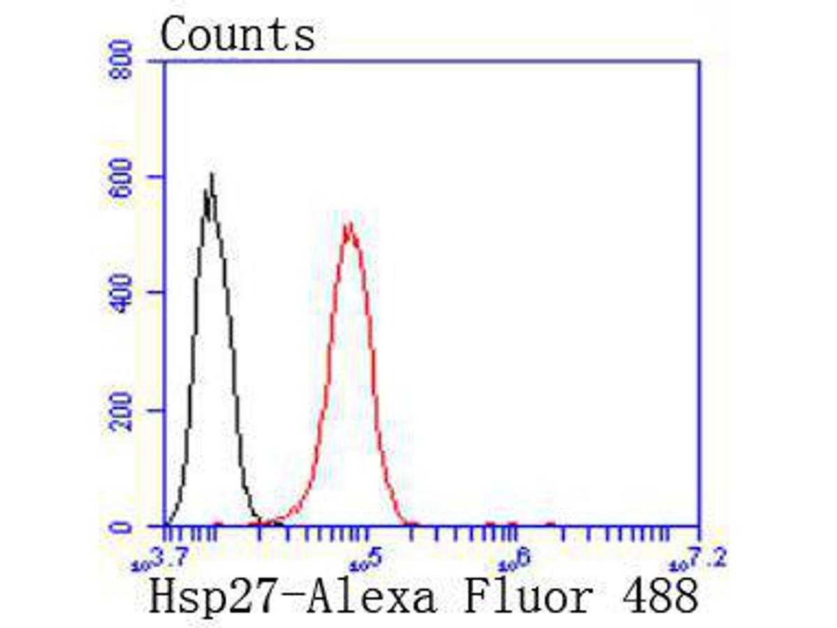 Flow cytometric analysis of Hsp27 was done on Hela cells. The cells were fixed, permeabilized and stained with the primary antibody (SLM-52757R, 1/50) (red). After incubation of the primary antibody at room temperature for an hour, the cells were stained with a Alexa Fluor 488-conjugated Goat anti-Rabbit IgG Secondary antibody at 1/1000 dilution for 30 minutes.Unlabelled sample was used as a control (cells without incubation with primary antibody; black).
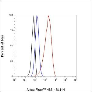 Cell line: HeLa
Fixative: 4% Paraformaldehyde
Permeabilization: 90% Methanol
Primary Ab dilution: 1:50
Secondary Ab: Goat anti Rabbit IgG
Unlabelled control: The cell without incubation with primary antibody and secondary antibody (Black line).
Isotype control: Rabbit monoclonal IgG (Blue line).
Comment: Line red is the positive signal for SLM-52757R
Cartpieces
Totalgoods,subtotals:¥Checkout
References (0)
No References
Bought notes(bought amounts latest0)
No one bought this product
User Comment(Total0User Comment Num)
- No comment


 +86 571 56623320
 +86 18668110335

