
Rabbit Anti-Lamin B1 antibody
lamin B1; LMB1; LMN; LMN2; LMNB 1; LMNB; LMNB1; MGC111419; LMNB1_HUMAN; Lamin-B1.
View History [Clear]
Details
Product Name [KO validated anti] Lamin B1 Chinese Name 核纤层蛋白B抗体(The nucleus膜Maker) Alias lamin B1; LMB1; LMN; LMN2; LMNB 1; LMNB; LMNB1; MGC111419; LMNB1_HUMAN; Lamin-B1. Research Area Cell biology Chromatin and nuclear signals Signal transduction Apoptosis Cell type markers Immunogen Species Rabbit Clonality Polyclonal React Species Human, Mouse, Rat, Applications WB=1:500-2000 IHC-P=1:50-200 IHC-F=1:50-200 IF=1:50-200 (Paraffin sections need antigen repair)
not yet tested in other applications.
optimal dilutions/concentrations should be determined by the end user.Theoretical molecular weight 66kDa Cellular localization The nucleus Form Liquid Concentration 1mg/ml immunogen Recombinant human Lamin B1: 397-586/586 Lsotype IgG Purification affinity purified by Protein A Buffer Solution 0.01M TBS(pH7.4) with 1% BSA, 0.03% Proclin300 and 50% Glycerol. Storage Store at -20 °C for one year. Avoid repeated freeze/thaw cycles. The lyophilized antibody is stable at room temperature for at least one month and for greater than a year when kept at -20°C. When reconstituted in sterile pH 7.4 0.01M PBS or diluent of antibody the antibody is stable for at least two weeks at 2-4 °C. Attention This product as supplied is intended for research use only, not for use in human, therapeutic or diagnostic applications. PubMed PubMed Product Detail The nuclear lamina consists of a two-dimensional matrix of proteins located next to the inner nuclear membrane. The lamin family of proteins make up the matrix and are highly conserved in evolution. During mitosis, the lamina matrix is reversibly disassembled as the lamin proteins are phosphorylated. Lamin proteins are thought to be involved in nuclear stability, chromatin structure and gene expression. Vertebrate lamins consist of two types, A and B. This gene encodes one of the two B type proteins, B1. Alternative splicing results in transcript variants and a duplication of this gene is associated with autosomal dominant adult-onset leukodystrophy (ADLD). [provided by RefSeq, Oct 2010].
Function:
Lamins are components of the nuclear lamina, a fibrous layer on the nucleoplasmic side of the inner nuclear membrane, which is thought to provide a framework for the nuclear envelope and may also interact with chromatin.
Subunit:
Homodimer. Interacts with lamin-associated polypeptides IA, IB and 2.
Subcellular Location:
Nucleus inner membrane; Lipid-anchor; Nucleoplasmic side.
Post-translational modifications:
B-type lamins undergo a series of modifications, such as farnesylation and phosphorylation. Increased phosphorylation of the lamins occurs before envelope disintegration and probably plays a role in regulating lamin associations.
DISEASE:
Defects in LMNB1 are the cause of leukodystrophy demyelinating autosomal dominant adult-onset (ADLD) [MIM:169500]. ADLD is a slowly progressive and fatal demyelinating leukodystrophy, presenting in the fourth or fifth decade of life. Clinically characterized by early autonomic abnormalities, pyramidal and cerebellar dysfunction, and symmetric demyelination of the CNS. It differs from multiple sclerosis and other demyelinating disorders in that neuropathology shows preservation of oligodendroglia in the presence of subtotal demyelination and lack of astrogliosis.
Similarity:
Belongs to the intermediate filament family.
SWISS:
P20700
Gene ID:
4001
Database links:Entrez Gene: 396223 Chicken
Entrez Gene: 4001 Human
Entrez Gene: 16906 Mouse
Omim: 150340 Human
SwissProt: P14731 Chicken
SwissProt: P20700 Human
SwissProt: P14733 Mouse
Unigene: 89497 Human
Unigene: 4105 Mouse
Unigene: 11362 Rat
核膜Maker(Nuclear Envelope Marker) 核纤层蛋白(Lamin) 是紧贴核内膜的一层厚度为20~50nm的纤维蛋白层或纤维网络。核纤层与细胞质骨架、核骨架连成一个整体,一般认为核纤层将核被膜和染色质提供了结构支架。有学者研究认为:lamin蛋白与Apoptosis及衰老有关联,它包括:核纤层蛋白A、核纤层蛋白B、核纤层蛋白C几个不同亚型的蛋白。Product Picture 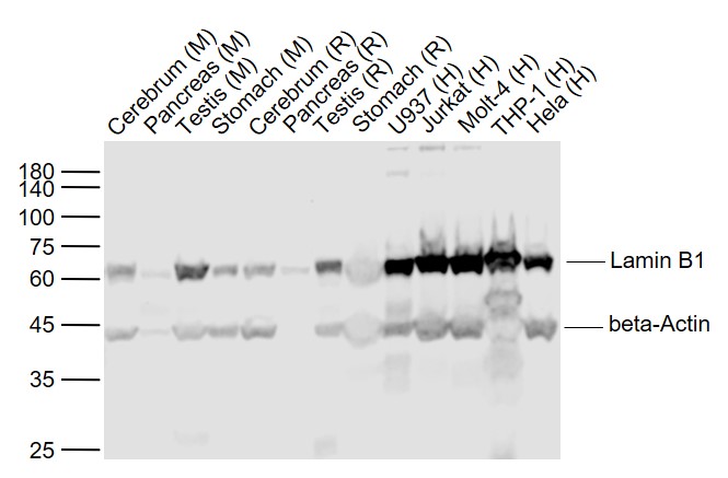 Sample:
Lane 1: Cerebrum (Mouse) Lysate at 40 ug
Lane 2: Pancreas (Mouse) Lysate at 40 ug
Lane 3: Testis (Mouse) Lysate at 40 ug
Lane 4: Stomach (Mouse) Lysate at 40 ug
Lane 5: Cerebrum (Rat) Lysate at 40 ug
Lane 7: Testis (Rat) Lysate at 40 ug
Lane 8: Stomach (Rat) Lysate at 40 ug
Lane 9: U937 (Human) Cell Lysate at 30 ug
Lane 10: Jurkat (Human) Cell Lysate at 30 ug
Lane 11: Molt-4 (Human) Cell Lysate at 30 ug
Lane 12: THP-1 (Human) Cell Lysate at 30 ug
Lane 13: Hela (Human) Cell Lysate at 30 ug
Primary:
Anti- Lamin B1 (SL55118R) at 1/1000 dilution
Anti-beta-Actin (SL0061R) at 1/2000 dilution
Secondary: IRDye800CW Goat Anti-Rabbit IgG at 1/20000 dilution
Predicted band size: 67-70 kD
Observed band size: 65 kD
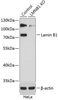 Sample:
Lane 1: Hela (Human) Cell Lysate at 25 ug
Lane 2: Lamin B1 knockout (KO) Hela (Human) Cell Lysate at 25 ug
Primary: Anti-Lamin B1 (SL55118R) at 1/1000 dilution
Secondary: HRP Goat Anti-Rabbit IgG (H+L) at 1:10000 dilution
Predicted band size: 67-70 kD
Observed band size: 72 kD
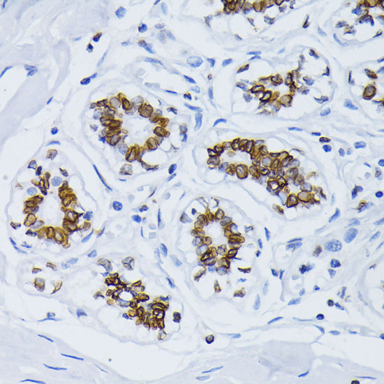 Paraformaldehyde-fixed, paraffin embedded (human breast); Antigen retrieval by boiling in sodium citrate buffer (pH6.0) for 15min; Block endogenous peroxidase by 3% hydrogen peroxide for 20 minutes; Blocking buffer (normal goat serum) at 37°C for 30min; Antibody incubation with (Lamin B1) Polyclonal Antibody, Unconjugated (SL55118R) at 1:100 overnight at 4°C, followed by operating according to SP Kit(Rabbit) (sp-0023) instructionsand DAB staining.
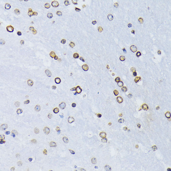 Paraformaldehyde-fixed, paraffin embedded (mouse brain); Antigen retrieval by boiling in sodium citrate buffer (pH6.0) for 15min; Block endogenous peroxidase by 3% hydrogen peroxide for 20 minutes; Blocking buffer (normal goat serum) at 37°C for 30min; Antibody incubation with (Lamin B1) Polyclonal Antibody, Unconjugated (SL55118R) at 1:100 overnight at 4°C, followed by operating according to SP Kit(Rabbit) (sp-0023) instructionsand DAB staining.
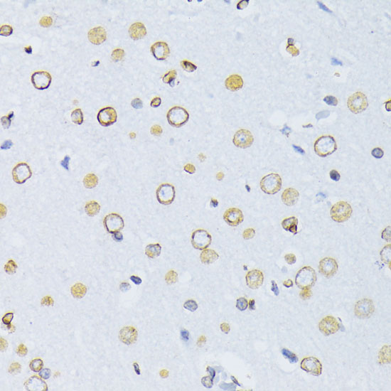 Paraformaldehyde-fixed, paraffin embedded (rat brain); Antigen retrieval by boiling in sodium citrate buffer (pH6.0) for 15min; Block endogenous peroxidase by 3% hydrogen peroxide for 20 minutes; Blocking buffer (normal goat serum) at 37°C for 30min; Antibody incubation with (Lamin B1) Polyclonal Antibody, Unconjugated (SL55118R) at 1:100 overnight at 4°C, followed by operating according to SP Kit(Rabbit) (sp-0023) instructionsand DAB staining.
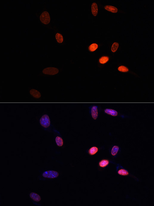 NIH/3T3 cell; 4% Paraformaldehyde-fixed; Triton X-100 at room temperature for 20 min; Blocking buffer (normal goat serum, C-0005) at 37°C for 20 min; Antibody incubation with (KO Validated)Lamin B1 polyclonal Antibody, Unconjugated (SL55118R) 1:100, 90 minutes at 37°C; followed by a conjugated Goat Anti-Rabbit IgG antibody at 37°C for 90 minutes, DAPI (blue, C02-04002) was used to stain the cell nuclei.
Cartpieces
Totalgoods,subtotals:¥Checkout
References (0)
No References
Bought notes(bought amounts latest0)
No one bought this product
User Comment(Total0User Comment Num)
- No comment


 +86 571 56623320
 +86 18668110335

