
Rabbit Anti-HDAC1 antibody
HD 1; HD1; HDAC 1; hdac1: histone deacetylase 1; Histone deacetylase 1 (HD1); Histone deacetylase 1; RPD3 (reduced potassium dependency yeast homolog) like 1; RPD3 (reduced potassium dependency); RPD3; RPD3L1; DKFZp686H12203; GON 10; HDAC1_HUMAN.
View History [Clear]
Details
Product Name [KO validated anti] HDAC1 Chinese Name 组蛋白去乙酰化酶1抗体 Alias HD 1; HD1; HDAC 1; hdac1: histone deacetylase 1; Histone deacetylase 1 (HD1); Histone deacetylase 1; RPD3 (reduced potassium dependency yeast homolog) like 1; RPD3 (reduced potassium dependency); RPD3; RPD3L1; DKFZp686H12203; GON 10; HDAC1_HUMAN. Research Area Tumour immunology Signal transduction Apoptosis transcriptional regulatory factor Immunogen Species Rabbit Clonality Polyclonal React Species Human, Rat, (predicted: Mouse, ) Applications WB=1:500-2000 IP=1:50-100 IHC-P=1:100-200 IHC-F=1:100-200 ICC=1:100 IF=1:50-100 (Paraffin sections need antigen repair)
not yet tested in other applications.
optimal dilutions/concentrations should be determined by the end user.Theoretical molecular weight 55kDa Cellular localization The nucleus Form Liquid Concentration 1mg/ml immunogen Recombinant human HDAC1 : 393-482/482 Lsotype IgG Purification affinity purified by Protein A Buffer Solution 0.01M TBS(pH7.4) with 1% BSA, 0.03% Proclin300 and 50% Glycerol. Storage Store at -20 °C for one year. Avoid repeated freeze/thaw cycles. The lyophilized antibody is stable at room temperature for at least one month and for greater than a year when kept at -20°C. When reconstituted in sterile pH 7.4 0.01M PBS or diluent of antibody the antibody is stable for at least two weeks at 2-4 °C. Attention This product as supplied is intended for research use only, not for use in human, therapeutic or diagnostic applications. PubMed PubMed Product Detail Histone acetylation and deacetylation, catalyzed by multisubunit complexes, play a key role in the regulation of eukaryotic gene expression. The protein encoded by this gene belongs to the histone deacetylase/acuc/apha family and is a component of the histone deacetylase complex. It also interacts with retinoblastoma tumor-suppressor protein and this complex is a key element in the control of cell proliferation and differentiation. Together with metastasis-associated protein-2, it deacetylates p53 and modulates its effect on cell growth and apoptosis. [provided by RefSeq, Jul 2008]
Function:
Responsible for the deacetylation of lysine residues on the N-terminal part of the core histones (H2A, H2B, H3 and H4). Histone deacetylation gives a tag for epigenetic repression and plays an important role in transcriptional regulation, cell cycle progression and developmental events. Histone deacetylases act via the formation of large multiprotein complexes. Deacetylates SP proteins, SP1 and SP3, and regulates their function. Component of the BRG1-RB1-HDAC1 complex, which negatively regulates the CREST-mediated transcription in resting neurons. Upon calcium stimulation, HDAC1 is released from the complex and CREBBP is recruited, which facilitates transcriptional activation. Deacetylates TSHZ3 and regulates its transcriptional repressor activity. Deacetylates 'Lys-310' in RELA and thereby inhibits the transcriptional activity of NF-kappa-B. Component a RCOR/GFI/KDM1A/HDAC complex that suppresses, via histone deacetylase (HDAC) recruitment, a number of genes implicated in multilineage blood cell development.
Subcellular Location:
Nucleus.
Tissue Specificity:
Ubiquitous, with higher levels in heart, pancreas and testis, and lower levels in kidney and brain.
Post-translational modifications:
Sumoylated on Lys-444 and Lys-476; which promotes enzymatic activity. Desumoylated by SENP1.
Phosphorylation on Ser-421 and Ser-423 promotes enzymatic activity and interactions with NuRD and SIN3 complexes. Phosphorylated by CDK5.
Ubiquitinated by CHFR, leading to its degradation by the proteasome. Ubiquitinated by KCTD11, leading to proteasomal degradation.
Similarity:
Belongs to the histone deacetylase family. HD type 1 subfamily.
SWISS:
Q13547
Gene ID:
3065
Database links:
Entrez Gene: 3065 Human
Entrez Gene: 433759 Mouse
Omim: 601241 Human
SwissProt: Q13547 Human
SwissProt: O09106 Mouse
Unigene: 88556 Human
Unigene: 202504 Mouse
Unigene: 391033 Mouse
Unigene: 1863 Rat
Product Picture 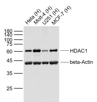 Sample:
Lane 1: Hela (Human) Cell Lysate at 30 ug
Lane 2: Molt-4 (Human) Cell Lysate at 30 ug
Lane 3: U251 (Human) Cell Lysate at 30 ug
Lane 4: MCF-7 (Human) Cell Lysate at 30 ug
Primary:
Anti- HDAC1 (SL55093R) at 1/1000 dilution
Anti- beta-Actin (SL0061R) at 1/2000 dilution
Secondary: IRDye800CW Goat Anti-Rabbit IgG at 1/20000 dilution
Predicted band size: 62 kD
Observed band size: 60 kD
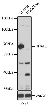 Sample:
Lane 1: 293T (Human) Cell Lysate at 25 ug
Lane 2: HDAC1 knockout (KO) 293T (Human) Cell Lysate at 25 ug
Primary: Anti-HDAC1 (SL55093R) at 1/1000 dilution
Secondary: HRP Goat Anti-Rabbit IgG (H+L) at 1:10000 dilution
Predicted band size: 62 kD
Observed band size: 65 kD
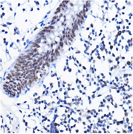 Paraformaldehyde-fixed, paraffin embedded (human colon); Antigen retrieval by boiling in sodium citrate buffer (pH6.0) for 15min; Block endogenous peroxidase by 3% hydrogen peroxide for 20 minutes; Blocking buffer (normal goat serum) at 37°C for 30min; Antibody incubation with (HDAC1) Polyclonal Antibody, Unconjugated (SL55093R) at 1:50 overnight at 4°C, followed by operating according to SP Kit(Rabbit) (sp-0023) instructionsand DAB staining.
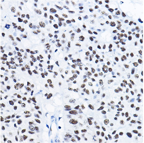 Paraformaldehyde-fixed, paraffin embedded (human lung cancer); Antigen retrieval by boiling in sodium citrate buffer (pH6.0) for 15min; Block endogenous peroxidase by 3% hydrogen peroxide for 20 minutes; Blocking buffer (normal goat serum) at 37°C for 30min; Antibody incubation with (HDAC1) Polyclonal Antibody, Unconjugated (SL55093R) at 1:50 overnight at 4°C, followed by operating according to SP Kit(Rabbit) (sp-0023) instructionsand DAB staining.
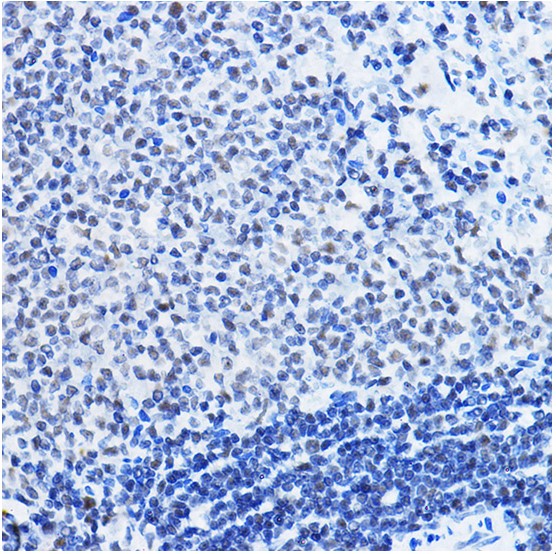 Paraformaldehyde-fixed, paraffin embedded (rat spleen); Antigen retrieval by boiling in sodium citrate buffer (pH6.0) for 15min; Block endogenous peroxidase by 3% hydrogen peroxide for 20 minutes; Blocking buffer (normal goat serum) at 37°C for 30min; Antibody incubation with (HDAC1) Polyclonal Antibody, Unconjugated (SL55093R) at 1:50 overnight at 4°C, followed by operating according to SP Kit(Rabbit) (sp-0023) instructionsand DAB staining.
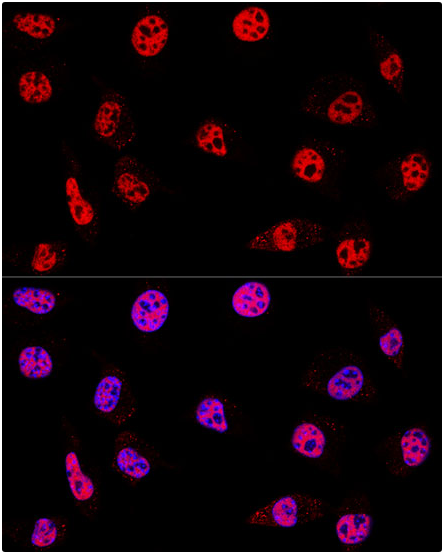 L929 cell; 4% Paraformaldehyde-fixed; Triton X-100 at room temperature for 20 min; Blocking buffer (normal goat serum, C-0005) at 37°C for 20 min; Antibody incubation with (KO Validated)HDAC1 polyclonal Antibody, Unconjugated (SL55093R) 1:200, 90 minutes at 37°C; followed by a conjugated Goat Anti-Rabbit IgG antibody at 37°C for 90 minutes, DAPI (blue, C02-04002) was used to stain the cell nuclei.
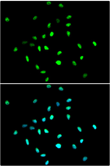 A549 cell; 4% Paraformaldehyde-fixed; Triton X-100 at room temperature for 20 min; Blocking buffer (normal goat serum, C-0005) at 37°C for 20 min; Antibody incubation with (KO Validated)HDAC1 polyclonal Antibody, Unconjugated (SL55093R) 1:100, 90 minutes at 37°C; followed by a conjugated Goat Anti-Rabbit IgG antibody at 37°C for 90 minutes, DAPI (blue, C02-04002) was used to stain the cell nuclei.
Cartpieces
Totalgoods,subtotals:¥Checkout
References (0)
No References
Bought notes(bought amounts latest0)
No one bought this product
User Comment(Total0User Comment Num)
- No comment


 +86 571 56623320
 +86 18668110335

