
Rabbit Anti-Vimentin antibody
VIM; FLJ36605; OTTHUMP00000019224; VIM; VIME_HUMAN; Vimentin.
View History [Clear]
Details
Product Name [KO validated anti] Vimentin Chinese Name 波形蛋白抗体 Alias VIM; FLJ36605; OTTHUMP00000019224; VIM; VIME_HUMAN; Vimentin. literatures Research Area Tumour Cell biology immunology Signal transduction Stem cells Cytoskeleton TumourCell biologyMaker Immunogen Species Rabbit Clonality Polyclonal React Species Human, Mouse, Rabbit, (predicted: Rat, Dog, Pig, Cow, Horse, ) Applications WB=1:500-2000 ELISA=1:5000-10000 IHC-F=1:100-500 Flow-Cyt=1μg/Test ICC=1:100 IF=1:100-500 (Paraffin sections need antigen repair)
not yet tested in other applications.
optimal dilutions/concentrations should be determined by the end user.Theoretical molecular weight 51kDa Cellular localization cytoplasmic Form Liquid Concentration 1mg/ml immunogen KLH conjugated synthetic peptide derived from human Vimentin: 371-466/466 Lsotype IgG Purification affinity purified by Protein A Buffer Solution 0.01M TBS(pH7.4) with 1% BSA, 0.03% Proclin300 and 50% Glycerol. Storage Shipped at 4℃. Store at -20 °C for one year. Avoid repeated freeze/thaw cycles. Attention This product as supplied is intended for research use only, not for use in human, therapeutic or diagnostic applications. PubMed PubMed Product Detail This gene encodes a member of the intermediate filament family. Intermediate filamentents, along with microtubules and actin microfilaments, make up the cytoskeleton. The protein encoded by this gene is responsible for maintaining cell shape, integrity of the cytoplasm, and stabilizing cytoskeletal interactions. It is also involved in the immune response, and controls the transport of low-density lipoprotein (LDL)-derived cholesterol from a lysosome to the site of esterification. It functions as an organizer of a number of critical proteins involved in attachment, migration, and cell signaling. Mutations in this gene causes a dominant, pulverulent cataract.[provided by RefSeq, Jun 2009]
Function:
Vimentins are class-III intermediate filaments found in various non-epithelial cells, especially mesenchymal cells. Vimentin is attached to the nucleus, endoplasmic reticulum, and mitochondria, either laterally or terminally. Involved with LARP6 in the stabilization of type I collagen mRNAs for CO1A1 and CO1A2. Subunit : Homopolymer assembled from elementary dimers. Interacts with HCV core protein. Interacts with LGSN and SYNM. Interacts (via rod region) with PLEC (via CH 1 domain) (By similarity). Interacts with SLC6A4. Interacts with STK33. Interacts with LARP6. Interacts with RAB8B (By similarity).
Subcellular Location:
Cytoplasm.
Tissue Specificity:
Highly expressed in fibroblasts, some expression in T- and B-lymphocytes, and little or no expression in Burkitt's lymphoma cell lines. Expressed in many hormone-independent mammary carcinoma cell lines.
Post-translational modifications:
Filament disassembly during mitosis is promoted by phosphorylation at Ser-55 as well as by nestin. One of the most prominent phosphoproteins in various cells of mesenchymal origin. Phosphorylation is enhanced during cell division, at which time vimentin filaments are significantly reorganized. Phosphorylation by PKN1 inhibits the formation of filaments. Phosphorylated at Ser-56 by CDK5 during neutrophil secretion in the cytoplasm. Phosphorylated by STK33.
Similarity:
Belongs to the intermediate filament family.
SWISS:
P08670
Gene ID:
7431
Database links:Entrez Gene: 7431 Human
Entrez Gene: 22352 Mouse
Omim: 193060 Human
SwissProt: P08670 Human
SwissProt: P20152 Mouse
Unigene: 455493 Human
Product Picture 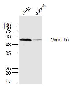 Sample:
A549(Human) Cell Lysate at 30 ug
Jurkat(Human) Cell Lysate at 30 ug
Primary: Anti-Vimentin (SL8533R) at 1/1000 dilution
Secondary: IRDye800CW Goat Anti-Rabbit IgG at 1/20000 dilution
Predicted band size: 51 kD
Observed band size: 53 kD
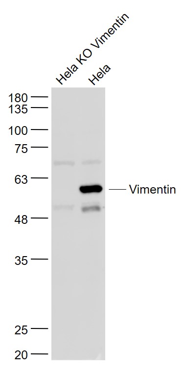 Sample:
Hela KO Vimentin (Human) Cell Lysate at 30 ug
Hela(Human) Cell Lysate at 30 ug
Primary: Anti- Vimentin (SL8533R) at 1/1000 dilution
Secondary: IRDye800CW Goat Anti-Rabbit IgG at 1/20000 dilution
Predicted band size: 51 kD
Observed band size: 57 kD
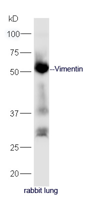 Protein: lung(rabbit) lysate at 40ug;
Primary: rabbit Anti-Vimentin (SL8533R) at 1:300;
Secondary: HRP conjugated Goat-Anti-rabbit IgG(SL0295G-HRP) at 1: 5000;
Predicted band size: 51 kD
Observed band size: 51 kD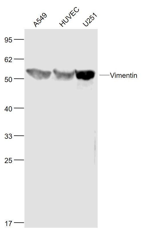 Sample:
A549(Human) Cell Lysate at 30 ug
HUVEC(Human) Cell Lysate at 30 ug
U251(Human) Cell Lysate at 30 ug
Primary: Anti-Vimentin (SL8533R) at 1/1000 dilution
Secondary: IRDye800CW Goat Anti-Rabbit IgG at 1/20000 dilution
Predicted band size: 53 kD
Observed band size: 53 kD
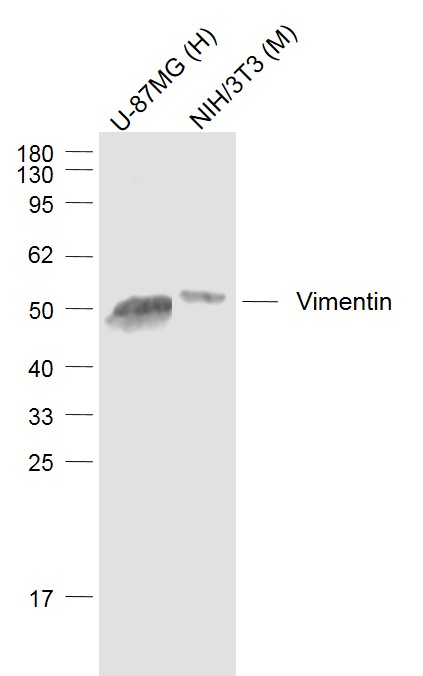 Sample:
U-87MG (Human) Cell Lysate at 30 ug
NIH/3T3 (Mouse) Cell Lysate at 30 ug
Primary: Anti-Vimentin (SL8533R) at 1/1000 dilution
Secondary: IRDye800CW Goat Anti-Rabbit IgG at 1/20000 dilution
Predicted band size: 51 kD
Observed band size: 53 kD
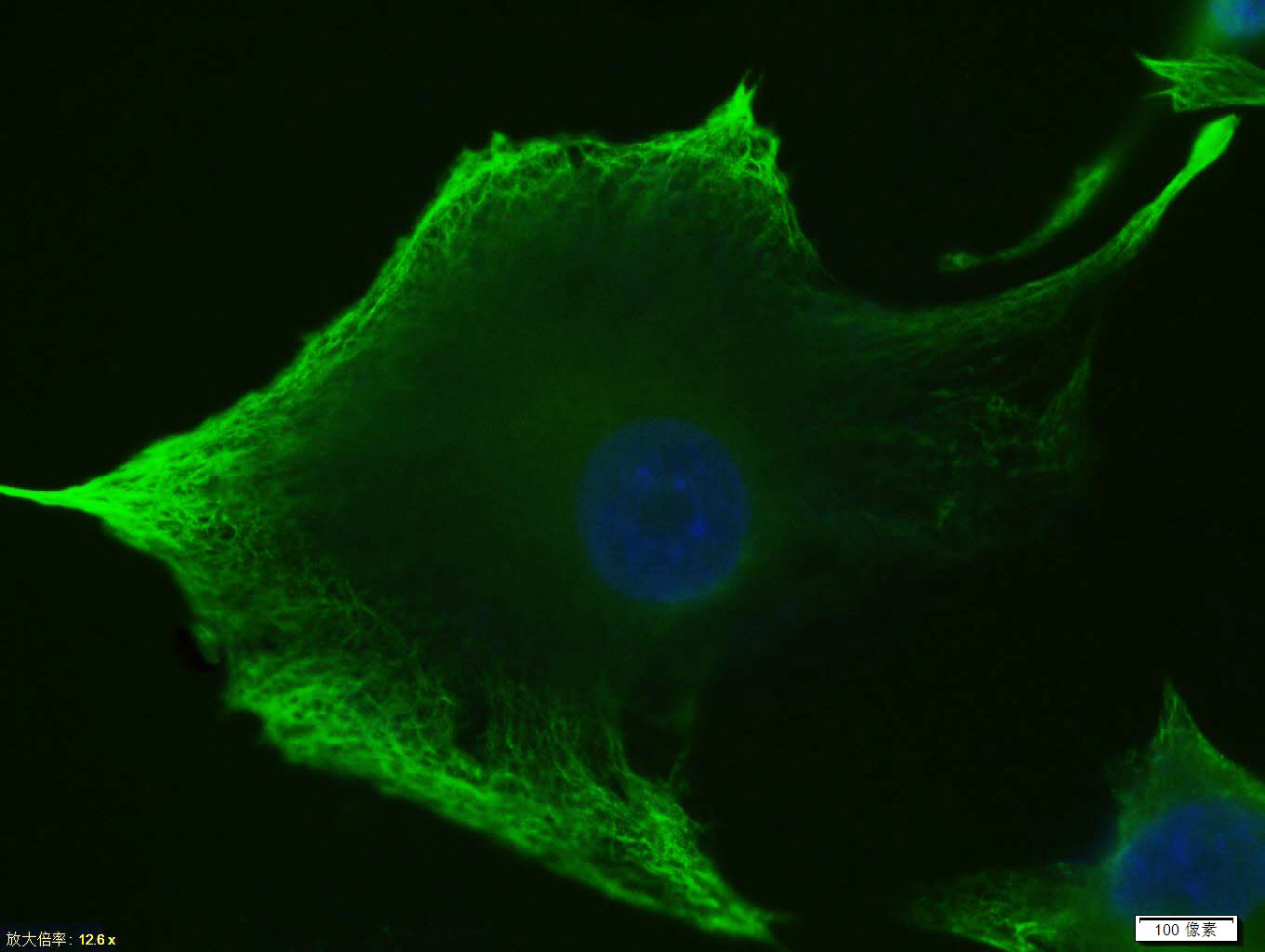 Tissue/cell: U-87MG cell; 4% Paraformaldehyde-fixed; Triton X-100 at room temperature for 20 min; Blocking buffer (normal goat serum, C-0005) at 37°C for 20 min; Antibody incubation with (Vimentin) Polyclonal Antibody, Unconjugated (SL8533R)antibody (SL0295G-FITC) at 37°C for 90 minutes, DAPI (blue, C02-04002) was used to stain the cell nuclei.
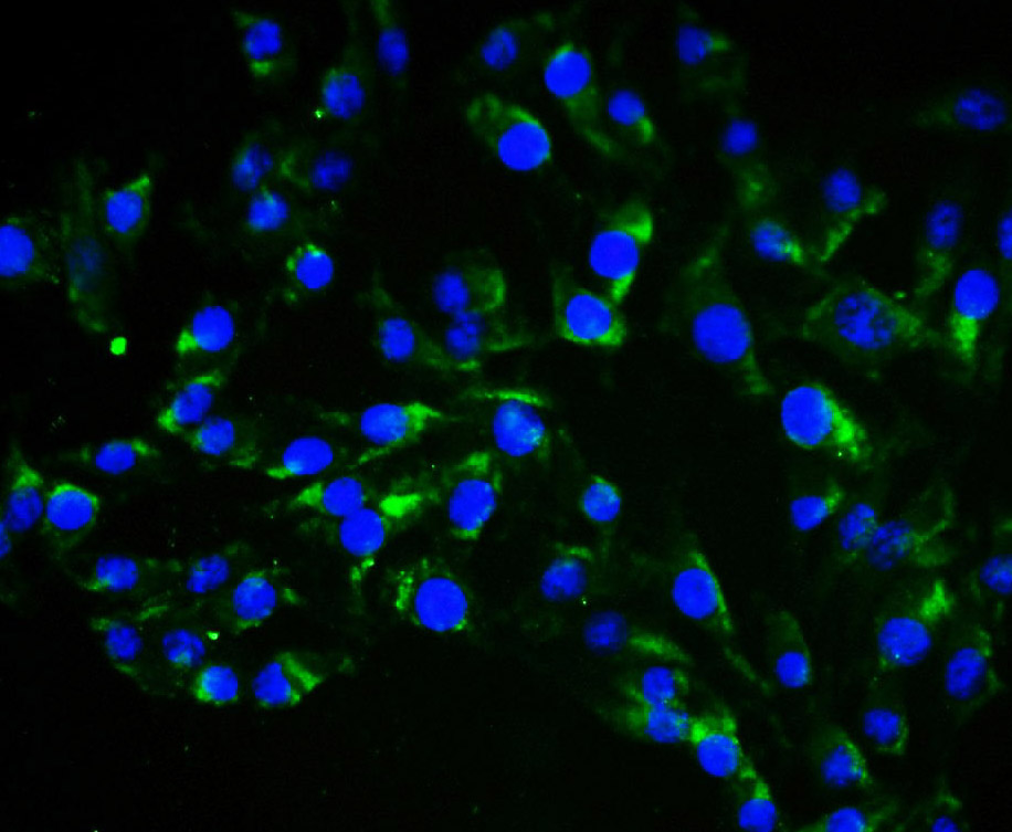 Tissue/cell: endothelial cells of umbilical artery;4% Paraformaldehyde-fixed;
Blocking buffer (normal goat serum,C-0005) at 37℃ for 20 min;
Incubation: Anti-Vimentin Polyclonal Antibody, Alexa Fluor 488 conjugated(SL8533R-A488) 1:100, 60 minutes at 37℃. DAPI(5ug/ml,blue,C-0033) was used to stain the cell nuclei
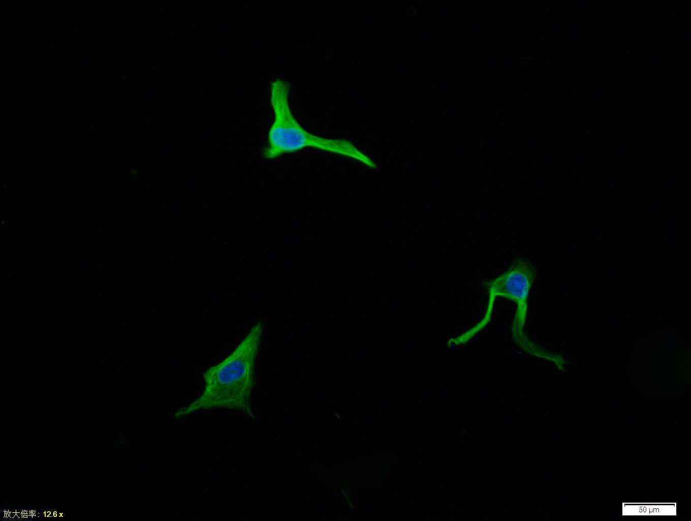 Tissue/cell: U-87MG cell; 4% Paraformaldehyde-fixed; Triton X-100 at room temperature for 20 min; Blocking buffer (normal goat serum, C-0005) at 37°C for 20 min; Antibody incubation with (Vimentin) polyclonal Antibody, Unconjugated (SL8533R) 1:100, 90 minutes at 37°C; followed by a conjugated Goat Anti-Rabbit IgG antibody at 37°C for 90 minutes, DAPI (blue, C02-04002) was used to stain the cell nuclei.
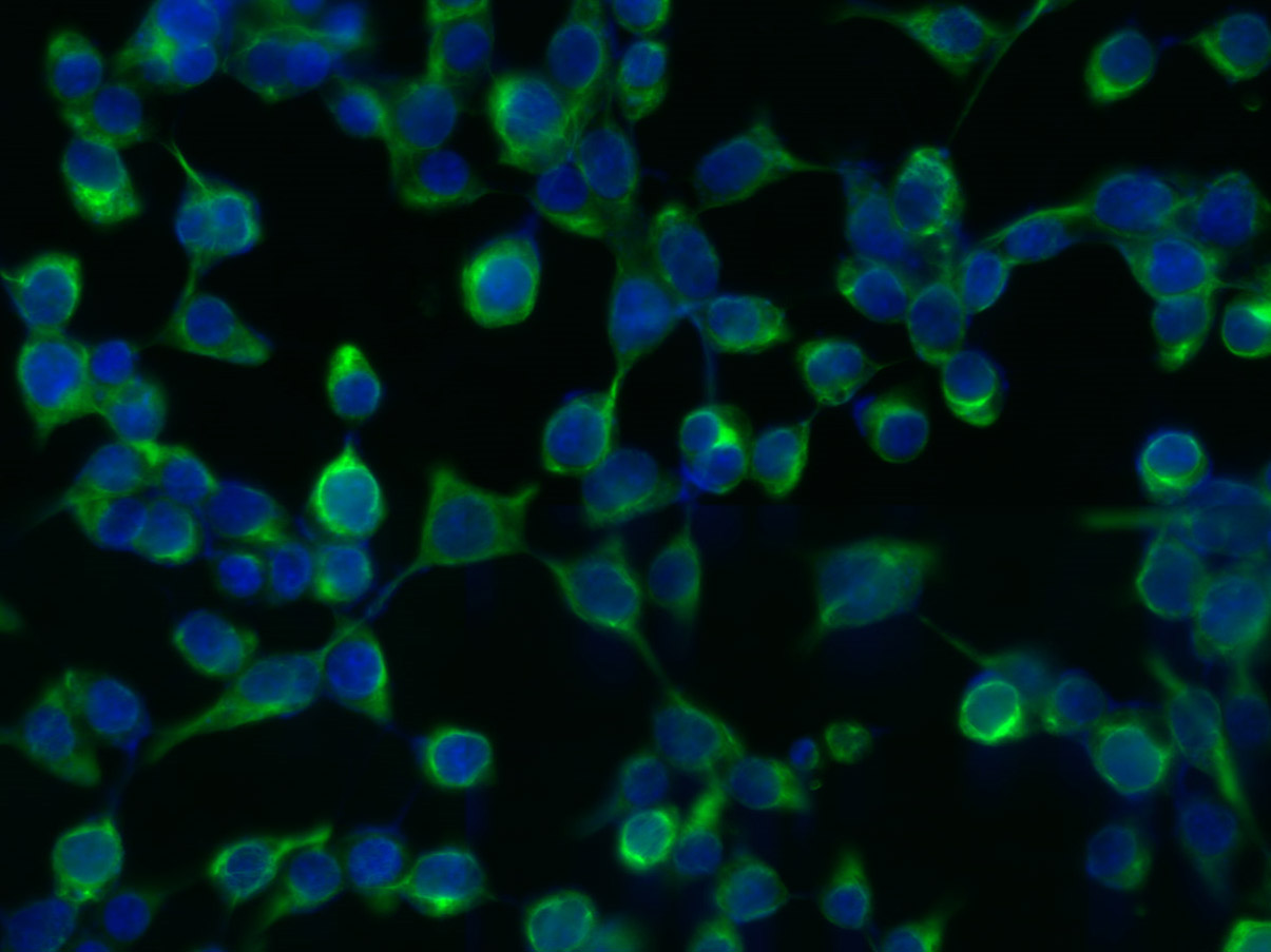 Tissue/cell: 293T cell; 4% Paraformaldehyde-fixed; Triton X-100 at room temperature for 20 min; Blocking buffer (normal goat serum, C-0005) at 37°C for 20 min; Antibody incubation with (Vimentin) Polyclonal Antibody, Unconjugated (SL8533R) 1:200, 2 hours at 37°C; followed by a conjugated Goat Anti-Rabbit IgG antibody (SL0295G-FITC) at 37°C for 90 minutes, DAPI (5ug/ml, blue, C-0033) was used to stain the cell nuclei.
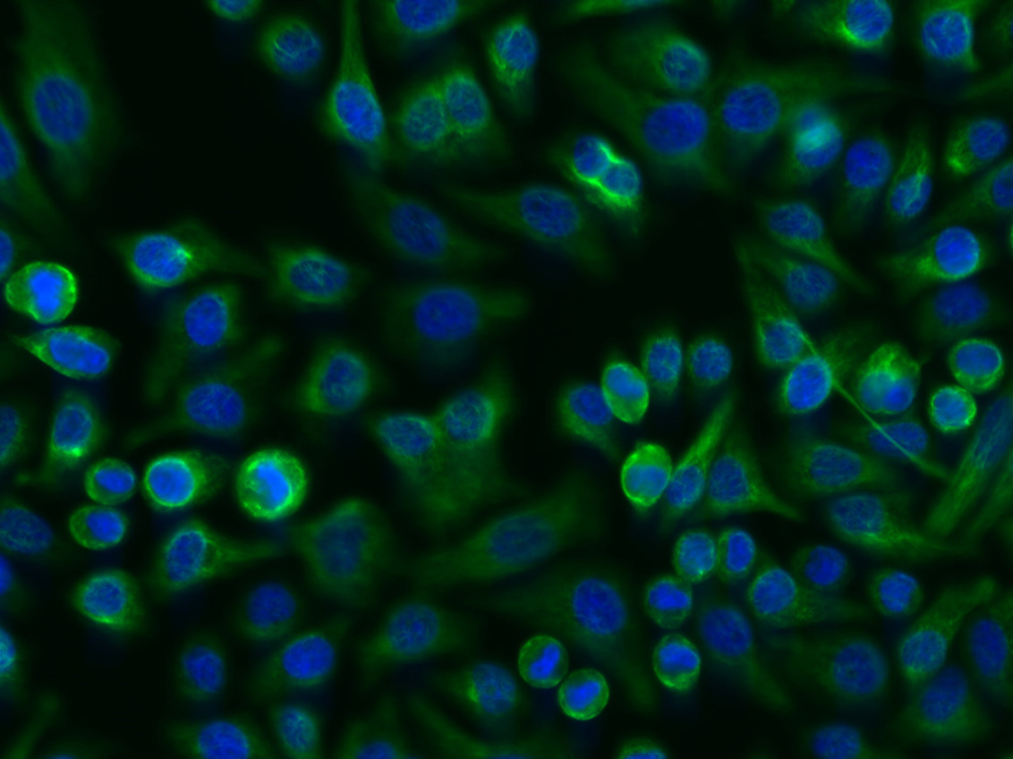 Tissue/cell: FHC cell; 4% Paraformaldehyde-fixed; Triton X-100 at room temperature for 20 min; Blocking buffer (normal goat serum, C-0005) at 37°C for 20 min; Antibody incubation with (Vimentin) Polyclonal Antibody, Unconjugated (SL8533R) 1:200, 2 hours at 37°C; followed by a conjugated Goat Anti-Rabbit IgG antibody (SL0295G-FITC) at 37°C for 90 minutes, DAPI (5ug/ml, blue, C-0033) was used to stain the cell nuclei.
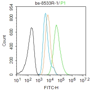 Blank control:A549.
Primary Antibody (green line): Rabbit Anti-Vimentin antibody (SL8533R)
Dilution: 1μg /10^6 cells;
Isotype Control Antibody (orange line): Rabbit IgG .
Secondary Antibody : Goat anti-rabbit IgG-AF488
Dilution: 1μg /test.
Protocol
The cells were fixed with 4% PFA (10min at room temperature)and then permeabilized with 90% ice-cold methanol for 20 min at -20℃. The cells were then incubated in 5%BSA to block non-specific protein-protein interactions for 30 min at room temperature .Cells stained with Primary Antibody for 30 min at room temperature. The secondary antibody used for 40 min at room temperature. Acquisition of 20,000 events was performed.
Cartpieces
Totalgoods,subtotals:¥Checkout
References (0)
No References
Bought notes(bought amounts latest0)
No one bought this product
User Comment(Total0User Comment Num)
- No comment


 +86 571 56623320
 +86 18668110335

