
Rabbit Anti-FOXO3A antibody
AF6q21; AF6q21 protein; DKFZp781A0677; FKHR2; FKHRL 1; FKHRL1; FKHRL1P2; Forkhead (Drosophila) homolog (rhabdomyosarcoma) like 1; Forkhead box O3A; Forkhead box protein O3A; Forkhead Drosophila homolog of in rhabdomyosarcoma like 1; Forkhead in rhabdomyos
View History [Clear]
Details
Product Name FOXO3A Chinese Name 叉头蛋白O3A抗体 Alias AF6q21; AF6q21 protein; DKFZp781A0677; FKHR2; FKHRL 1; FKHRL1; FKHRL1P2; Forkhead (Drosophila) homolog (rhabdomyosarcoma) like 1; Forkhead box O3A; Forkhead box protein O3A; Forkhead Drosophila homolog of in rhabdomyosarcoma like 1; Forkhead in rhabdomyosarcoma like 1; FOX O3A; FOXO2; MGC12739; MGC31925; FOXO3_HUMAN Research Area Tumour immunology transcriptional regulatory factor Immunogen Species Rabbit Clonality Polyclonal React Species Human, (predicted: Mouse, Rat, Pig, Cow, Sheep, ) Applications Flow-Cyt=1ug/Test
not yet tested in other applications.
optimal dilutions/concentrations should be determined by the end user.Theoretical molecular weight 74kDa Cellular localization The nucleus cytoplasmic Form Liquid Concentration 1mg/ml immunogen KLH conjugated synthetic peptide derived from human FOXO3A: 521-620/673 Lsotype IgG Purification affinity purified by Protein A Buffer Solution 0.01M TBS(pH7.4) with 1% BSA, 0.03% Proclin300 and 50% Glycerol. Storage Shipped at 4℃. Store at -20 °C for one year. Avoid repeated freeze/thaw cycles. Attention This product as supplied is intended for research use only, not for use in human, therapeutic or diagnostic applications. PubMed PubMed Product Detail This gene belongs to the forkhead family of transcription factors which are characterized by a distinct forkhead domain. This gene likely functions as a trigger for apoptosis through expression of genes necessary for cell death. Translocation of this gene with the MLL gene is associated with secondary acute leukemia. Alternatively spliced transcript variants encoding the same protein have been observed. [provided by RefSeq, Jul 2008]
Function:
Transcriptional activator which triggers apoptosis in the absence of survival factors, including neuronal cell death upon oxidative stress. Recognizes and binds to the DNA sequence 5'-[AG]TAAA[TC]A-3'. Participates in post-transcriptional regulation of MYC: following phosphorylation by MAPKAPK5, promotes induction of miR-34b and miR-34c expression, 2 post-transcriptional regulators of MYC that bind to the 3'UTR of MYC transcript and prevent its translation.
Subunit:
Interacts with YWHAB/14-3-3-beta and YWHAZ/14-3-3-zeta, which are required for cytosolic sequestration. Upon oxidative stress, interacts with STK4/MST1, which disrupts interaction with YWHAB/14-3-3-beta and leads to nuclear translocation. Interacts with PIM1.
Subcellular Location:
Cytoplasm, cytosol. Nucleus. Note=Translocates to the nucleus upon oxidative stress and in the absence of survival factors.
Tissue Specificity:
Ubiquitous.
Post-translational modifications:
: In the presence of survival factors such as IGF-1, phosphorylated on Thr-32 and Ser-253 by AKT1/PKB. This phosphorylated form then interacts with 14-3-3 proteins and is retained in the cytoplasm. Survival factor withdrawal induces dephosphorylation and promotes translocation to the nucleus where the dephosphorylated protein induces transcription of target genes and triggers apoptosis. Although AKT1/PKB doesn't appear to phosphorylate Ser-315 directly, it may activate other kinases that trigger phosphorylation at this residue. Phosphorylated by STK4/MST1 on Ser-209 upon oxidative stress, which leads to dissociation from YWHAB/14-3-3-beta and nuclear translocation. Phosphorylated by PIM1. Phosphorylation by AMPK leads to the activation of transcriptional activity without affecting subcellular localization. Phosphorylation by MAPKAPK5 promotes nuclear localization and DNA-binding, leading to induction of miR-34b and miR-34c expression, 2 post-transcriptional regulators of MYC that bind to the 3'UTR of MYC transcript and prevent its translation.
DISEASE:
Note=A chromosomal aberration involving FOXO3 is found in secondary acute leukemias. Translocation t(6;11)(q21;q23) with MLL/HRX.
Similarity:
Contains 1 fork-head DNA-binding domain.
SWISS:
O43524
Gene ID:
2309
Database links:Entrez Gene: 2309 Human
Entrez Gene: 56484 Mouse
Omim: 602681 Human
SwissProt: O43524 Human
SwissProt: Q9WVH4 Mouse
Unigene: 220950 Human
Unigene: 338613 Mouse
Unigene: 24593 Rat
Product Picture 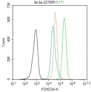 Blank control:A431.
Primary Antibody (green line): Rabbit Anti-FOXO3A antibody (SL22765R)
Dilution: 1μg /10^6 cells;
Isotype Control Antibody (orange line): Rabbit IgG .
Secondary Antibody : Goat anti-rabbit IgG-FITC
Dilution: 0.5μg /test.
Protocol
The cells were fixed with 4% PFA (10min at room temperature)and then permeabilized with 90% ice-cold methanol for 20 min at-20℃. The cells were then incubated in 5%BSA to block non-specific protein-protein interactions for 30 min at room temperature .Cells stained with Primary Antibody for 30 min at room temperature. The secondary antibody used for 40 min at room temperature. Acquisition of 20,000 events was performed.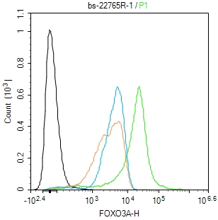 Blank control:THP-1.
Primary Antibody (green line): Rabbit Anti-FOXO3A antibody (SL22765R)
Dilution: 1μg /10^6 cells;
Isotype Control Antibody (orange line): Rabbit IgG .
Secondary Antibody : Goat anti-rabbit IgG-FITC
Dilution: 0.5μg /test.
Protocol
The cells were fixed with 4% PFA (10min at room temperature)and then permeabilized with 90% ice-cold methanol for 20 min at-20℃. The cells were then incubated in 5%BSA to block non-specific protein-protein interactions for 30 min at room temperature .Cells stained with Primary Antibody for 30 min at room temperature. The secondary antibody used for 40 min at room temperature. Acquisition of 20,000 events was performed.
Cartpieces
Totalgoods,subtotals:¥Checkout
References (0)
No References
Bought notes(bought amounts latest0)
No one bought this product
User Comment(Total0User Comment Num)
- No comment


 +86 571 56623320
 +86 18668110335

