
Rabbit Anti-Ferritin Heavy Chain antibody
Apoferritin; Cell proliferation inducing gene 15 protein; F HC; Ferritin H subunit; Ferritin heavy chain; Ferritin heavy polypeptide 1; FHC; FRIH; FTH 1; FTH; FTH1; FTH1 protein; FTHL 6; FTHL6; Iron overload autosomal dominant; MGC104426; OK/SW-cl.84; PIG
View History [Clear]
Details
Product Name Ferritin Heavy Chain Chinese Name 铁蛋白Recombinant rabbit monoclonal anti Alias Apoferritin; Cell proliferation inducing gene 15 protein; F HC; Ferritin H subunit; Ferritin heavy chain; Ferritin heavy polypeptide 1; FHC; FRIH; FTH 1; FTH; FTH1; FTH1 protein; FTHL 6; FTHL6; Iron overload autosomal dominant; MGC104426; OK/SW-cl.84; PIG 15; PIG15; Placenta immunoregulatory factor; PLIF; Proliferation inducing gene 15 protein; Proliferation inducing protein 15; FRIH_HUMAN. Research Area Cardiovascular Cell biology immunology Neurobiology Immunogen Species Rabbit Clonality Monoclonal React Species (predicted: Human, Mouse, Rat, ) Applications WB=1:500-1000 IHC-P=1:100-500 ICC=1:150-200 (Paraffin sections need antigen repair)
not yet tested in other applications.
optimal dilutions/concentrations should be determined by the end user.Theoretical molecular weight 20kDa Cellular localization The nucleus cytoplasmic Form Liquid Concentration 1mg/ml immunogen KLH conjugated synthetic peptide derived from human Ferritin Heavy Chain Lsotype IgG Purification affinity purified by Protein A Buffer Solution 0.01M TBS(pH7.4) with 1% BSA, 0.03% Proclin300 and 50% Glycerol. Storage Shipped at 4℃. Store at -20 °C for one year. Avoid repeated freeze/thaw cycles. Attention This product as supplied is intended for research use only, not for use in human, therapeutic or diagnostic applications. PubMed PubMed Product Detail This gene encodes the heavy subunit of ferritin, the major intracellular iron storage protein in prokaryotes and eukaryotes. It is composed of 24 subunits of the heavy and light ferritin chains. Variation in ferritin subunit composition may affect the rates of iron uptake and release in different tissues. A major function of ferritin is the storage of iron in a soluble and nontoxic state. Defects in ferritin proteins are associated with several neurodegenerative diseases. This gene has multiple pseudogenes. Several alternatively spliced transcript variants have been observed, but their biological validity has not been determined. [provided by RefSeq, Jul 2008].
Function:
Stores iron in a soluble, non-toxic, readily available form. Important for iron homeostasis. Has ferroxidase activity. Iron is taken up in the ferrous form and deposited as ferric hydroxides after oxidation. Also plays a role in delivery of iron to cells. Mediates iron uptake in capsule cells of the developing kidney.
Subunit:
Oligomer of 24 subunits. There are two types of subunits: L (light) chain and H (heavy) chain. The major chain can be light or heavy, depending on the species and tissue type. The functional molecule forms a roughly spherical shell with a diameter of 12 nm and contains a central cavity into which the insoluble mineral iron core is deposited.
Subcellular Location:
Cytoplasmic.
Tissue Specificity:
In human liver the heavy chain is the major chain.
Similarity:
Belongs to the ferritin family.
Contains 1 ferritin-like diiron domain.
SWISS:
P02794
Gene ID:
2495
Database links:Entrez Gene: 654516 Cat
Entrez Gene: 2495 Human
Entrez Gene: 14319 Mouse
Entrez Gene: 100173063 Orangutan
Omim: 134770 Human
SwissProt: P02794 Human
SwissProt: P09528 Mouse
SwissProt: Q5R8J7 Orangutan
Unigene: 524910 Human
Unigene: 645560 Human
Unigene: 1776 Mouse
Product Picture  Western blot analysis of Ferritin Heavy Chain on human liver tissue lysates. Proteins were transferred to a PVDF membrane and blocked with 5% BSA in PBS for 1 hour at room temperature. The primary antibody (SLM-54108R, 1/500) was used in 5% BSA at room temperature for 2 hours. Goat Anti-Rabbit IgG - HRP Secondary Antibody at 1:5,000 dilution was used for 1 hour at room temperature.
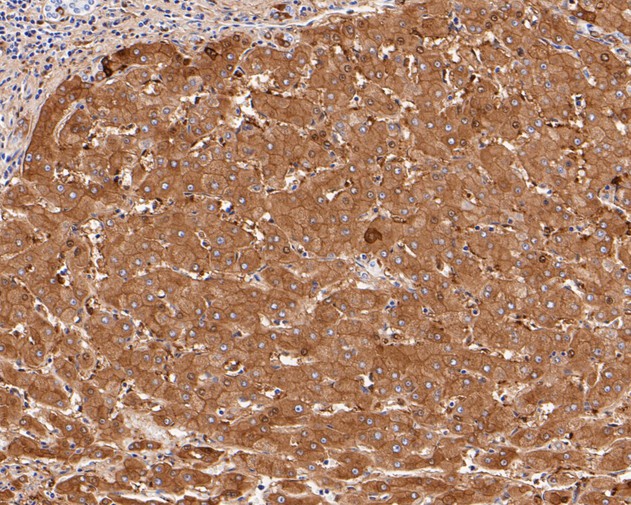 Immunohistochemical analysis of paraffin-embedded human liver tissue using anti-Ferritin Heavy Chain antibody. The section was pre-treated using heat mediated antigen retrieval with Tris-EDTA buffer (pH 8.0-8.4) for 20 minutes.The tissues were blocked in 5% BSA for 30 minutes at room temperature, washed with ddH2O and PBS, and then probed with the primary antibody (SLM-54108R, 1/50) for 30 minutes at room temperature. The detection was performed using an HRP conjugated compact polymer system. DAB was used as the chromogen. Tissues were counterstained with hematoxylin and mounted with DPX.
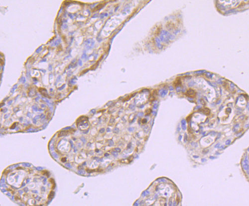 Immunohistochemical analysis of paraffin-embedded human liver tissue using anti-Ferritin Heavy Chain antibody. The section was pre-treated using heat mediated antigen retrieval with Tris-EDTA buffer (pH 8.0-8.4) for 20 minutes.The tissues were blocked in 5% BSA for 30 minutes at room temperature, washed with ddH2O and PBS, and then probed with the primary antibody (SLM-54108R, 1/50) for 30 minutes at room temperature. The detection was performed using an HRP conjugated compact polymer system. DAB was used as the chromogen. Tissues were counterstained with hematoxylin and mounted with DPX.
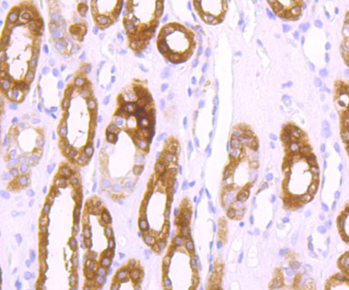 Immunohistochemical analysis of paraffin-embedded human placenta tissue using anti-Ferritin Heavy Chain antibody. The section was pre-treated using heat mediated antigen retrieval with Tris-EDTA buffer (pH 8.0-8.4) for 20 minutes.The tissues were blocked in 5% BSA for 30 minutes at room temperature, washed with ddH2O and PBS, and then probed with the primary antibody (SLM-54108R, 1/50) for 30 minutes at room temperature. The detection was performed using an HRP conjugated compact polymer system. DAB was used as the chromogen. Tissues were counterstained with hematoxylin and mounted with DPX.
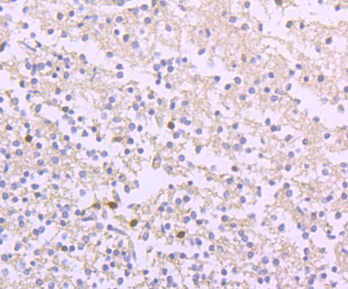 Immunohistochemical analysis of paraffin-embedded human brain tissue using anti-Ferritin Heavy Chain antibody. The section was pre-treated using heat mediated antigen retrieval with Tris-EDTA buffer (pH 8.0-8.4) for 20 minutes.The tissues were blocked in 5% BSA for 30 minutes at room temperature, washed with ddH2O and PBS, and then probed with the primary antibody (SLM-54108R, 1/50) for 30 minutes at room temperature. The detection was performed using an HRP conjugated compact polymer system. DAB was used as the chromogen. Tissues were counterstained with hematoxylin and mounted with DPX. .06
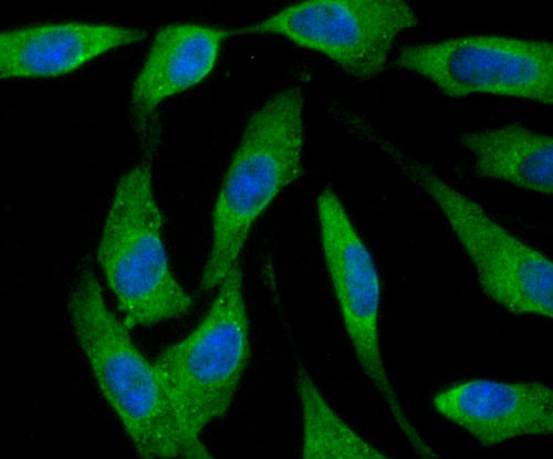 Immunohistochemical analysis of paraffin-embedded human brain tissue using anti-Ferritin Heavy Chain antibody. The section was pre-treated using heat mediated antigen retrieval with Tris-EDTA buffer (pH 8.0-8.4) for 20 minutes.The tissues were blocked in 5% BSA for 30 minutes at room temperature, washed with ddH2O and PBS, and then probed with the primary antibody (SLM-54108R, 1/50) for 30 minutes at room temperature. The detection was performed using an HRP conjugated compact polymer system. DAB was used as the chromogen. Tissues were counterstained with hematoxylin and mounted with DPX.Immunohistochemical analysis of paraffin-embedded human brain tissue using anti-Ferritin Heavy Chain antibody. The section was pre-treated using heat mediated antigen retrieval with Tris-EDTA buffer (pH 8.0-8.4) for 20 minutes.The tissues were blocked in 5% BSA for 30 minutes at room temperature, washed with ddH2O and PBS, and then probed with the primary antibody (SLM-54108R, 1/50) for 30 minutes at room temperature. The detection was performed using an HRP conjugated compact polymer system. DAB was used as the chromogen. Tissues were counterstained with hematoxylin and mounted with DPX.
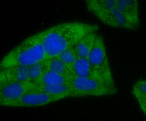 ICC staining of Ferritin Heavy Chain in Hela cells (green). Formalin fixed cells were permeabilized with 0.1% Triton X-100 in TBS for 10 minutes at room temperature and blocked with 1% Blocker BSA for 15 minutes at room temperature. Cells were probed with the primary antibody (SLM-54108R, 1/50) for 1 hour at room temperature, washed with PBS. Alexa Fluor®488 Goat anti-Rabbit IgG was used as the secondary antibody at 1/1,000 dilution. The nuclear counter stain is DAPI (blue).
 ICC staining of Ferritin Heavy Chain in A549 cells (green). Formalin fixed cells were permeabilized with 0.1% Triton X-100 in TBS for 10 minutes at room temperature and blocked with 1% Blocker BSA for 15 minutes at room temperature. Cells were probed with the primary antibody (SLM-54108R, 1/50) for 1 hour at room temperature, washed with PBS. Alexa Fluor®488 Goat anti-Rabbit IgG was used as the secondary antibody at 1/1,000 dilution. The nuclear counter stain is DAPI (blue).
Cartpieces
Totalgoods,subtotals:¥Checkout
References (0)
No References
Bought notes(bought amounts latest0)
No one bought this product
User Comment(Total0User Comment Num)
- No comment


 +86 571 56623320
 +86 18668110335

