
Rabbit Anti-PDPK1 antibody
3 Phosphoinositide Dependent Protein Kinase 1; hPDK 1; hPDK 1; PDK 1; PkB kinase; PkB kinase like gene 1; PkB like 1; PDPK1_HUMAN.
View History [Clear]
Details
Product Name PDPK1 Chinese Name 3磷酸肌醇依赖性蛋白激酶1Recombinant rabbit monoclonal anti Alias 3 Phosphoinositide Dependent Protein Kinase 1; hPDK 1; hPDK 1; PDK 1; PkB kinase; PkB kinase like gene 1; PkB like 1; PDPK1_HUMAN. Research Area Tumour Cell biology immunology Chromatin and nuclear signals Stem cells Cyclin transcriptional regulatory factor Immunogen Species Rabbit Clonality Monoclonal Clone NO. P1B6 React Species Human, (predicted: Mouse, ) Applications IHC-P=1:100 ICC=1:50 IF=1:50-100 (Paraffin sections need antigen repair)
not yet tested in other applications.
optimal dilutions/concentrations should be determined by the end user.Theoretical molecular weight 61kDa Cellular localization cytoplasmic The cell membrane Form Liquid Concentration 1mg/ml immunogen KLH conjugated synthetic peptide derived from human PDPK1 Lsotype IgG Purification affinity purified by Protein A Buffer Solution 0.01M TBS(pH7.4) with 1% BSA, 0.03% Proclin300 and 50% Glycerol. Storage Shipped at 4℃. Store at -20 °C for one year. Avoid repeated freeze/thaw cycles. Attention This product as supplied is intended for research use only, not for use in human, therapeutic or diagnostic applications. PubMed PubMed Product Detail Enables 3-phosphoinositide-dependent protein kinase activity; phospholipase activator activity; and phospholipase binding activity. Involved in several processes, including cell surface receptor signaling pathway; regulation of protein kinase activity; and regulation of signal transduction. Acts upstream of or within intracellular signal transduction. Located in cell projection; cytosol; and plasma membrane. Implicated in prostate cancer. Biomarker of lung non-small cell carcinoma. [provided by Alliance of Genome Resources, Nov 2021]
SWISS:
O15530
Gene ID:
5170
Product Picture 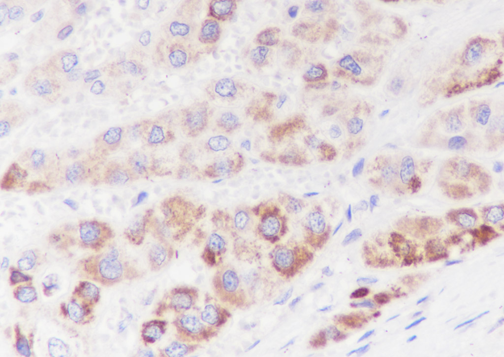 Tissue:Human liver cancer
Section type: Formalin fixed & Paraffin -embedded section
Retrieval method: High temperature and high pressure
Retrieval buffer: Tris/EDTA buffer, pH 9.0 Primary ab dilution: 1:100
Primary ab incubation condition: 1 hour at room temperature
Counter stain: Hematoxylin
Comment: Color brown is the positive signal for SLM-60302R
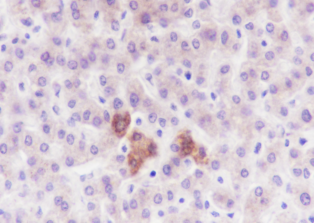 Tissue:Human liver
Section type: Formalin fixed & Paraffin -embedded section
Retrieval method: High temperature and high pressure
Retrieval buffer: Tris/EDTA buffer, pH 9.0 Primary ab dilution: 1:100
Primary ab incubation condition: 1 hour at room temperature
Counter stain: Hematoxylin
Comment: Color brown is the positive signal for SLM-60302R
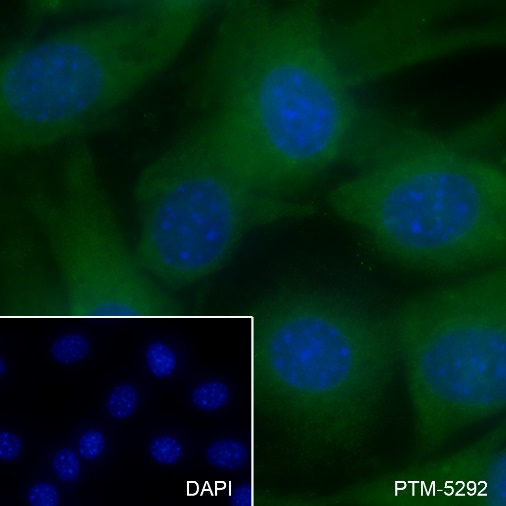 Cell line: NIH3T3
Fixative: 4% Paraformaldehyde
Permeabilization: 0.1% TritonX-100
Primary ab dilution: 1:50
Primary incubation condition: 4°C overnight
Secondary ab: Goat Anti-Rabbit IgG
Nuclear counter stain: DAPI (Blue)
Comment: Color green is the positive signal for SLM-60302R
Cartpieces
Totalgoods,subtotals:¥Checkout
References (0)
No References
Bought notes(bought amounts latest0)
No one bought this product
User Comment(Total0User Comment Num)
- No comment


 +86 571 56623320
 +86 18668110335

