
Rabbit Anti-LYPD3 antibody
C4.4A; FLJ10525; GPI-anchored metastasis-associated protein C4.4A homolog; GPI-anchored metastasis-associated protein homolog; Ly6/PLAUR domain-containing protein 3; LYPD3_HUMAN; Matrigel-induced gene C4 protein; MIG-C4; SHINC3.
View History [Clear]
Details
Product Name LYPD3 Chinese Name C4.4A/LYPD3Recombinant rabbit monoclonal anti Alias C4.4A; FLJ10525; GPI-anchored metastasis-associated protein C4.4A homolog; GPI-anchored metastasis-associated protein homolog; Ly6/PLAUR domain-containing protein 3; LYPD3_HUMAN; Matrigel-induced gene C4 protein; MIG-C4; SHINC3. Research Area Tumour Cell type markers Immunogen Species Rabbit Clonality Monoclonal Clone NO. C4F10 React Species Human, Applications WB=1:500-1000 IHC-P=1:1000 ICC=1:50 IF=1:50-100 (Paraffin sections need antigen repair)
not yet tested in other applications.
optimal dilutions/concentrations should be determined by the end user.Theoretical molecular weight 33kDa Cellular localization The cell membrane Form Liquid Concentration 1mg/ml immunogen KLH conjugated synthetic peptide derived from human LYPD3 Lsotype IgG Purification affinity purified by Protein A Buffer Solution 0.01M TBS(pH7.4) with 1% BSA, 0.03% Proclin300 and 50% Glycerol. Storage Shipped at 4℃. Store at -20 °C for one year. Avoid repeated freeze/thaw cycles. Attention This product as supplied is intended for research use only, not for use in human, therapeutic or diagnostic applications. PubMed PubMed Product Detail Predicted to enable laminin binding activity. Predicted to act upstream of or within cell-matrix adhesion. Located in extracellular space. [provided by Alliance of Genome Resources, Nov 2021]
SWISS:
O95274
Gene ID:
27076
Database links:Entrez Gene: 27076 Human
SwissProt: O95274 Human
Product Picture 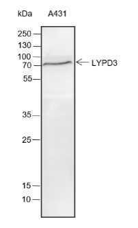 Blocking buffer: 5% NFDM/TBST
Primary ab dilution: 1:2000
Primary ab incubation condition: 2 hours at
room temperature
Lysate: A431
Protein loading quantity: 20 μg
Exposure time: 30 s
Predicted MW: 36 kDa
Observed MW: 70 kDa
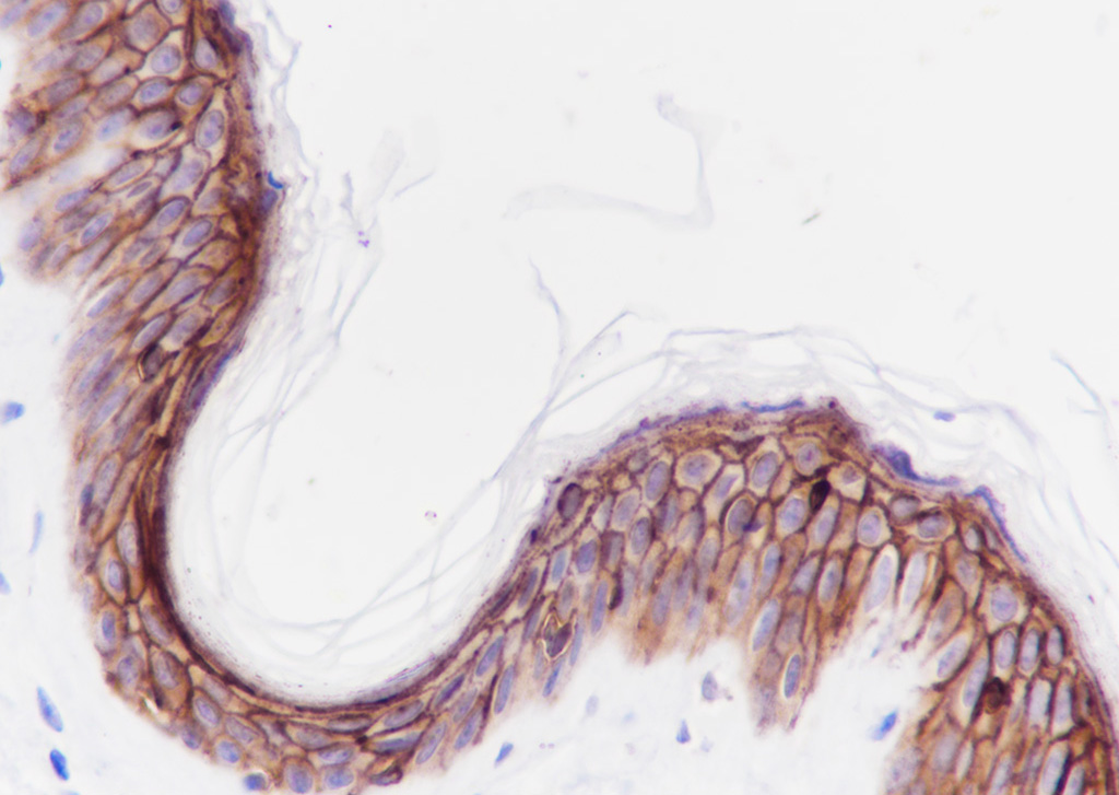 Tissue: Human skin
Section type: Formalin fixed & Paraffin -embedded section
Retrieval method: High temperature and high pressure
Retrieval buffer: Tris/EDTA buffer, pH 9.0 Primary ab dilution: 1:1000
Primary ab incubation condition: 1 hour at room temperature
Counter stain: Hematoxylin
Comment: Color brown is the positive signal for SLM-60286R
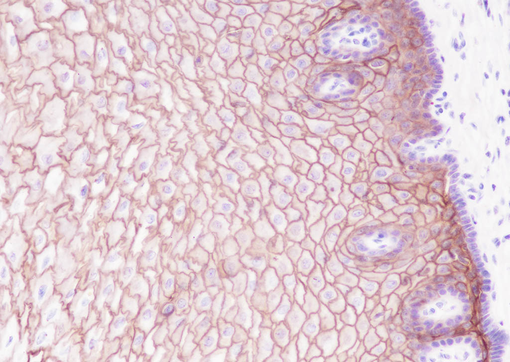 Tissue: Human esophagus
Section type: Formalin fixed & Paraffin -embedded section
Retrieval method: High temperature and high pressure
Retrieval buffer: Tris/EDTA buffer, pH 9.0 Primary ab dilution: 1:1000
Primary ab incubation condition: 1 hour at room temperature
Counter stain: Hematoxylin
Comment: Color brown is the positive signal for SLM-60286R
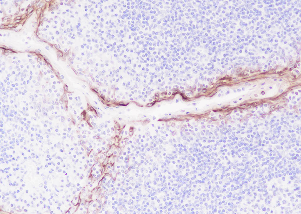 Tissue: Human tonsil
Section type: Formalin fixed & Paraffin -embedded section
Retrieval method: High temperature and high pressure
Retrieval buffer: Tris/EDTA buffer, pH 9.0 Primary ab dilution: 1:1000
Primary ab incubation condition: 1 hour at room temperature
Counter stain: Hematoxylin
Comment: Color brown is the positive signal for SLM-60286R
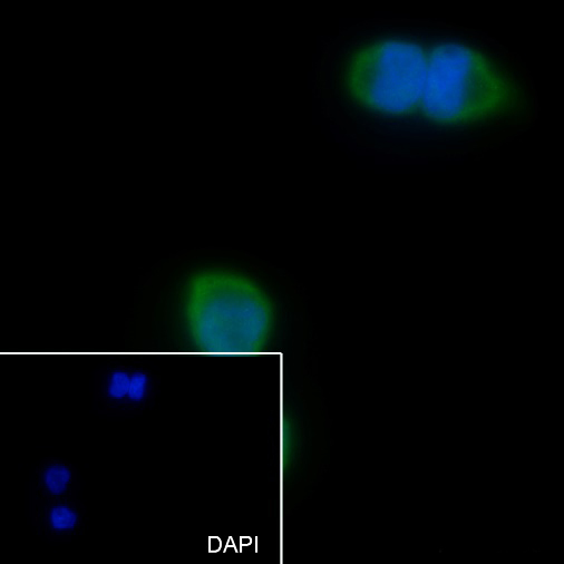 Cell line: A431
Fixative: 4% Paraformaldehyde
Permeabilization: 0.1% TritonX-100
Primary ab dilution: 1:50
Primary incubation condition: 4°C overnight
Secondary ab: Goat Anti-Rabbit IgG
Nuclear counter stain: DAPI (Blue)
Comment: Color green is the positive signal for SLM-60286R
Cartpieces
Totalgoods,subtotals:¥Checkout
References (0)
No References
Bought notes(bought amounts latest0)
No one bought this product
User Comment(Total0User Comment Num)
- No comment


 +86 571 56623320
 +86 18668110335

