
Rabbit Anti-Keratin 6 antibody
Keratin, type II cytoskeletal 6C; KRT6C; Cytokeratin-6C; CK-6C; Cytokeratin-6E; CK-6E; Keratin K6h; Keratin-6C; K6C; Type-II keratin Kb12; KRT6E; K2C6C_HUMAN
View History [Clear]
Details
Product Name Keratin 6 Chinese Name 细胞角蛋白6兔单克隆抗体 Alias Keratin, type II cytoskeletal 6C; KRT6C; Cytokeratin-6C; CK-6C; Cytokeratin-6E; CK-6E; Keratin K6h; Keratin-6C; K6C; Type-II keratin Kb12; KRT6E; K2C6C_HUMAN Research Area Tumour Signal transduction Immunogen Species Rabbit Clonality Monoclonal Clone NO. B12C5 React Species Human, Mouse, Rat, Applications WB=1:500-2000 IHC-P=1:100-500 ICC=1:20-100 IF=1:50 (Paraffin sections need antigen repair)
not yet tested in other applications.
optimal dilutions/concentrations should be determined by the end user.Theoretical molecular weight 60kDa Cellular localization cytoplasmic Form Liquid Concentration 1mg/ml immunogen KLH conjugated synthetic peptide derived from human Keratin 6 Lsotype IgG Purification affinity purified by Protein A Buffer Solution 0.01M TBS(pH7.4) with 1% BSA, 0.03% Proclin300 and 50% Glycerol. Storage Shipped at 4℃. Store at -20 °C for one year. Avoid repeated freeze/thaw cycles. Attention This product as supplied is intended for research use only, not for use in human, therapeutic or diagnostic applications. PubMed PubMed Product Detail Keratins are intermediate filament proteins responsible for the structural integrity of epithelial cells and are subdivided into epithelial keratins and hair keratins. The type II keratins are clustered in a region of chromosome 12q13. [provided by RefSeq, Jul 2009]
Function:
There are at least six isoforms of human type II keratin-6 (K6).
There are two types of cytoskeletal and microfibrillar keratin, I (acidic) and II (neutral to basic) (40-55 and 56-70 kDa, respectively).
Subunit:
Heterodimer of a type I and a type II keratin. KRT6 isomers associate with KRT16 and/or KRT17. Interacts with TCHP.
Tissue Specificity:
Constitutively expressed in distinct types of epithelia such as those in oral mucosa, esophagus, papillae of tongue and hair follicle outer root sheath.
DISEASE:
Defects in KRT6A are a cause of pachyonychia congenital type 1 (PC1) [MIM:167200]; also known as Jadassohn-Lewandowsky syndrome. PC1 is an autosomal dominant ectodermal dysplasia characterized by hypertrophic nail dystrophy resulting in onchyogryposis (thickening and increase in curvature of the nail), palmoplantar keratoderma, follicular hyperkeratosis, and oral leukokeratosis. Hyperhidrosis of the hands and feet is usually present.
Similarity:
Belongs to the intermediate filament family.
SWISS:
P48668
Gene ID:
286887
Database links:Entrez Gene: 286887 Human
Entrez Gene: 16687 Mouse
SwissProt: P48668 Human
SwissProt: P50446 Mouse
Product Picture 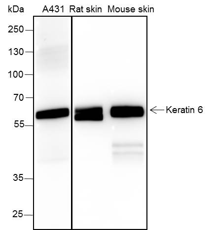 Blocking buffer: 5% NFDM/TBST
Primary ab dilution: 1:2000
Primary ab incubation condition: 2 hours at
room temperature
Lysate: A431, Rat skin, Mouse skin
Protein loading quantity: 20 μg
Exposure time: 15 s
Predicted MW: 60 kDa
Observed MW: 56 kDa
 Paraformaldehyde-fixed, paraffin embedded (human breast); Antigen retrieval by boiling in sodium citrate buffer (pH6.0) for 15min; Block endogenous peroxidase by 3% hydrogen peroxide for 20 minutes; Blocking buffer (normal goat serum) at 37°C for 30min; Antibody incubation with (Keratin 6) Monoclonal Antibody, Unconjugated (SLM-60235R) at 1:200 overnight at 4°C, followed by operating according to SP Kit(Rabbit) (sp-0023) instructionsand DAB staining.
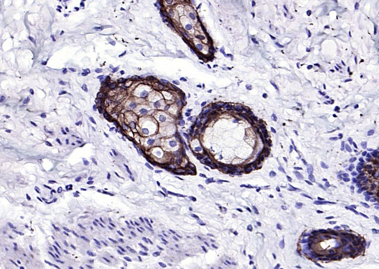
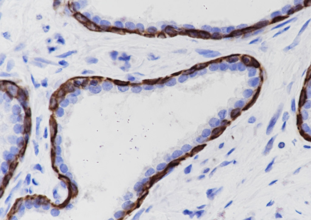 Tissue: Human prostate hyperplasia
Section type: Formalin fixed & Paraffin -embedded section
Retrieval method: High temperature and high pressure
Retrieval buffer: Tris/EDTA buffer, pH 9.0 Primary ab dilution: 1:1000
Primary ab incubation condition: 1 hour at room temperature
Counter stain: Hematoxylin
Comment: Color brown is the positive signal for SLM-60235R
 Tissue: Human breast
Section type: Formalin fixed & Paraffin -embedded section
Retrieval method: High temperature and high pressure
Retrieval buffer: Tris/EDTA buffer, pH 9.0 Primary ab dilution: 1:1000
Primary ab incubation condition: 1 hour at room temperature
Counter stain: Hematoxylin
Comment: Color brown is the positive signal for SLM-60235R
 Paraformaldehyde-fixed, paraffin embedded (human esophageal); Antigen retrieval by boiling in sodium citrate buffer (pH6.0) for 15min; Block endogenous peroxidase by 3% hydrogen peroxide for 20 minutes; Blocking buffer (normal goat serum) at 37°C for 30min; Antibody incubation with (Keratin 6) Monoclonal Antibody, Unconjugated (SLM-60235R) at 1:200 overnight at 4°C, followed by operating according to SP Kit(Rabbit) (sp-0023) instructionsand DAB staining.
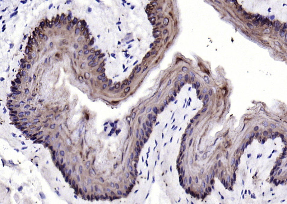 Paraformaldehyde-fixed, paraffin embedded (mouse esophageal); Antigen retrieval by boiling in sodium citrate buffer (pH6.0) for 15min; Block endogenous peroxidase by 3% hydrogen peroxide for 20 minutes; Blocking buffer (normal goat serum) at 37°C for 30min; Antibody incubation with (Keratin 6) Monoclonal Antibody, Unconjugated (SLM-60235R) at 1:200 overnight at 4°C, followed by operating according to SP Kit(Rabbit) (sp-0023) instructionsand DAB staining.
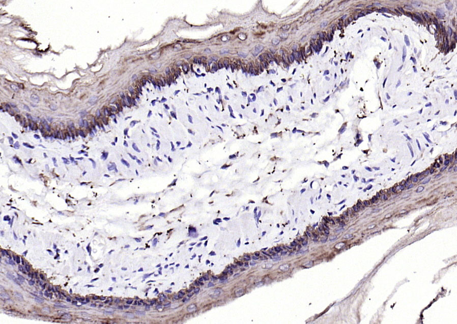 Paraformaldehyde-fixed, paraffin embedded (rat esophageal); Antigen retrieval by boiling in sodium citrate buffer (pH6.0) for 15min; Block endogenous peroxidase by 3% hydrogen peroxide for 20 minutes; Blocking buffer (normal goat serum) at 37°C for 30min; Antibody incubation with (Keratin 6) Monoclonal Antibody, Unconjugated (SLM-60235R) at 1:200 overnight at 4°C, followed by operating according to SP Kit(Rabbit) (sp-0023) instructionsand DAB staining.
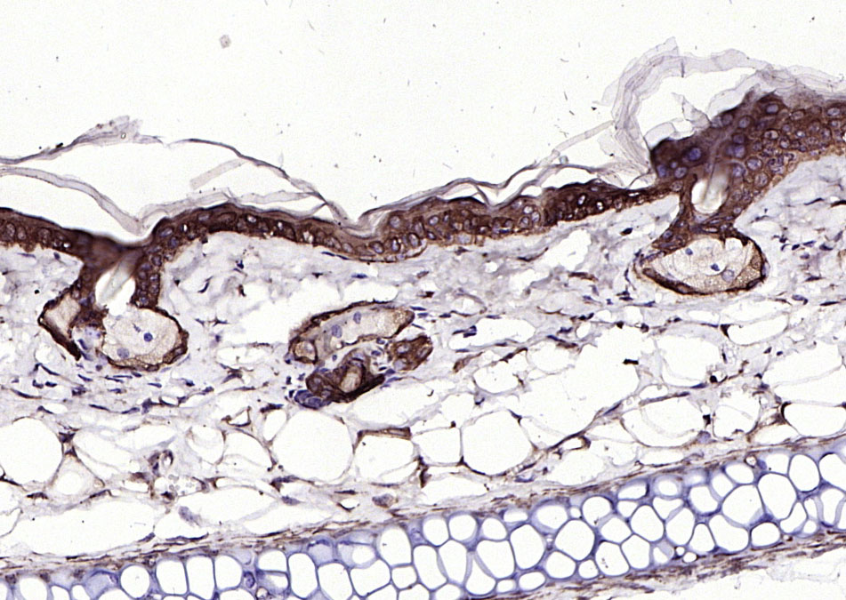 Paraformaldehyde-fixed, paraffin embedded (mouse skin); Antigen retrieval by boiling in sodium citrate buffer (pH6.0) for 15min; Block endogenous peroxidase by 3% hydrogen peroxide for 20 minutes; Blocking buffer (normal goat serum) at 37°C for 30min; Antibody incubation with (Keratin 6) Monoclonal Antibody, Unconjugated (SLM-60235R) at 1:200 overnight at 4°C, followed by operating according to SP Kit(Rabbit) (sp-0023) instructionsand DAB staining.
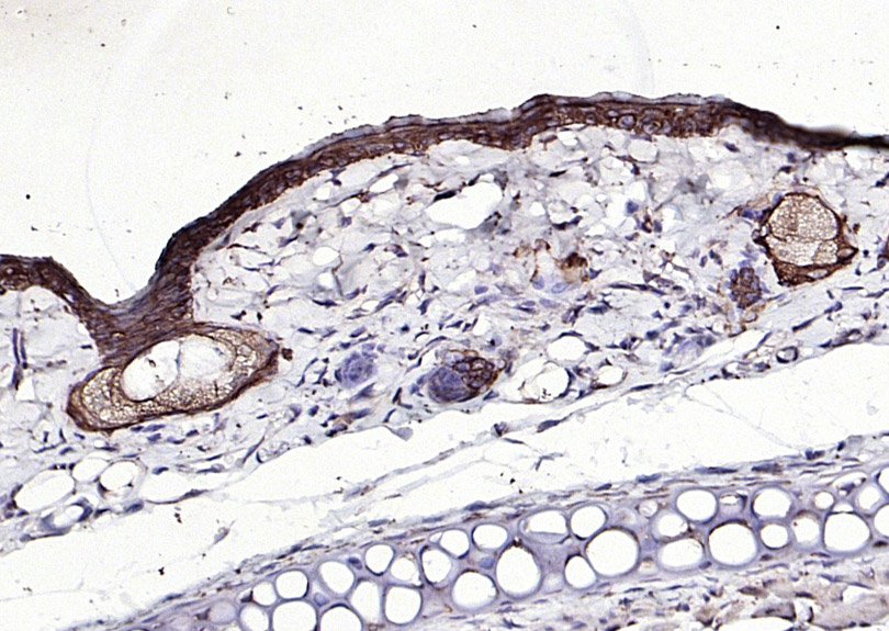 Paraformaldehyde-fixed, paraffin embedded (rat skin); Antigen retrieval by boiling in sodium citrate buffer (pH6.0) for 15min; Block endogenous peroxidase by 3% hydrogen peroxide for 20 minutes; Blocking buffer (normal goat serum) at 37°C for 30min; Antibody incubation with (Keratin 6) Monoclonal Antibody, Unconjugated (SLM-60235R) at 1:200 overnight at 4°C, followed by operating according to SP Kit(Rabbit) (sp-0023) instructionsand DAB staining.
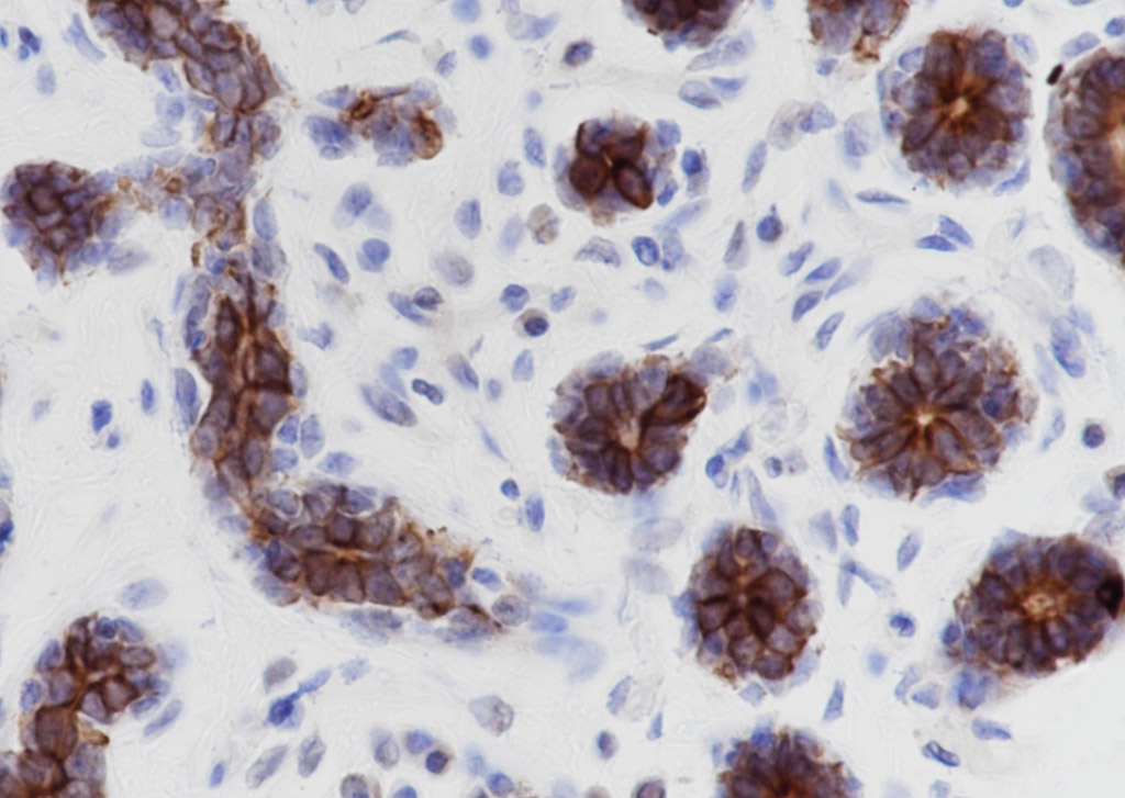
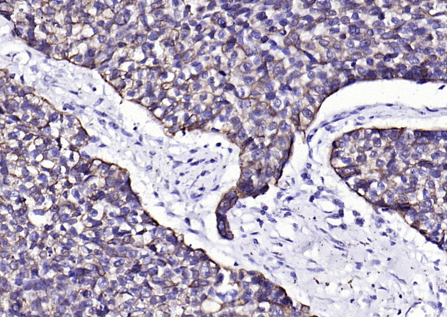
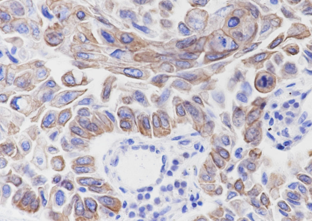 Tissue: Human lung squamous cell carcinoma
Section type: Formalin fixed & Paraffin -embedded section
Retrieval method: High temperature and high pressure
Retrieval buffer: Tris/EDTA buffer, pH 9.0 Primary ab dilution: 1:1000
Primary incubation condition: 1 hour at room temperature
Counter stain: Hematoxylin
Comment: Color brown is the positive signal for SLM-60235R
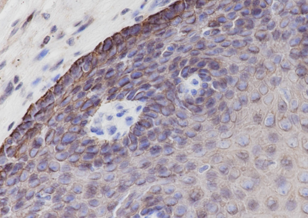 Tissue: Human esophagus
Section type: Formalin fixed & Paraffin -embedded section
Retrieval method: High temperature and high pressure
Retrieval buffer: Tris/EDTA buffer, pH 9.0 Primary ab dilution: 1:1000
Primary incubation condition: 1 hour at room temperature
Counter stain: Hematoxylin
Comment: Color brown is the positive signal for SLM-60235R
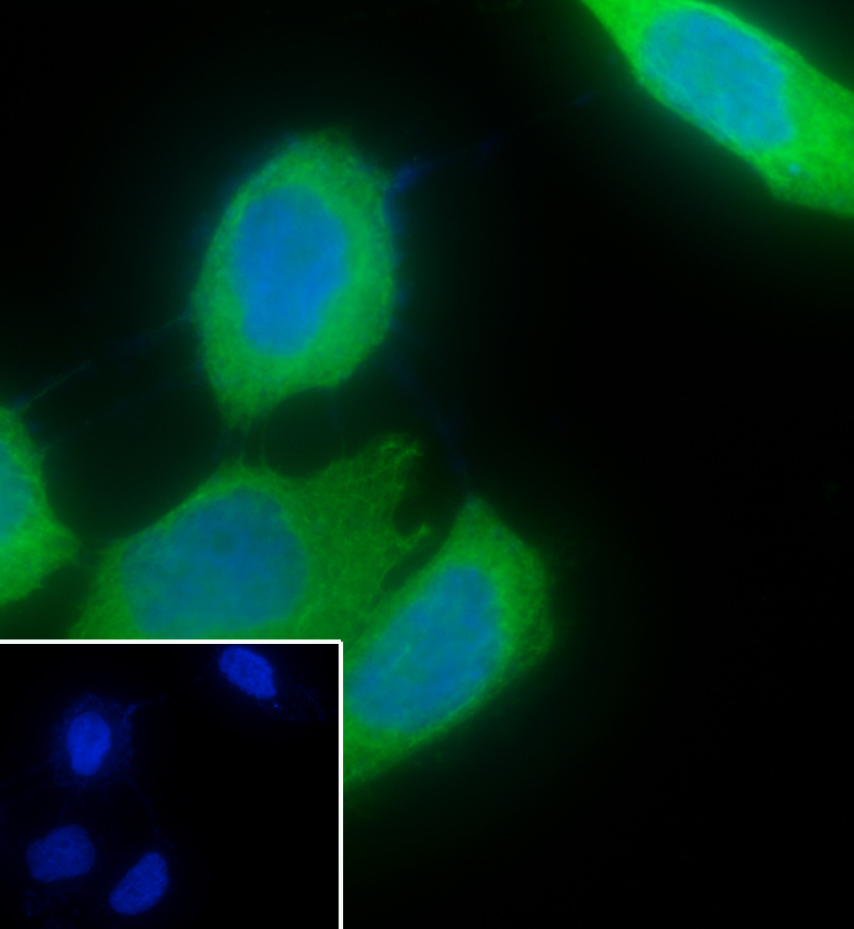 Cell line: HaCat
Fixative: 4% Paraformaldehyde
Permeabilization: 0.1% TritonX-100
Primary ab dilution: 1:50
Primary incubation condition: 4°C overnight
Nuclear counter stain: DAPI (Blue)
Comment: Color green is the positive signal for SLM-60235R
Cartpieces
Totalgoods,subtotals:¥Checkout
References (0)
No References
Bought notes(bought amounts latest0)
No one bought this product
User Comment(Total0User Comment Num)
- No comment


 +86 571 56623320
 +86 18668110335

