
Rabbit Anti-MSH6 antibody
DNA mismatch repair protein MSH6; G/T mismatch binding protein; GTBP; GTMBP; HNPCC 5; HNPCC5; HSAP; MSH 6; mutS (E. coli) homolog 6; MutS alpha 160 kDa subunit; mutS homolog 6; p160; Sperm associated protein; MSH6_HUMAN; MutS Protein Homolog 6.
View History [Clear]
Details
Product Name MSH6 Chinese Name 错配修复蛋白6Recombinant rabbit monoclonal anti Alias DNA mismatch repair protein MSH6; G/T mismatch binding protein; GTBP; GTMBP; HNPCC 5; HNPCC5; HSAP; MSH 6; mutS (E. coli) homolog 6; MutS alpha 160 kDa subunit; mutS homolog 6; p160; Sperm associated protein; MSH6_HUMAN; MutS Protein Homolog 6. Research Area Chromatin and nuclear signals Immunogen Species Rabbit Clonality Monoclonal Clone NO. M5F6 React Species Human, Mouse, Rat, Applications WB=1:500-2000 IHC-P=1:200-1:1000 ICC=1:100 IF=1:50-100 (Paraffin sections need antigen repair)
not yet tested in other applications.
optimal dilutions/concentrations should be determined by the end user.Theoretical molecular weight 153kDa Cellular localization The nucleus Form Liquid Concentration 1mg/ml immunogen KLH conjugated synthetic peptide derived from human MSH6 Lsotype IgG Purification affinity purified by Protein A Buffer Solution 0.01M TBS(pH7.4) with 1% BSA, 0.03% Proclin300 and 50% Glycerol. Storage Shipped at 4℃. Store at -20 °C for one year. Avoid repeated freeze/thaw cycles. Attention This product as supplied is intended for research use only, not for use in human, therapeutic or diagnostic applications. PubMed PubMed Product Detail This gene encodes a member of the DNA mismatch repair MutS family. In E. coli, the MutS protein helps in the recognition of mismatched nucleotides prior to their repair. A highly conserved region of approximately 150 aa, called the Walker-A adenine nucleotide binding motif, exists in MutS homologs. The encoded protein heterodimerizes with MSH2 to form a mismatch recognition complex that functions as a bidirectional molecular switch that exchanges ADP and ATP as DNA mismatches are bound and dissociated. Mutations in this gene may be associated with hereditary nonpolyposis colon cancer, colorectal cancer, and endometrial cancer. Transcripts variants encoding different isoforms have been described. [provided by RefSeq, Jul 2013]
SWISS:
P52701
Gene ID:
2956
Product Picture 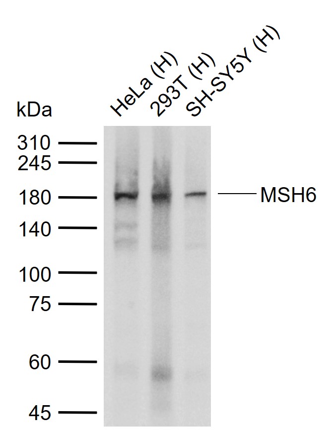 Sample:
Lane 1: Human HeLa cell lysates
Lane 2: Human 293T cell lysates
Lane 3: Human SH-SY5Y cell lysates
Primary: Anti-MSH6 (SLM-60220R) at 1/4000 dilution
Secondary: IRDye800CW Goat Anti-Rabbit IgG at 1/20000 dilution
Predicted band size: 153 kDa
Observed band size: 180 kDa
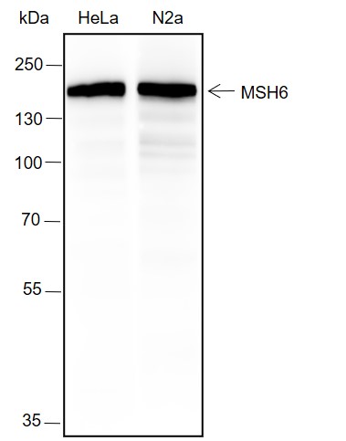 Blocking buffer: 5% NFDM/TBST
Primary ab dilution: 1:4000
Primary ab incubation condition: 2 hours at room temperature
Lysate: HeLa, N2a
Protein loading quantity: 20 μg
Exposure time: 15 s
Predicted MW: 150 kDa
Observed MW: 170 kDa
 Tissue: Human colon
Section type: Formalin fixed & Paraffin -embedded section
Retrieval method: High temperature and high pressure
Retrieval buffer: Tris/EDTA buffer, pH 9.0 Primary ab dilution: 1:1000
Primary ab incubation condition: 1 hour at room temperature
Counter stain: Hematoxylin
Comment: Color brown is the positive signal for SLM-60220R
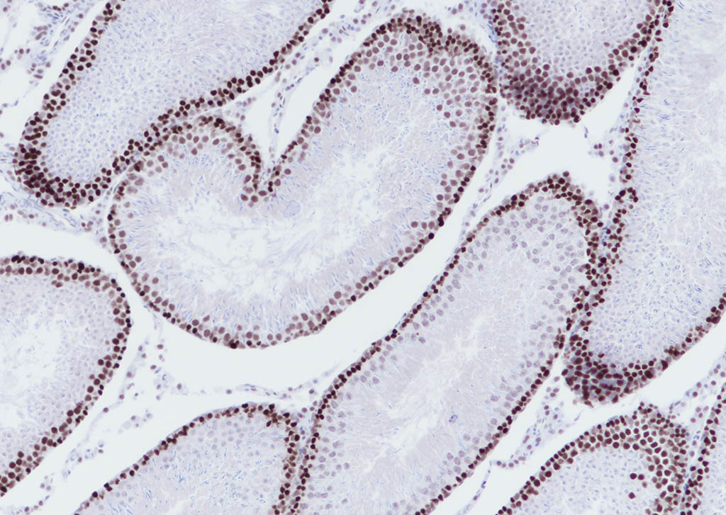 Tissue: Rat testis
Section type: Formalin fixed & Paraffin -embedded section
Retrieval method: High temperature and high pressure
Retrieval buffer: Tris/EDTA buffer, pH 9.0 Primary ab dilution: 1:1000
Primary ab incubation condition: 1 hour at room temperature
Counter stain: Hematoxylin
Comment: Color brown is the positive signal for SLM-60220R
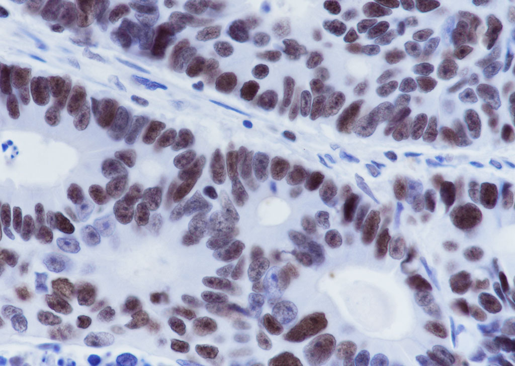 Tissue: Human colon cancer
Section type: Formalin fixed & Paraffin -embedded section
Retrieval method: High temperature and high pressure
Retrieval buffer: Tris/EDTA buffer, pH 9.0 Primary ab dilution: 1:1000
Primary ab incubation condition: 1 hour at room temperature
Counter stain: Hematoxylin
Comment: Color brown is the positive signal for SLM-60220R
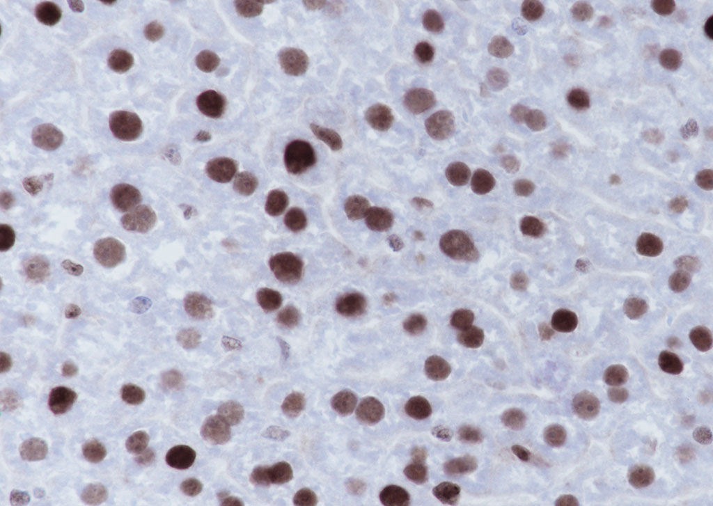 Tissue: Mouse liver
Section type: Formalin fixed & Paraffin -embedded section
Retrieval method: High temperature and high pressure
Retrieval buffer: Tris/EDTA buffer, pH 9.0 Primary ab dilution: 1:1000
Primary ab incubation condition: 1 hour at room temperature
Counter stain: Hematoxylin
Comment: Color brown is the positive signal for SLM-60220R
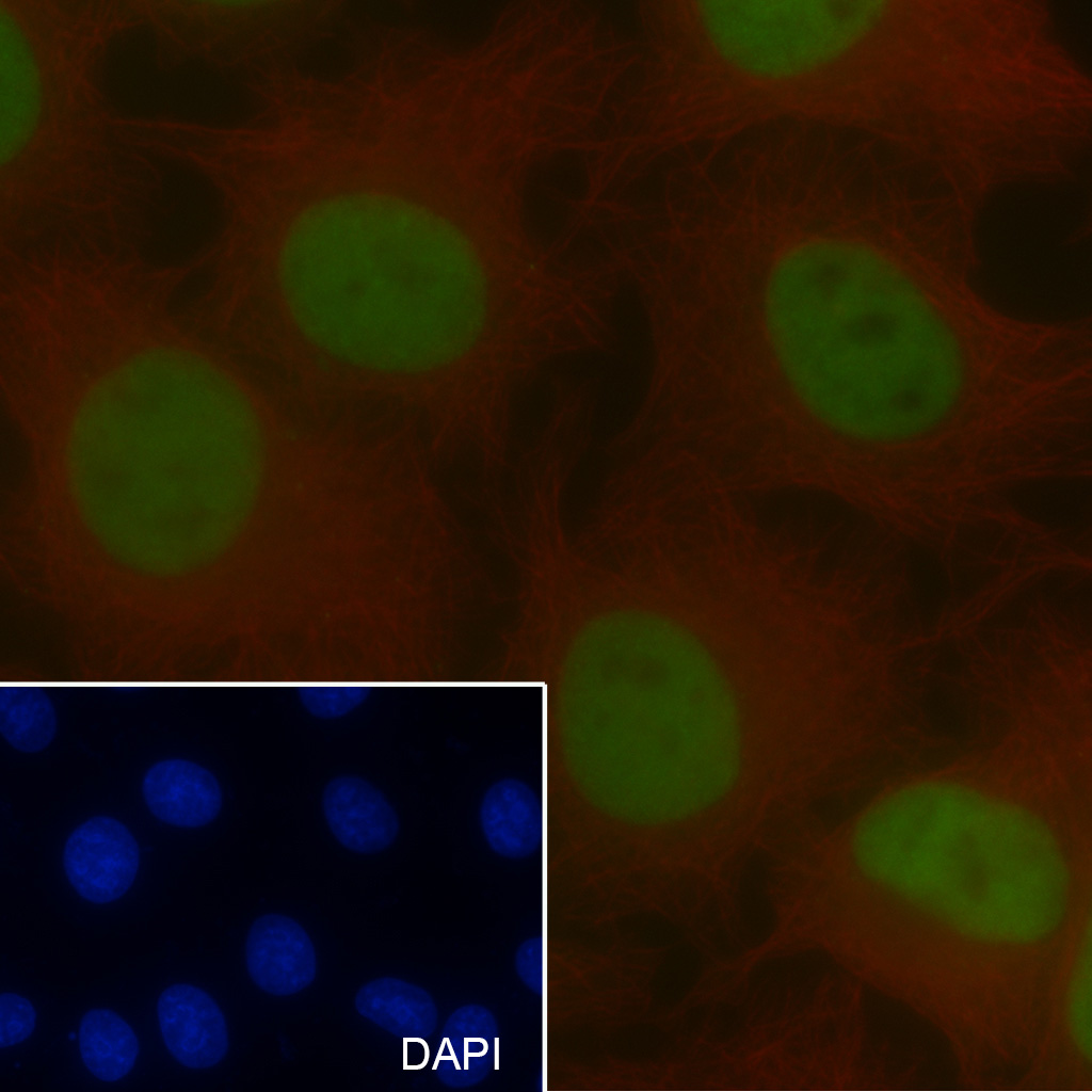 Cell line: HeLa Fixative: 4% Paraformaldehyde Permeabilization: 0.1% TritonX-100 Primary ab dilution: 1:100 Primary incubation condition: 1 hour at room temperature Nuclear counter stain: DAPI (Blue) Counter stain: Tubulin (Red) Comment: Color green is the positive signal for SLM-60220R
Cartpieces
Totalgoods,subtotals:¥Checkout
References (0)
No References
Bought notes(bought amounts latest0)
No one bought this product
User Comment(Total0User Comment Num)
- No comment


 +86 571 56623320
 +86 18668110335

