
Rabbit Anti-phospho-GSK3 Alpha + Beta (Tyr279+Tyr216)antibody
Phospho-GSK3(alpha+beta)(Y216+Y279); GSK3B(Phospho-Tyr279+Tyr216); GSK3B(Phospho-Y279/Y216); p-GSK-3 Beta(Tyr279+Tyr216); p-GSK-3 beta(Y279/Y216); Glycogen synthase kinase 3 beta; GSK 3 beta; GSK 3B; GSK3B; GSK3B protein; GSK3beta isoform; GSK3 beta; Glyc
View History [Clear]
Details
Product Name phospho-GSK3 Alpha + Beta (Tyr279+Tyr216) Chinese Name 磷酸化糖原合酶激酶3α/βRecombinant rabbit monoclonal anti Alias Phospho-GSK3(alpha+beta)(Y216+Y279); GSK3B(Phospho-Tyr279+Tyr216); GSK3B(Phospho-Y279/Y216); p-GSK-3 Beta(Tyr279+Tyr216); p-GSK-3 beta(Y279/Y216); Glycogen synthase kinase 3 beta; GSK 3 beta; GSK 3B; GSK3B; GSK3B protein; GSK3beta isoform; GSK3 beta; Glycogen synthase kinase-3 beta; GSK-3 beta; Glycogen synthase kinase-3 alpha; GSK3A; GSK-3 alpha; Serine/threonine-protein kinase GSK3A; GSK3A_HUMAN; GSK3B_HUMAN. GSK 3β; GSK 3 β; GSK-3β; GSK3β; GSK 3α; GSK 3 α; GSK-3α; GSK3α; Research Area Cell biology immunology Neurobiology Signal transduction Apoptosis transcriptional regulatory factor Kinases and Phosphatases Immunogen Species Rabbit Clonality Monoclonal Clone NO. 5G8 React Species Human, Mouse, Rat, Applications WB=1:500-2000 IHC-P=1:50-200 ICC=1:50 (Paraffin sections need antigen repair)
not yet tested in other applications.
optimal dilutions/concentrations should be determined by the end user.Theoretical molecular weight 47/51kDa Cellular localization The nucleus cytoplasmic The cell membrane Form Liquid Concentration 1mg/ml immunogen KLH conjugated Synthesised phosphopeptide derived from human GSK-3 Beta around the phosphorylation site of Tyr216 Lsotype IgG Purification affinity purified by Protein A Buffer Solution 0.01M TBS(pH7.4) with 1% BSA, 0.03% Proclin300 and 50% Glycerol. Storage Shipped at 4℃. Store at -20 °C for one year. Avoid repeated freeze/thaw cycles. Attention This product as supplied is intended for research use only, not for use in human, therapeutic or diagnostic applications. PubMed PubMed Product Detail Glycogen synthase kinase 3 (GSK3) is a proline directed serine threonine kinase that was initially identified as a phosphorylating and inactivating glycogen synthase, a key enzyme in glycogen metabolism. Since then, it has been shown to be involved in the regulation of a diverse array of cellular functions, including protein synthesis, cell proliferation, cell differentiation, microtubule assembly/disassembly, and apoptosis. GSK3s substrate specificity is unique in that phosphorylation of substrate only occurs if a phosphoserine or phosphotyrosine is present four residues C terminal to the site of GSK phosphorylation. There exists two isoforms of GSK3, alpha and beta, and they show a high degree of amino acid homology. The two isoforms of GSK3 are strictly regulated via phosphorylation. Phosphorylation of GSK3 beta on Ser9 (Ser21 in GSK3 alpha) by protein kinase B (PKB) causes its inactivation is the primary mechanism responsible for growth factor inhibition of this kinase. Activation of GSK3 beta is dependent upon the phosphorylation of Tyr216 (Tyr279 in GSK3 alpha). Upon activation, it has been shown to phosphorylate a number of different cellular proteins, including p53, c-Myc, c-Jun, heat shock factor 1 (HSF1), and cyclin D1. GSK3 beta also has been shown to phosphorylate aberrant sites on the microtubule associated protein tau, which is critical for the progression of Alzheimer's disease. GSK3B is involved in energy metabolism, neuronal cell development, and body pattern formation.
Function:
Constitutively active protein kinase that acts as a negative regulator in the hormonal control of glucose homeostasis, Wnt signaling and regulation of transcription factors and microtubules, by phosphorylating and inactivating glycogen synthase (GYS1 or GYS2), EIF2B, CTNNB1/beta-catenin, APC, AXIN1, JUN, NFATC1/NFATC, MAPT/TAU and MACF1. Requires primed phosphorylation of the majority of its substrates. In skeletal muscle, contributes to insulin regulation of glycogen synthesis by phosphorylating and inhibiting GYS1 activity and hence glycogen synthesis. May also mediate the development of insulin resistance by regulating activation of transcription factors. Regulates protein synthesis by controlling the activity of initiation factor 2B (EIF2BE/EIF2B5) in the same manner as glycogen synthase. In Wnt signaling, GSK3B forms a multimeric complex with APC, AXIN1 and CTNNB1/beta-catenin and phosphorylates the N-terminus of CTNNB1 leading to its degradation mediated by ubiquitin/proteasomes. Phosphorylates JUN at sites proximal to its DNA-binding domain, thereby reducing its affinity for DNA. Phosphorylates NFATC1/NFATC on conserved serine residues promoting NFATC1/NFATC nuclear export, shutting off NFATC1/NFATC gene regulation, and thereby opposing the action of calcineurin. Phosphorylates MAPT/TAU on 'Thr-548', decreasing significantly MAPT/TAU ability to bind and stabilize microtubules. MAPT/TAU is the principal component of neurofibrillary tangles in Alzheimer disease. Plays an important role in ERBB2-dependent stabilization of microtubules at the cell cortex. Phosphorylates MACF1, inhibiting its binding to microtubules which is critical for its role in bulge stem cell migration and skin wound repair. Probably regulates NF-kappa-B (NFKB1) at the transcriptional level and is required for the NF-kappa-B-mediated anti-apoptotic response to TNF-alpha (TNF/TNFA). Negatively regulates replication in pancreatic beta-cells, resulting in apoptosis, loss of beta-cells and diabetes. Phosphorylates MUC1 in breast cancer cells, decreasing the interaction of MUC1 with CTNNB1/beta-catenin. Is necessary for the establishment of neuronal polarity and axon outgrowth. Phosphorylates MARK2, leading to inhibit its activity. Phosphorylates SIK1 at 'Thr-182', leading to sustain its activity.
Subunit:
Monomer. Interacts with ARRB2 and DISC1. Interacts with CABYR, MMP2, MUC1, NIN and PRUNE Interacts with AXIN1; the interaction mediates hyperphosphorylation of CTNNB1 leading to its ubiquitination and destruction. Interacts with and phosphorylates SNAI1. Interacts with DNM1L (via a C-terminal domain). Found in a complex composed of MACF1, APC, AXIN1, CTNNB1 and GSK3B.
Subcellular Location:
Cytoplasm. Nucleus. Cell membrane. Note=The phosphorylated form shows localization to cytoplasm and cell membrane. The MEMO1-RHOA-DIAPH1 signaling pathway controls localization of the phosophorylated form to the cell membrane.
Tissue Specificity:
Expressed in testis, thymus, prostate and ovary and weakly expressed in lung, brain and kidney.
Post-translational modifications:
Phosphorylated by AKT1 and ILK1. Upon insulin-mediated signaling, the activated PKB/AKT1 protein kinase phosphorylates and desactivates GSK3B, resulting in the dephosphorylation and activation of GYS1. Activated by phosphorylation at Tyr-216.
Similarity:
Belongs to the protein kinase superfamily. CMGC Ser/Thr protein kinase family. GSK-3 subfamily.
Contains 1 protein kinase domain.
SWISS:
P49841
Gene ID:
2932
Database links:Entrez Gene: 2932 Human
Entrez Gene: 56637 Mouse
Omim: 605004 Human
SwissProt: P49841 Human
SwissProt: Q9WV60 Mouse
Unigene: 445733 Human
Unigene: 394930 Mouse
Product Picture 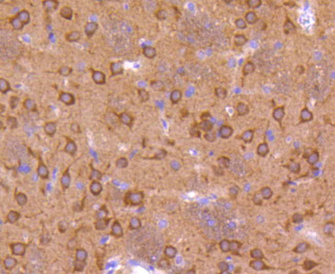 Paraformaldehyde-fixed, paraffin embedded (rat brain); Antigen retrieval by boiling in sodium citrate buffer (pH6.0) for 15min; Block endogenous peroxidase by 3% hydrogen peroxide for 20 minutes; Blocking buffer (normal goat serum) at 37°C for 30min; Antibody incubation with (phospho-GSK3 Alpha + Beta (Tyr279+Tyr216) ) Monoclonal Antibody, Unconjugated (SLM-54505R) at 1:50 overnight at 4°C, followed by operating according to SP Kit(Rabbit) (sp-0023) instructionsand DAB staining.
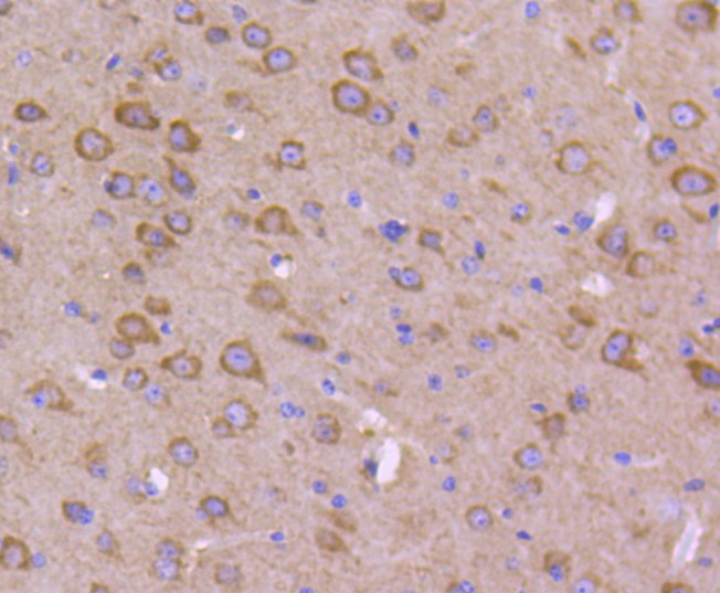 Paraformaldehyde-fixed, paraffin embedded (mouse brain); Antigen retrieval by boiling in sodium citrate buffer (pH6.0) for 15min; Block endogenous peroxidase by 3% hydrogen peroxide for 20 minutes; Blocking buffer (normal goat serum) at 37°C for 30min; Antibody incubation with (phospho-GSK3 Alpha + Beta (Tyr279+Tyr216) ) Monoclonal Antibody, Unconjugated (SLM-54505R) at 1:50 overnight at 4°C, followed by operating according to SP Kit(Rabbit) (sp-0023) instructionsand DAB staining.
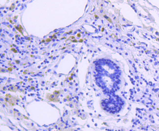 Paraformaldehyde-fixed, paraffin embedded (human breast carcinoma); Antigen retrieval by boiling in sodium citrate buffer (pH6.0) for 15min; Block endogenous peroxidase by 3% hydrogen peroxide for 20 minutes; Blocking buffer (normal goat serum) at 37°C for 30min; Antibody incubation with (phospho-GSK3 Alpha + Beta (Tyr279+Tyr216) ) Monoclonal Antibody, Unconjugated (SLM-54505R) at 1:50 overnight at 4°C, followed by operating according to SP Kit(Rabbit) (sp-0023) instructionsand DAB staining.
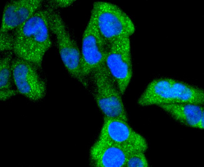 Hela cell; 4% Paraformaldehyde-fixed; Triton X-100 at room temperature for 20 min; Blocking buffer (normal goat serum, C-0005) at 37°C for 20 min; Antibody incubation with (Phospho-GSK3(alpha+beta)(Y216+Y279)) monoclonal Antibody, Unconjugated (SLM-54505R) 1:50, 90 minutes at 37°C; followed by a conjugated Goat Anti-Rabbit IgG antibody at 37°C for 90 minutes, DAPI (blue, C02-04002) was used to stain the cell nuclei.
 A431 cell; 4% Paraformaldehyde-fixed; Triton X-100 at room temperature for 20 min; Blocking buffer (normal goat serum, C-0005) at 37°C for 20 min; Antibody incubation with (Phospho-GSK3(alpha+beta)(Y216+Y279)) monoclonal Antibody, Unconjugated (SLM-54505R) 1:50, 90 minutes at 37°C; followed by a conjugated Goat Anti-Rabbit IgG antibody at 37°C for 90 minutes, DAPI (blue, C02-04002) was used to stain the cell nuclei.A431 cell; 4% Paraformaldehyde-fixed; Triton X-100 at room temperature for 20 min; Blocking buffer (normal goat serum, C-0005) at 37°C for 20 min; Antibody incubation with (Phospho-GSK3(alpha+beta)(Y216+Y279)) monoclonal Antibody, Unconjugated (SLM-54505R) 1:50, 90 minutes at 37°C; followed by a conjugated Goat Anti-Rabbit IgG antibody at 37°C for 90 minutes, DAPI (blue, C02-04002) was used to stain the cell nuclei.
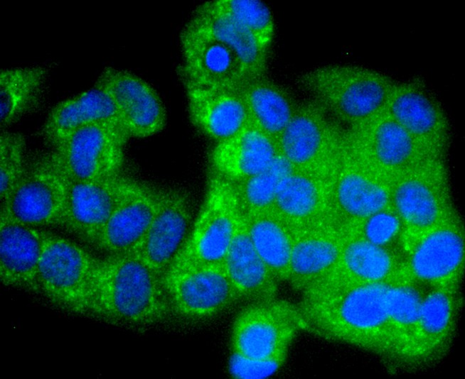 MCF-7 cell; 4% Paraformaldehyde-fixed; Triton X-100 at room temperature for 20 min; Blocking buffer (normal goat serum, C-0005) at 37°C for 20 min; Antibody incubation with (WWW) monoclonal Antibody, Unconjugated (SLM-54505R) 1:50, 90 minutes at 37°C; followed by a conjugated Goat Anti-Rabbit IgG antibody at 37°C for 90 minutes, DAPI (blue, C02-04002) was used to stain the cell nuclei.
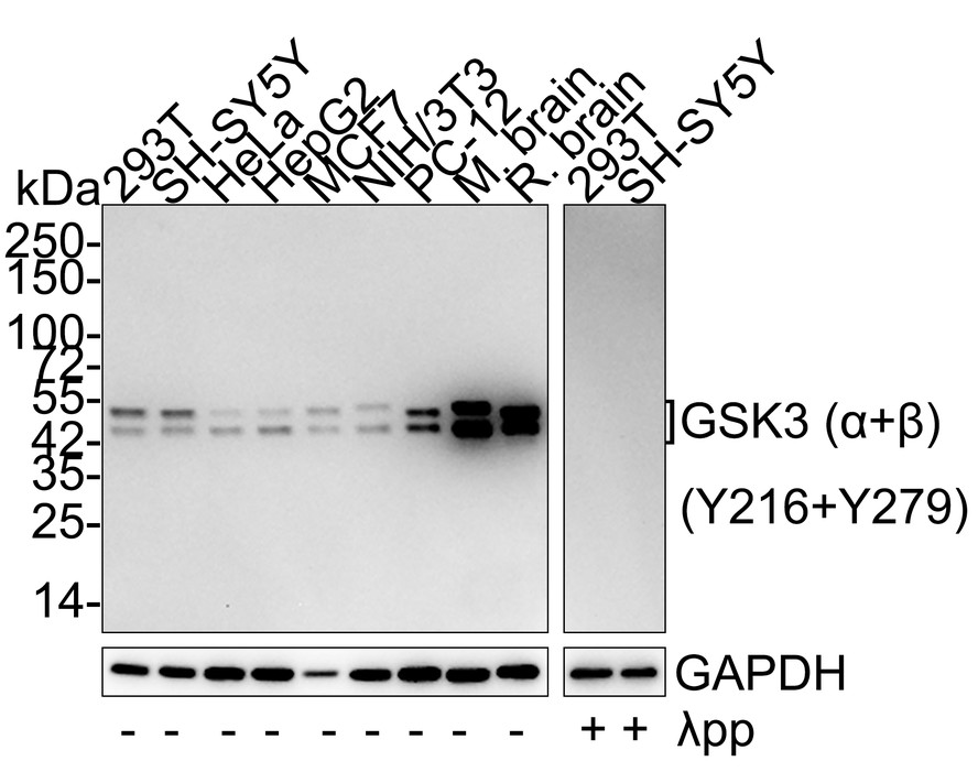 Western blot analysis of Phospho-GSK3 (alpha+beta) (Y216+Y279) on different lysates with Rabbit anti-Phospho-GSK3 (alpha+beta) (Y216+Y279) antibody at 1/5,000 dilution. Lane 1: 293T cell lysate Lane 2: SH-SY5Y cell lysate Lane 3: HeLa cell lysate Lane 4: HepG2 cell lysate Lane 5: MCF7 cell lysate Lane 6: NIH/3T3 cell lysate Lane 7: PC-12 cell lysate Lane 8: Mouse brain tissue lysate Lane 9: Rat brain tissue lysate Lane 10: 293T treated with λpp for 1 hour cell lysate Lane 11: SH-SY5Y treated with λpp for 1 hour cell lysate Predicted band size: 47/51 kDa Observed band size: 47/51 kDa
Cartpieces
Totalgoods,subtotals:¥Checkout
References (0)
No References
Bought notes(bought amounts latest0)
No one bought this product
User Comment(Total0User Comment Num)
- No comment


 +86 571 56623320
 +86 18668110335

