
Rabbit Anti-GEF H1 antibody
Lbcl1; Lfc; ARHG2; ARHG2_HUMAN; ARHGEF 2; ARHGEF-2; ARHGEF2; GEF; GEF H1; GEF-H1; GEFH1; Guanine nucleotide exchange factor H1; LFP40; Microtubule-regulated Rho-GEF; P40 antibody Proliferating cell nucleolar antigen p40; Protein GEF-H1; Rho guanine nucleo
View History [Clear]
Details
Product Name GEF H1 Chinese Name Rho鸟苷酸交换因子2Recombinant rabbit monoclonal anti Alias Lbcl1; Lfc; ARHG2; ARHG2_HUMAN; ARHGEF 2; ARHGEF-2; ARHGEF2; GEF; GEF H1; GEF-H1; GEFH1; Guanine nucleotide exchange factor H1; LFP40; Microtubule-regulated Rho-GEF; P40 antibody Proliferating cell nucleolar antigen p40; Protein GEF-H1; Rho guanine nucleotide exchange factor 2; rho/rac guanine nucleotide exchange factor (GEF) 2; rho/rac guanine nucleotide exchange factor 2; rho/rac guanine nucleotide exchange factor. Research Area Tumour immunology Signal transduction transcriptional regulatory factor Immunogen Species Rabbit Clonality Monoclonal Clone NO. 9D4 React Species Human, Mouse, Rat, Applications WB=1:500 IHC-P=1:50-200 IHC-F=1:50-200 ICC=1:50 IF=1:50-200 (Paraffin sections need antigen repair)
not yet tested in other applications.
optimal dilutions/concentrations should be determined by the end user.Theoretical molecular weight 112kDa Cellular localization cytoplasmic The cell membrane Form Liquid Concentration 1mg/ml immunogen KLH conjugated synthetic peptide derived from human GEF H1 Lsotype IgG Purification affinity purified by Protein A Buffer Solution 0.01M TBS(pH7.4) with 1% BSA, 0.03% Proclin300 and 50% Glycerol. Storage Shipped at 4℃. Store at -20 °C for one year. Avoid repeated freeze/thaw cycles. Attention This product as supplied is intended for research use only, not for use in human, therapeutic or diagnostic applications. PubMed PubMed Product Detail Activates Rho-GTPases by promoting the exchange of GDP for GTP. May be involved in epithelial barrier permeability, cell motility and polarization, dendritic spine morphology, antigen presentation, leukemic cell differentiation, cell cycle regulation, and cancer. Binds Rac-GTPases, but does not seem to promote nucleotide exchange activity toward Rac-GTPases, which was uniquely reported in PubMed:9857026. May stimulate instead the cortical activity of Rac. Inactive toward CDC42, TC10, or Ras-GTPases. Forms an intracellular sensing system along with NOD1 for the detection of microbial effectors during cell invasion by pathogens. Required for RHOA and RIP2 dependent NF-kappaB signaling pathways activation upon S.flexneri cell invasion. Involved not only in sensing peptidoglycan (PGN)-derived muropeptides through NOD1 that is independent of its GEF activity, but also in the activation of NF-kappaB by Shigella effector proteins (IpgB2 and OspB) which requires its GEF activity and the activation of RhoA.
Function:
Activates Rho-GTPases by promoting the exchange of GDP for GTP. May be involved in epithelial barrier permeability, cell motility and polarization, dendritic spine morphology, antigen presentation, leukemic cell differentiation, cell cycle regulation, and cancer. Binds Rac-GTPases, but does not seem to promote nucleotide exchange activity toward Rac-GTPases, which was uniquely reported in PubMed:9857026. May stimulate instead the cortical activity of Rac. Inactive toward CDC42, TC10, or Ras-GTPases. Forms an intracellular sensing system along with NOD1 for the detection of microbial effectors during cell invasion by pathogens. Required for RHOA and RIP2 dependent NF-kappaB signaling pathways activation upon S.flexneri cell invasion. Involved not only in sensing peptidoglycan (PGN)-derived muropeptides through NOD1 that is independent of its GEF activity, but also in the activation of NF-kappaB by Shigella effector proteins (IpgB2 and OspB) which requires its GEF activity and the activation of RhoA.
Subunit:
Interacts with 14-3-3 zeta; when phosphorylated at Ser-886. Interacts with the kinases PAK4, AURKA and MAPK1. Interacts with RHOA and RAC1. Interacts with NOD1. Interacts (via the N-terminal zinc finger) with CAPN6 (via domain II).
Subcellular Location:
Cytoplasm. Cell junction, tight junction. Golgi apparatus. Cytoplasm, cytoskeleton, spindle. Cell projection, ruffle membrane. Note=Localizes to the tips of cortical microtubules of the mitotic spindle during cell division, and is further released upon microtubule depolymerization. Recruited into membrane ruffles induced by S.flexneri at tight junctions of polarized epithelial cells.
Post-translational modifications:
Phosphorylation of Ser-886 by PAK1 induces binding to protein 14-3-3 zeta, promoting its relocation to microtubules and the inhibition of its activity.
Phosphorylated by STK6 and CDK1 during mitosis, which negatively regulates its activity. Phosphorylation by MAPK1 or MAPK3 increases nucleotide exchange activity. Phosphorylation by PAK4 releases GEF-H1 from the microtubules.
Similarity:
Contains 1 DH (DBL-homology) domain.
Contains 1 PH domain.
Contains 1 phorbol-ester/DAG-type zinc finger.
SWISS:
Q92974
Gene ID:
9181
Database links:Entrez Gene: 9181 Human
Entrez Gene: 16800 Mouse
Omim: 607560 Human
SwissProt: Q92974 Human
SwissProt: Q60875 Mouse
Unigene: 516790 Human
Unigene: 239329 Mouse
Unigene: 482396 Mouse
Unigene: 12255 Rat
Product Picture 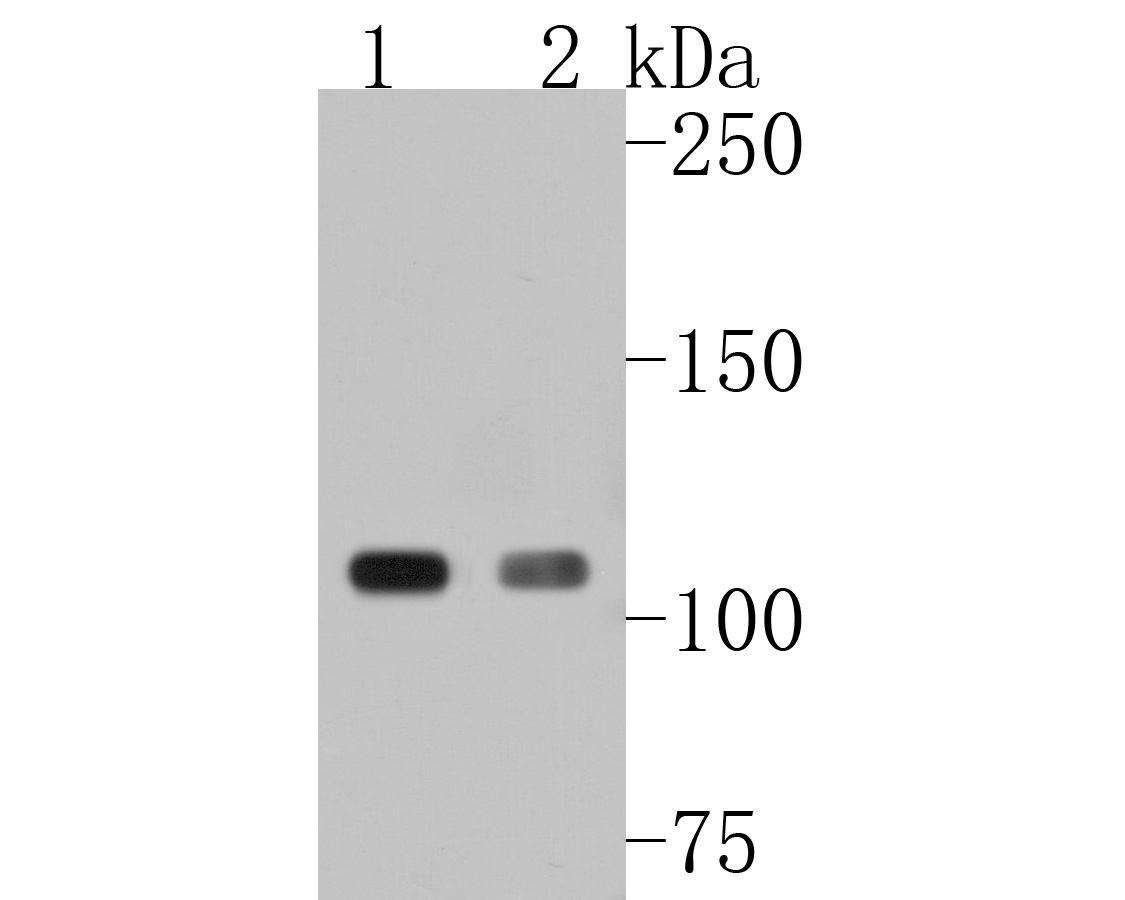 Sample:
Lane 1: 293T cell lysate
Lane 2: A431 cell lysate
Primary: Anti-GEF H1 (SLM-54369R) at 1:500 dilution
Secondary: Goat Anti-Rabbit IgG - HRP at 1:5000 dilution
Predicted band size: 112 kD
Observed band size: 112 kD
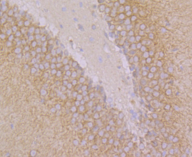 Paraformaldehyde-fixed, paraffin embedded (rat brain); Antigen retrieval by boiling in sodium citrate buffer (pH6.0) for 15min; Block endogenous peroxidase by 3% hydrogen peroxide for 20 minutes; Blocking buffer (normal goat serum) at 37°C for 30min; Antibody incubation with (GEF H1) Monoclonal Antibody, Unconjugated (SLM-54369R) at 1:50 overnight at 4°C, followed by operating according to SP Kit(Rabbit) (sp-0023) instructionsand DAB staining.
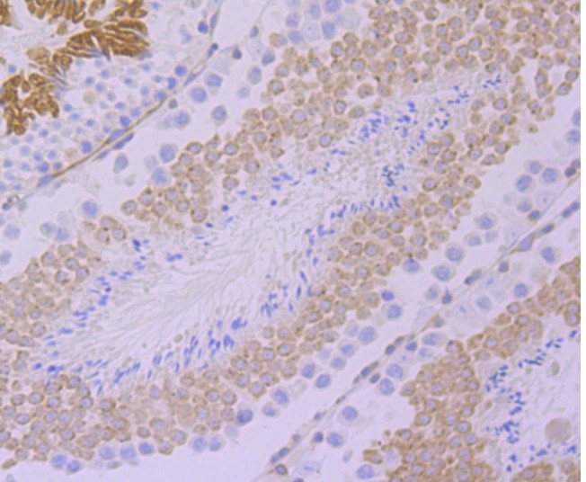 Paraformaldehyde-fixed, paraffin embedded (mouse testis); Antigen retrieval by boiling in sodium citrate buffer (pH6.0) for 15min; Block endogenous peroxidase by 3% hydrogen peroxide for 20 minutes; Blocking buffer (normal goat serum) at 37°C for 30min; Antibody incubation with (GEF H1) Monoclonal Antibody, Unconjugated (SLM-54369R) at 1:50 overnight at 4°C, followed by operating according to SP Kit(Rabbit) (sp-0023) instructionsand DAB staining.
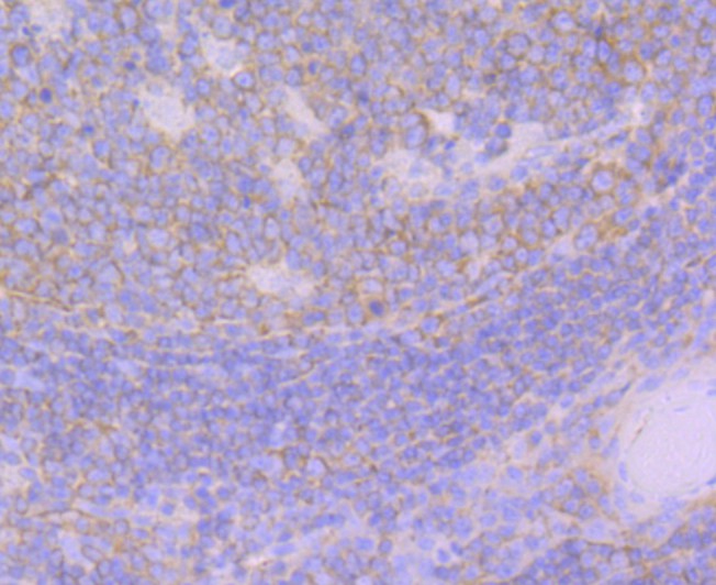 Paraformaldehyde-fixed, paraffin embedded (human tonsil); Antigen retrieval by boiling in sodium citrate buffer (pH6.0) for 15min; Block endogenous peroxidase by 3% hydrogen peroxide for 20 minutes; Blocking buffer (normal goat serum) at 37°C for 30min; Antibody incubation with (GEF H1) Monoclonal Antibody, Unconjugated (SLM-54369R) at 1:50 overnight at 4°C, followed by operating according to SP Kit(Rabbit) (sp-0023) instructionsand DAB staining.
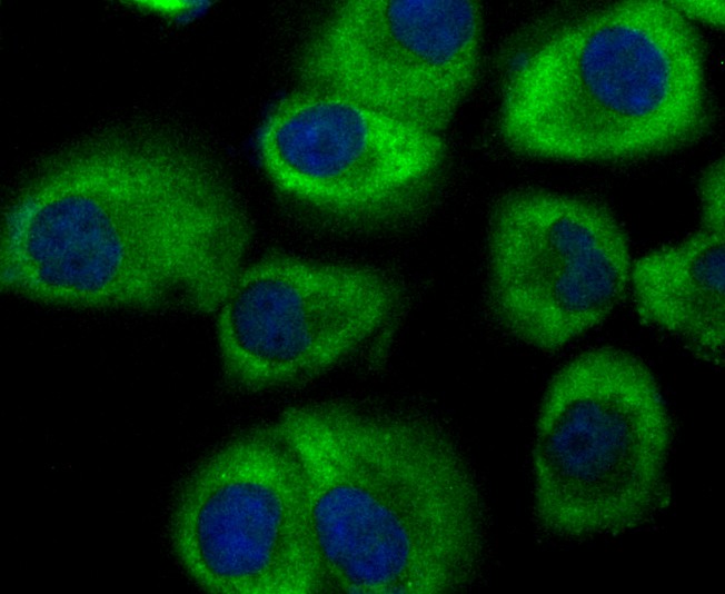 HUVEC cell; 4% Paraformaldehyde-fixed; Triton X-100 at room temperature for 20 min; Blocking buffer (normal goat serum, C-0005) at 37°C for 20 min; Antibody incubation with (GEF H1) monoclonal Antibody, Unconjugated (SLM-54369R) 1:50, 90 minutes at 37°C; followed by a conjugated Goat Anti-Rabbit IgG antibody at 37°C for 90 minutes, DAPI (blue, C02-04002) was used to stain the cell nuclei.
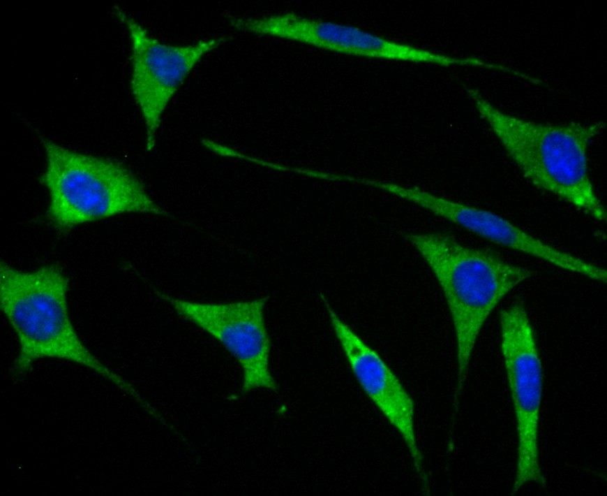 SH-SY5Y cell; 4% Paraformaldehyde-fixed; Triton X-100 at room temperature for 20 min; Blocking buffer (normal goat serum, C-0005) at 37°C for 20 min; Antibody incubation with (GEF H1) monoclonal Antibody, Unconjugated (SLM-54369R) 1:50, 90 minutes at 37°C; followed by a conjugated Goat Anti-Rabbit IgG antibody at 37°C for 90 minutes, DAPI (blue, C02-04002) was used to stain the cell nuclei.
Cartpieces
Totalgoods,subtotals:¥Checkout
References (0)
No References
Bought notes(bought amounts latest0)
No one bought this product
User Comment(Total0User Comment Num)
- No comment


 +86 571 56623320
 +86 18668110335

