
Rabbit Anti-ARP3 antibody
Actin-like protein 3; Actin-related protein 3; ACTR3; ARP3 actin-related protein 3 homolog; ARP3_HUMAN.
View History [Clear]
Details
Product Name ARP3 Chinese Name Cytoskeleton肌动蛋白样蛋白3抗体 Alias Actin-like protein 3; Actin-related protein 3; ACTR3; ARP3 actin-related protein 3 homolog; ARP3_HUMAN. Research Area Cell biology Cell adhesion molecule Cytoskeleton Immunogen Species Rabbit Clonality Monoclonal Clone NO. 1A1 React Species Human, Mouse, Rat, Applications WB=1:500-1000 IHC-P=1:50-200 Flow-Cyt=1:50-100 ICC=1:50-100 IF=1:50-100 (Paraffin sections need antigen repair)
not yet tested in other applications.
optimal dilutions/concentrations should be determined by the end user.Theoretical molecular weight 47kDa Form Liquid Concentration 1mg/ml immunogen Recombinant protein of C-terminal human Arp3. Lsotype IgG Purification affinity purified by Protein A Buffer Solution 0.01M TBS(pH7.4) with 1% BSA, 0.03% Proclin300 and 50% Glycerol. Storage Shipped at 4℃. Store at -20 °C for one year. Avoid repeated freeze/thaw cycles. Attention This product as supplied is intended for research use only, not for use in human, therapeutic or diagnostic applications. PubMed PubMed Product Detail Actin polymerization is required for a variety of cell functions, including chemotaxis, cell migration, cell adhesion, and platelet activation. Cells trigger actin polymerization through either the de novo nucleation of filaments from monomeric actin, the severing of existing filaments to create uncapped barbed ends, or the uncapping existing barbed ends. The nucleation of actin is a rate-limiting and unfavorable reaction in actin polymerization and therefore requires the involvement of the Arp2/3 complex, which helps create new filaments and promotes the end-to-side cross-linking of actin filaments into the branching meshwork. The Arp2/3 complex consists of the actin-related proteins Arp2 and Arp3, and various other accessory proteins. The Arp2/3 complex promotes actin nucleation by binding the pointed end of actin filaments, or by associating with the side of an existing filament, and nucleates growth in the barbed direction. In addition, the Arp2/3 complex also mediates actin cytoskeletal outgrowths that are regulated by the Rho family of small GTPases. In response to GTP-binding Cdc42, the Arp2/3 complex binds the Cdc42 substrates, namely the WASP proteins, and initiates the formation of lamellipodia and filopodia.
Function:
The Arp2/3 complex involved in Golgi polerization in NIH3T3 cells. In a different model it has been shown that the Arp2/3 complex is necessary for neutrophil chemotaxis and phagocytosis.
Subunit:
Component of the Arp2/3 complex composed of ARP2, ARP3, ARPC1B/p41-ARC, ARPC2/p34-ARC, ARPC3/p21-ARC, ARPC4/p20-ARC and ARPC5/p16-ARC. Interacts with WHDC1.
Subcellular Location:
Cytoplasm, cytoskeleton. Cell projection.
Similarity:
Belongs to the actin family. ARP3 subfamily.
SWISS:
P61158
Gene ID:
10096
Database links:Entrez Gene: 10096 Human
Omim: 604222 Human
SwissProt: P61158 Human
Product Picture 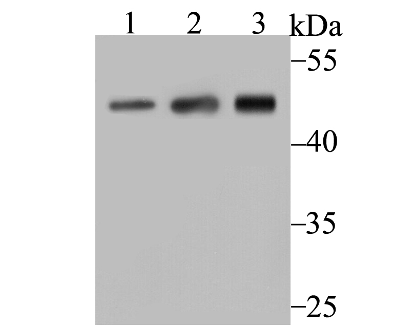 Sample:
Lane 1: A431 cell lysate
Lane 2: Mouse placenta tissue lysate
Lane 3: Mouse thymus tissue lysate
Primary: Anti-ARP3 (SLM-54337R) at 1:500 dilution
Secondary: Goat Anti-Rabbit IgG - HRP at 1:5000 dilution
Predicted band size: 47 kD
Observed band size: 50 kD
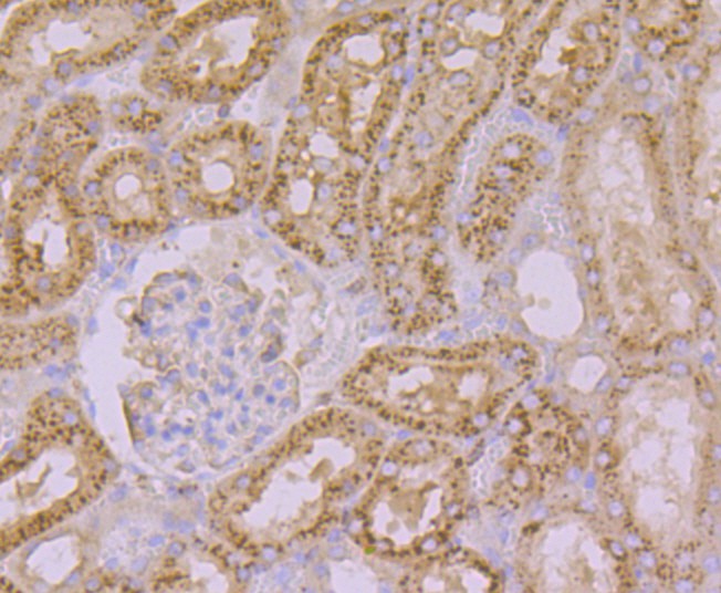 Paraformaldehyde-fixed, paraffin embedded (rat kidney); Antigen retrieval by boiling in sodium citrate buffer (pH6.0) for 15min; Block endogenous peroxidase by 3% hydrogen peroxide for 20 minutes; Blocking buffer (normal goat serum) at 37°C for 30min; Antibody incubation with (ARP3) Monoclonal Antibody, Unconjugated (SLM-54337R) at 1:50 overnight at 4°C, followed by operating according to SP Kit(Rabbit) (sp-0023) instructionsand DAB staining.
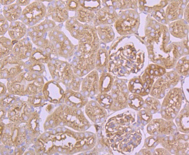 Paraformaldehyde-fixed, paraffin embedded (mouse kidney); Antigen retrieval by boiling in sodium citrate buffer (pH6.0) for 15min; Block endogenous peroxidase by 3% hydrogen peroxide for 20 minutes; Blocking buffer (normal goat serum) at 37°C for 30min; Antibody incubation with (ARP3) Monoclonal Antibody, Unconjugated (SLM-54337R) at 1:50 overnight at 4°C, followed by operating according to SP Kit(Rabbit) (sp-0023) instructionsand DAB staining.
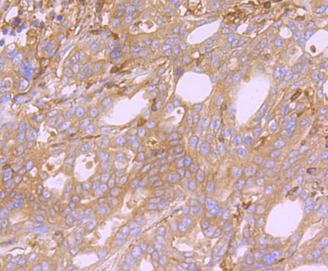 Paraformaldehyde-fixed, paraffin embedded (human stomach cancer); Antigen retrieval by boiling in sodium citrate buffer (pH6.0) for 15min; Block endogenous peroxidase by 3% hydrogen peroxide for 20 minutes; Blocking buffer (normal goat serum) at 37°C for 30min; Antibody incubation with (ARP3) Monoclonal Antibody, Unconjugated (SLM-54337R) at 1:50 overnight at 4°C, followed by operating according to SP Kit(Rabbit) (sp-0023) instructionsand DAB staining.
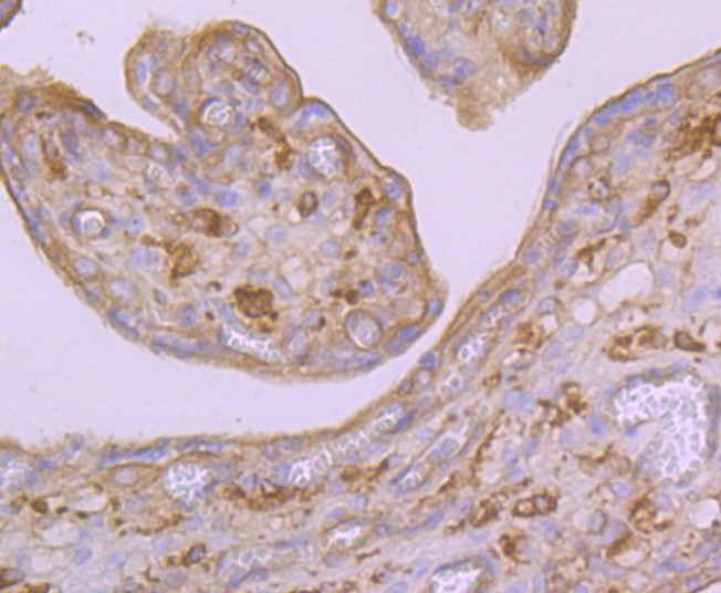 Paraformaldehyde-fixed, paraffin embedded (human placenta); Antigen retrieval by boiling in sodium citrate buffer (pH6.0) for 15min; Block endogenous peroxidase by 3% hydrogen peroxide for 20 minutes; Blocking buffer (normal goat serum) at 37°C for 30min; Antibody incubation with (ARP3) Monoclonal Antibody, Unconjugated (SLM-54337R) at 1:50 overnight at 4°C, followed by operating according to SP Kit(Rabbit) (sp-0023) instructionsand DAB staining.
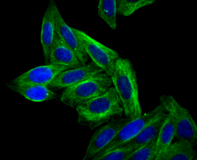 SiHa cell; 4% Paraformaldehyde-fixed; Triton X-100 at room temperature for 20 min; Blocking buffer (normal goat serum, C-0005) at 37°C for 20 min; Antibody incubation with (Arp3) monoclonal Antibody, Unconjugated (SLM-54337R) 1:50, 90 minutes at 37°C; followed by a conjugated Goat Anti-Rabbit IgG antibody at 37°C for 90 minutes, DAPI (blue, C02-04002) was used to stain the cell nuclei.
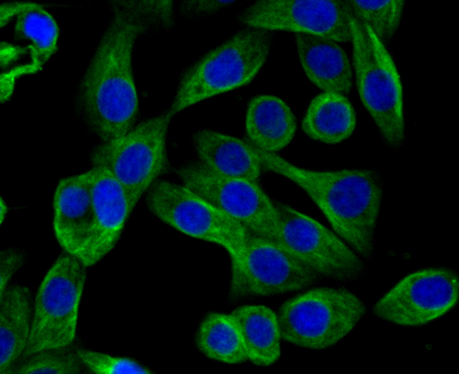 LOVO cell; 4% Paraformaldehyde-fixed; Triton X-100 at room temperature for 20 min; Blocking buffer (normal goat serum, C-0005) at 37°C for 20 min; Antibody incubation with (Arp3) monoclonal Antibody, Unconjugated (SLM-54337R) 1:50, 90 minutes at 37°C; followed by a conjugated Goat Anti-Rabbit IgG antibody at 37°C for 90 minutes, DAPI (blue, C02-04002) was used to stain the cell nuclei.
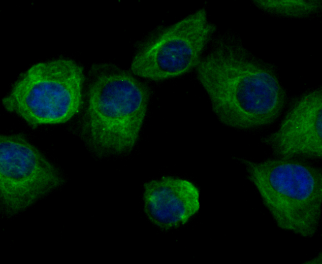 HUVEC cell; 4% Paraformaldehyde-fixed; Triton X-100 at room temperature for 20 min; Blocking buffer (normal goat serum, C-0005) at 37°C for 20 min; Antibody incubation with (Arp3) monoclonal Antibody, Unconjugated (SLM-54337R) 1:50, 90 minutes at 37°C; followed by a conjugated Goat Anti-Rabbit IgG antibody at 37°C for 90 minutes, DAPI (blue, C02-04002) was used to stain the cell nuclei.
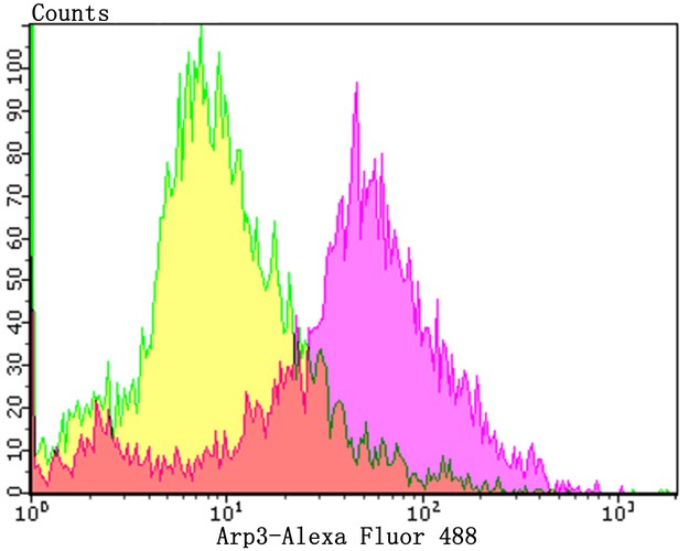 Blank control:HL-60.
Primary Antibody (green line): Rabbit Anti-Arp3 antibody (SLM-54337R)
Dilution: 1:100 cells;
Secondary Antibody : Goat anti-rabbit IgG-AF488
Dilution: 1:1000.
Protocol
The cells were fixed with 4% PFA (10min at room temperature)and then permeabilized with 0.1% PBST for 20 min at room temperature. The cells were then incubated in 5%BSA to block non-specific protein-protein interactions for 30 min at room temperature .Cells stained with Primary Antibody for 30 min at room temperature. The secondary antibody used for 40 min at room temperature. Acquisition of 20,000 events was performed.
Cartpieces
Totalgoods,subtotals:¥Checkout
References (0)
No References
Bought notes(bought amounts latest0)
No one bought this product
User Comment(Total0User Comment Num)
- No comment


 +86 571 56623320
 +86 18668110335

