
Rabbit Anti-BST2 antibody
Bone marrow stromal antigen 2; Bone marrow stromal cell antigen 2; Bone marrow stromal cell antigen; BST 2; BST-2; BST2_HUMAN; CD 317; CD317; CD317 antigen; HM1.24 antigen; NPC A 7; Tetherin.
View History [Clear]
Details
Product Name BST2 Chinese Name 骨髓基质Stem cells抗原2Recombinant rabbit monoclonal anti Alias Bone marrow stromal antigen 2; Bone marrow stromal cell antigen 2; Bone marrow stromal cell antigen; BST 2; BST-2; BST2_HUMAN; CD 317; CD317; CD317 antigen; HM1.24 antigen; NPC A 7; Tetherin. Research Area Tumour Cardiovascular Cell biology immunology Stem cells Cell Surface Molecule lymphocyte Immunogen Species Rabbit Clonality Monoclonal Clone NO. 1C3 React Species Human, Applications WB=1:300-500 IHC-P=1:50-200 IHC-F=1:50-200 Flow-Cyt=1:50 (Paraffin sections need antigen repair)
not yet tested in other applications.
optimal dilutions/concentrations should be determined by the end user.Theoretical molecular weight 18kDa Cellular localization cytoplasmic The cell membrane Form Liquid Concentration 1mg/ml immunogen KLH conjugated synthetic peptide derived from human BST2 Lsotype IgG Purification affinity purified by Protein A Buffer Solution 0.01M TBS(pH7.4) with 1% BSA, 0.03% Proclin300 and 50% Glycerol. Storage Shipped at 4℃. Store at -20 °C for one year. Avoid repeated freeze/thaw cycles. Attention This product as supplied is intended for research use only, not for use in human, therapeutic or diagnostic applications. PubMed PubMed Product Detail Bone marrow stromal cells act as regulators for B-cell growth and development through their surface molecules and cytokines. Bone marrow stromal antigen-2 (BST-2), also designated CD317 antigen, is a single- pass type II membrane protein. BST-2, which is expressed mainly on synovial cell lines and bone marrow stromal cell lines, is primarily expressed in liver, heart, placenta and lung tissues. BST-2 is thought to be involved in pre-B cell growth. It has been implicated in B cell activation in rheumatoid arthritis.
Function:
IFN-induced antiviral host restriction factor which efficiently blocks the release of diverse mammalian enveloped viruses by directly tethering nascent virions to the membranes of infected cells. Acts as a direct physical tether, holding virions to the cell membrane and linking virions to each other. The tethered virions can be internalized by endocytosis and subsequently degraded or they can remain on the cell surface. In either case, their spread as cell-free virions is restricted. Its target viruses belong to diverse families, including retroviridae: human immunodeficiency virus type 1 (HIV-1), human immunodeficiency virus type 2 (HIV-2), simian immunodeficiency viruses (SIVs), equine infectious anemia virus (EIAV), feline immunodeficiency virus (FIV), prototype foamy virus (PFV), Mason-Pfizer monkey virus (MPMV), human T-cell leukemia virus type 1 (HTLV-1), Rous sarcoma virus (RSV) and murine leukemia virus (MLV), flavivirideae: hepatitis C virus (HCV), filoviridae: ebola virus (EBOV) and marburg virus (MARV), arenaviridae: lassa virus (LASV) and machupo virus (MACV), herpesviridae: kaposis sarcoma-associated herpesvirus (KSHV), rhabdoviridae: vesicular stomatitis virus (VSV), orthomyxoviridae: influenza A virus, and paramyxoviridae: nipah virus. Can inhibit cell surface proteolytic activity of MMP14 causing decreased activation of MMP15 which results in inhibition of cell growth and migration. Can stimulate signaling by LILRA4/ILT7 and consequently provide negative feedback to the production of IFN by plasmacytoid dendritic cells in response to viral infection. Plays a role in the organization of the subapical actin cytoskeleton in polarized epithelial cells.
Subunit:
Parallel homodimer; disulfide-linked. May form homotetramers under reducing conditions. Dimerization is essential for its antiviral activity. Interacts (via cytoplasmic domain) with ARHGAP44 (By similarity). Interacts with MMP14 (via C-terminal cytoplasmic tail). Interacts with LILRA4/ILT7. Interacts (via transmembrane domain) with HIV-1 VPU (via transmembrane domain). Interacts with HIV-2 ENV and ebola GP protein.
Subcellular Location:
Golgi apparatus; trans-Golgi network. Cell membrane. Cell membrane. Late endosome. Targeted to late endosomes upon KSHV infection and subsequent ubiquitination. Targeted to the trans-Golgi network by viral VPU protein.
Tissue Specificity:
Predominantly expressed in liver, lung, heart and placenta. Lower levels in pancreas, kidney, skeletal muscle and brain. Overexpressed in multiple myeloma cells. Highly expressed during B-cell development, from pro-B precursors to plasma cells. Highly expressed on T-cells, monocytes, NK cells and dendritic cells (at protein level).
Post-translational modifications:
Monoubiquitinated by KSHV E3 ubiquitin-protein ligase K5, leading to its targeting to late endosomes and degradation.
The GPI anchor is essential for its antiviral activity.
Similarity:
Belongs to the tetherin family.
SWISS:
Q10589
Gene ID:
684
Database links:Entrez Gene: 684 Human
Omim: 600534 Human
SwissProt: Q10589 Human
Unigene: 118110 Human
Product Picture 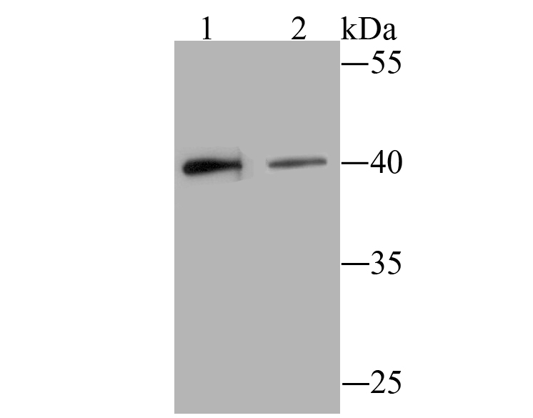 Sample:
Lane 1: Hela cell lysate
Lane 2: Siha cell lysatee
Primary: Anti-BST2 (SLM-54196R) at 1:500 dilution
Secondary: Goat Anti-Rabbit IgG - HRP at 1:5000 dilution
Predicted band size: 18 kD
Observed band size: 40 kD
 Paraformaldehyde-fixed, paraffin embedded (human breast carcinoma); Antigen retrieval by boiling in sodium citrate buffer (pH6.0) for 15min; Block endogenous peroxidase by 3% hydrogen peroxide for 20 minutes; Blocking buffer (normal goat serum) at 37°C for 30min; Antibody incubation with (BST2) Monoclonal Antibody, Unconjugated (SLM-54196R) at 1:200 overnight at 4°C, followed by operating according to SP Kit(Rabbit) (sp-0023) instructionsand DAB staining.
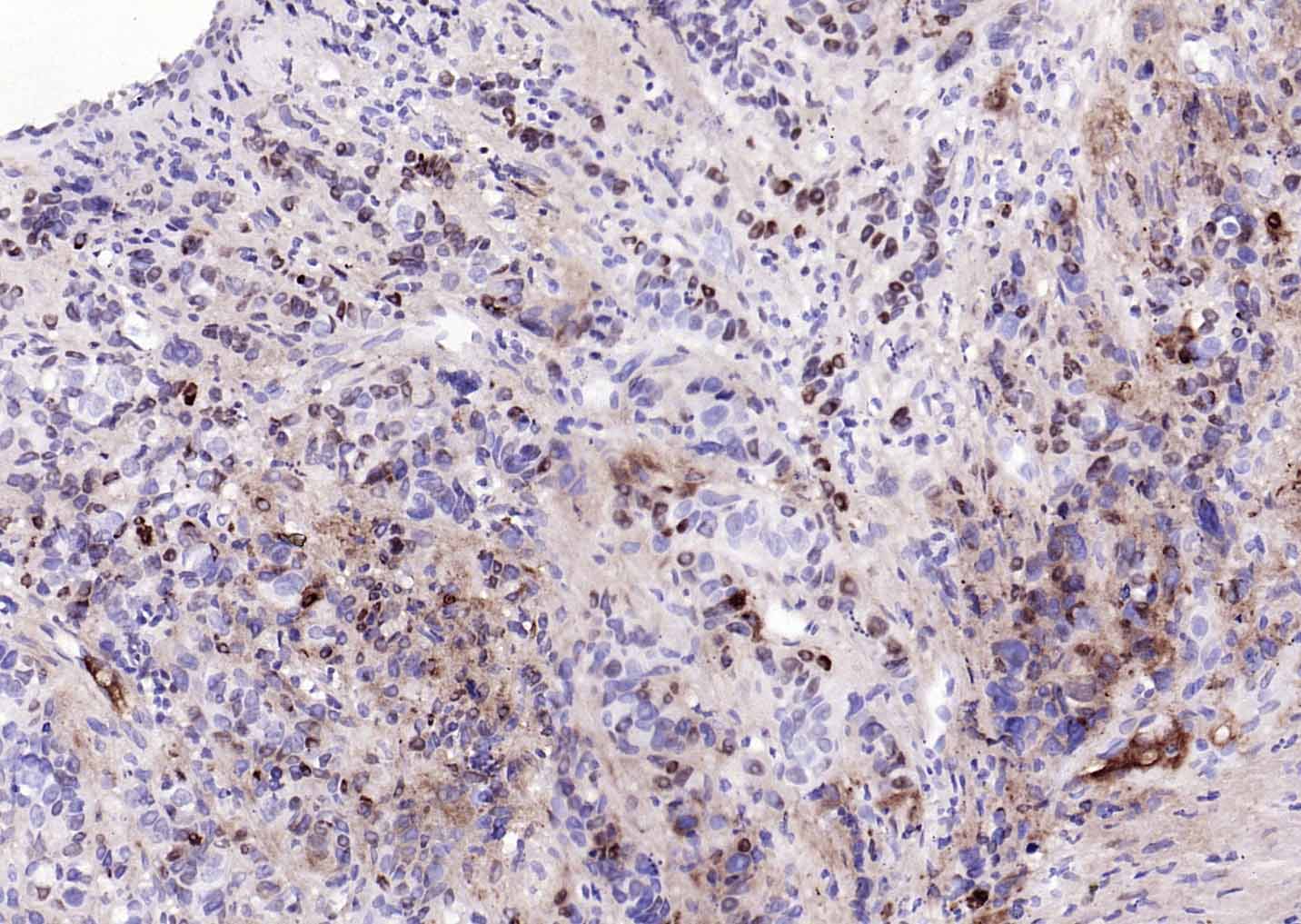
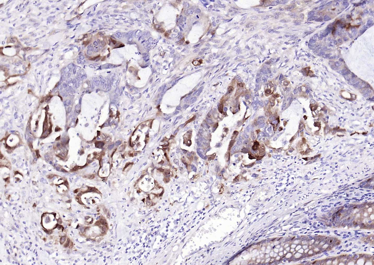 Paraformaldehyde-fixed, paraffin embedded (human colon carcinoma); Antigen retrieval by boiling in sodium citrate buffer (pH6.0) for 15min; Block endogenous peroxidase by 3% hydrogen peroxide for 20 minutes; Blocking buffer (normal goat serum) at 37°C for 30min; Antibody incubation with (BST2) Monoclonal Antibody, Unconjugated (SLM-54196R) at 1:200 overnight at 4°C, followed by operating according to SP Kit(Rabbit) (sp-0023) instructionsand DAB staining.
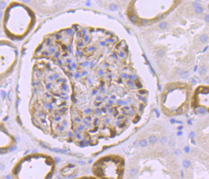 Paraformaldehyde-fixed, paraffin embedded (human kidney); Antigen retrieval by boiling in sodium citrate buffer (pH6.0) for 15min; Block endogenous peroxidase by 3% hydrogen peroxide for 20 minutes; Blocking buffer (normal goat serum) at 37°C for 30min; Antibody incubation with (BST2) Monoclonal Antibody, Unconjugated (SLM-54196R) at 1:50 overnight at 4°C, followed by operating according to SP Kit(Rabbit) (sp-0023) instructionsand DAB staining.
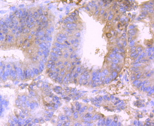 Paraformaldehyde-fixed, paraffin embedded (human colon cancer); Antigen retrieval by boiling in sodium citrate buffer (pH6.0) for 15min; Block endogenous peroxidase by 3% hydrogen peroxide for 20 minutes; Blocking buffer (normal goat serum) at 37°C for 30min; Antibody incubation with (BST2) Monoclonal Antibody, Unconjugated (SLM-54196R) at 1:50 overnight at 4°C, followed by operating according to SP Kit(Rabbit) (sp-0023) instructionsand DAB staining.
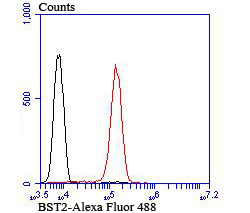 Blank control:Jurkat .
Primary Antibody (green line): Rabbit Anti-BST2 antibody (SLM-54196R)
Dilution: 1:50;
Isotype Control Antibody (orange line): Rabbit IgG .
Secondary Antibody : Goat anti-rabbit IgG-AF488
Dilution: 1:1000.
Protocol
The cells were fixed with 4% PFA (10min at room temperature)and then permeabilized with 90% ice-cold methanol for 20 min at-20℃. The cells were then incubated in 5%BSA to block non-specific protein-protein interactions for 30 min at room temperature .Cells stained with Primary Antibody for 30 min at room temperature. The secondary antibody used for 40 min at room temperature. Acquisition of 20,000 events was performed.
Cartpieces
Totalgoods,subtotals:¥Checkout
References (0)
No References
Bought notes(bought amounts latest0)
No one bought this product
User Comment(Total0User Comment Num)
- No comment


 +86 571 56623320
 +86 18668110335

