
Rabbit Anti-Histone H1.2 antibody
H1 histone family member 2 ;H1.a ;H12_HUMAN ;H1F2 ;H1s-1 ;HIST1H1C ;Histone 1 H1c ;Histone cluster 1 H1c ;Histone H1.2 ;Histone H1c ;Histone H1d ;Histone H1s-1 ;MGC3992 .
View History [Clear]
Details
Product Name Histone H1.2 Chinese Name 组蛋白H1.2Recombinant rabbit monoclonal anti Alias H1 histone family member 2 ; H1.a ; H12_HUMAN ; H1F2 ; H1s-1 ; HIST1H1C ; Histone 1 H1c ; Histone cluster 1 H1c ; Histone H1.2 ; Histone H1c ; Histone H1d ; Histone H1s-1 ; MGC3992 . Immunogen Species Rabbit Clonality Monoclonal Clone NO. 9C9 React Species Human, Mouse, Rat, Applications WB=1:500-2000 IHC-P=1:400-800 ICC=1:50 IF=1:50-100 (Paraffin sections need antigen repair)
not yet tested in other applications.
optimal dilutions/concentrations should be determined by the end user.Theoretical molecular weight 21kDa Cellular localization The nucleus Form Liquid Concentration 1mg/ml immunogen KLH conjugated synthetic peptide derived from human Histone H1.2 Lsotype IgG Purification affinity purified by Protein A Buffer Solution 0.01M TBS(pH7.4) with 1% BSA, 0.03% Proclin300 and 50% Glycerol. Storage Shipped at 4℃. Store at -20 °C for one year. Avoid repeated freeze/thaw cycles. Attention This product as supplied is intended for research use only, not for use in human, therapeutic or diagnostic applications. PubMed PubMed Product Detail Histones are basic nuclear proteins responsible for nucleosome structure of the chromosomal fiber in eukaryotes. Two molecules of each of the four core histones (H2A, H2B, H3, and H4) form an octamer, around which approximately 146 bp of DNA is wrapped in repeating units, called nucleosomes. The linker histone, H1, interacts with linker DNA between nucleosomes and functions in the compaction of chromatin into higher order structures. This gene is intronless and encodes a replication-dependent histone that is a member of the histone H1 family. Transcripts from this gene lack polyA tails but instead contain a palindromic termination element. This gene is found in the large histone gene cluster on chromosome 6. [provided by RefSeq, Aug 2015]
Function:
Histone H1 protein binds to linker DNA between nucleosomes forming the macromolecular structure known as the chromatin fiber. Histones H1 are necessary for the condensation of nucleosome chains into higher-order structured fibers. Acts also as a regulator of individual gene transcription through chromatin remodeling, nucleosome spacing and DNA methylation (By similarity).
Subcellular Location:
Nucleus.
Tissue Specificity:
Expressed in calcaneal tendon and 216 other tissues.
Post-translational modifications:
H1 histones are progressively phosphorylated during the cell cycle, becoming maximally phosphorylated during late G2 phase and M phase, and being dephosphorylated sharply thereafter.
Crotonylation (Kcr) is specifically present in male germ cells and marks testis-specific genes in post-meiotic cells, including X-linked genes that escape sex chromosome inactivation in haploid cells. Crotonylation marks active promoters and enhancers and confers resistance to transcriptional repressors. It is also associated with post-meiotically activated genes on autosomes.
Citrullination at Arg-54 (H1R54ci) by PADI4 takes place within the DNA-binding site of H1 and results in its displacement from chromatin and global chromatin decondensation, thereby promoting pluripotency and stem cell maintenance.
ADP-ribosylated on Ser-188 in response to DNA damage.
SWISS:
P16403
Gene ID:
3006
Database links:Entrez Gene: 3006 Human
Omim: 142710 Human
SwissProt: P16403 Human
Unigene: 7644 Human
Product Picture 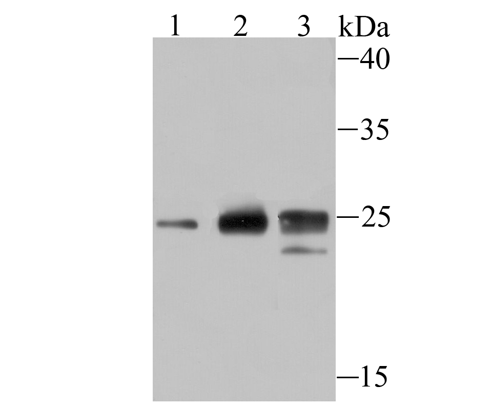 Sample:
Lane 1: Hela cell lysate
Lane 2: 293 cell lysate
Lane 3: MCF-7 cell lysate
Primary: Anti-Histone H1.2 (SLM-54176R) at 1:500 dilution
Secondary: Goat Anti-Rabbit IgG - HRP at 1:5000 dilution
Predicted band size: 21 kD
Observed band size: 25 kD
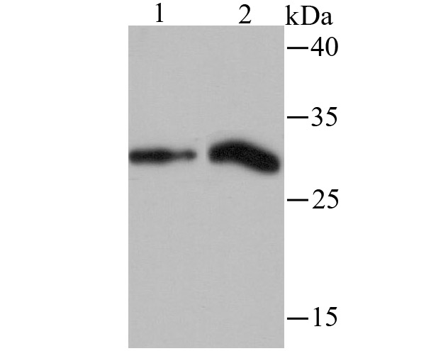 Sample:
Lane 1: rat liver tissue lysate
Lane 2: mouse lung tissue lysate
Primary: Anti-Histone H1.2 (SLM-54176R) at 1:500 dilution
Secondary: Goat Anti-Rabbit IgG - HRP at 1:5000 dilution
Predicted band size: 21 kD
Observed band size: 25 kD
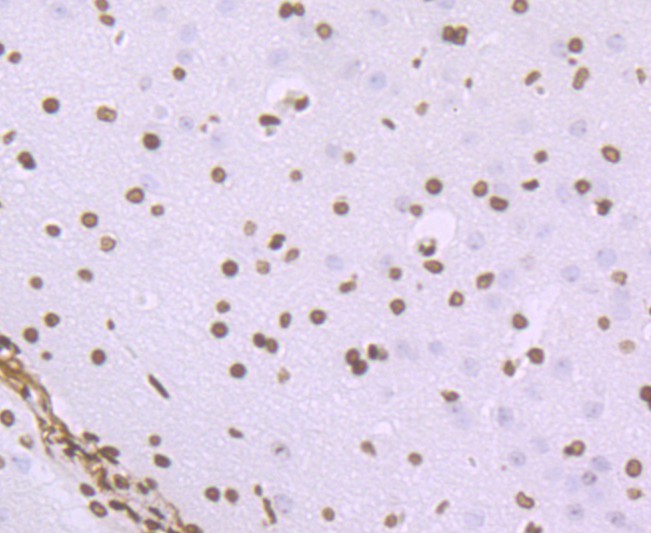 Paraformaldehyde-fixed, paraffin embedded (rat brain); Antigen retrieval by boiling in sodium citrate buffer (pH6.0) for 15min; Block endogenous peroxidase by 3% hydrogen peroxide for 20 minutes; Blocking buffer (normal goat serum) at 37°C for 30min; Antibody incubation with (Histone H1.2) Monoclonal Antibody, Unconjugated (SLM-54176R) at 1:50 overnight at 4°C, followed by operating according to SP Kit(Rabbit) (sp-0023) instructionsand DAB staining.
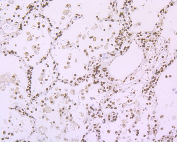 Paraformaldehyde-fixed, paraffin embedded (human lung carcinoma); Antigen retrieval by boiling in sodium citrate buffer (pH6.0) for 15min; Block endogenous peroxidase by 3% hydrogen peroxide for 20 minutes; Blocking buffer (normal goat serum) at 37°C for 30min; Antibody incubation with (Histone H1.2) Monoclonal Antibody, Unconjugated (SLM-54176R) at 1:50 overnight at 4°C, followed by operating according to SP Kit(Rabbit) (sp-0023) instructionsand DAB staining.
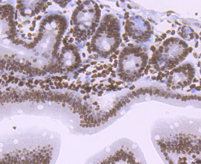 Paraformaldehyde-fixed, paraffin embedded (mouse colon); Antigen retrieval by boiling in sodium citrate buffer (pH6.0) for 15min; Block endogenous peroxidase by 3% hydrogen peroxide for 20 minutes; Blocking buffer (normal goat serum) at 37°C for 30min; Antibody incubation with (Histone H1.2) Monoclonal Antibody, Unconjugated (SLM-54176R) at 1:50 overnight at 4°C, followed by operating according to SP Kit(Rabbit) (sp-0023) instructionsand DAB staining.
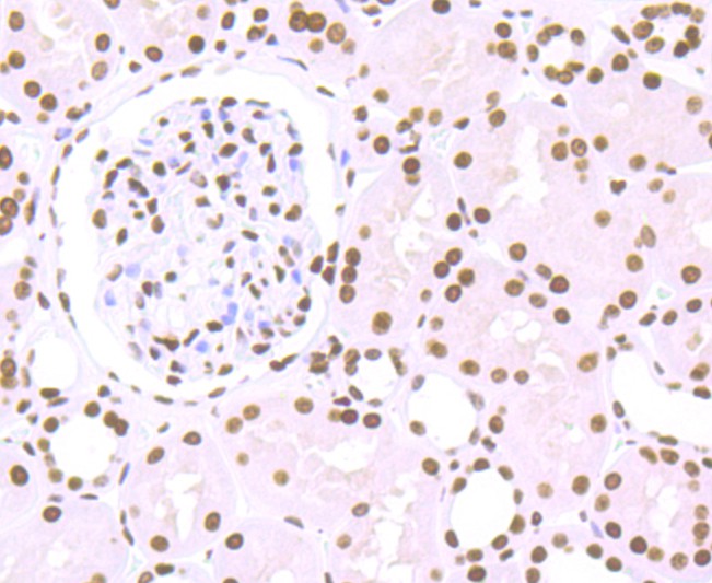 Paraformaldehyde-fixed, paraffin embedded (human kidney); Antigen retrieval by boiling in sodium citrate buffer (pH6.0) for 15min; Block endogenous peroxidase by 3% hydrogen peroxide for 20 minutes; Blocking buffer (normal goat serum) at 37°C for 30min; Antibody incubation with (Histone H1.2) Monoclonal Antibody, Unconjugated (SLM-54176R) at 1:50 overnight at 4°C, followed by operating according to SP Kit(Rabbit) (sp-0023) instructionsand DAB staining.
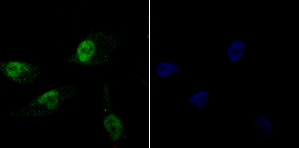 PC-3M cell; 4% Paraformaldehyde-fixed; Triton X-100 at room temperature for 20 min; Blocking buffer (normal goat serum, C-0005) at 37°C for 20 min; Antibody incubation with (Histone H1.2) monoclonal Antibody, Unconjugated (SLM-54176R) 1:100, 90 minutes at 37°C; followed by a conjugated Goat Anti-Rabbit IgG antibody at 37°C for 90 minutes, DAPI (blue, C02-04002) was used to stain the cell nuclei.
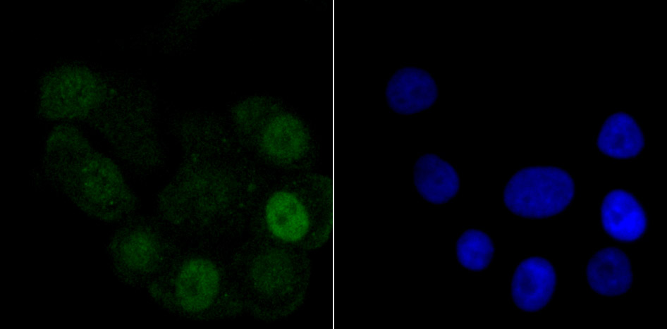 SK-Br-3 cell; 4% Paraformaldehyde-fixed; Triton X-100 at room temperature for 20 min; Blocking buffer (normal goat serum, C-0005) at 37°C for 20 min; Antibody incubation with (Histone H1.2) monoclonal Antibody, Unconjugated (SLM-54176R) 1:100, 90 minutes at 37°C; followed by a conjugated Goat Anti-Rabbit IgG antibody at 37°C for 90 minutes, DAPI (blue, C02-04002) was used to stain the cell nuclei.
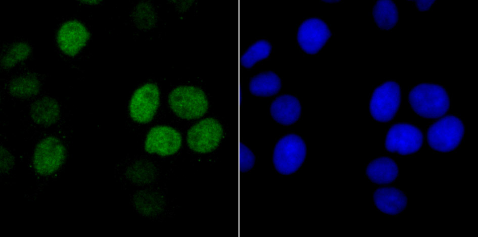 Hela cell; 4% Paraformaldehyde-fixed; Triton X-100 at room temperature for 20 min; Blocking buffer (normal goat serum, C-0005) at 37°C for 20 min; Antibody incubation with (Histone H1.2) monoclonal Antibody, Unconjugated (SLM-54176R) 1:100, 90 minutes at 37°C; followed by a conjugated Goat Anti-Rabbit IgG antibody at 37°C for 90 minutes, DAPI (blue, C02-04002) was used to stain the cell nuclei.
Cartpieces
Totalgoods,subtotals:¥Checkout
References (0)
No References
Bought notes(bought amounts latest0)
No one bought this product
User Comment(Total0User Comment Num)
- No comment


 +86 571 56623320
 +86 18668110335

