
Rabbit Anti-NRF1 antibody
erythroid derived 2; like 1; Locus control region factor 1; Locus control region-factor 1; NF-E2-related factor 1; NF2L1_HUMAN; NFE2 related factor 1; NFE2-related factor 1; NFE2L1; NRF1; NRF-1; NRF 1; Nuclear factor; Nuclear factor erythroid 2-related fa
View History [Clear]
Details
Product Name NRF1 Chinese Name 核呼吸因子-1Recombinant rabbit monoclonal anti Alias erythroid derived 2; like 1; Locus control region factor 1; Locus control region-factor 1; NF-E2-related factor 1; NF2L1_HUMAN; NFE2 related factor 1; NFE2-related factor 1; NFE2L1; NRF1; NRF-1; NRF 1; Nuclear factor; Nuclear factor erythroid 2-related factor 1; Nuclear factor erythroid derived 2 like 1; TCF-11; TCF11; Transcription factor 11; Transcription factor HBZ17; Transcription factor LCR F1; Transcription factor LCR-F1. Research Area Chromatin and nuclear signals transcriptional regulatory factor Synthesis and Degradation Mitochondrion Immunogen Species Rabbit Clonality Monoclonal Clone NO. 6G5 React Species Human, Mouse, Rat, Applications WB=1:500 IP=1:10-50 IHC-P=1:50-200 IHC-F=1:50-200 ICC=1:50 IF=1:50-200 (Paraffin sections need antigen repair)
not yet tested in other applications.
optimal dilutions/concentrations should be determined by the end user.Theoretical molecular weight 55kDa Cellular localization The nucleus Mitochondrion Form Liquid Concentration 1mg/ml immunogen KLH conjugated synthetic peptide derived from human NRF1 Lsotype IgG Purification affinity purified by Protein A Buffer Solution 0.01M TBS(pH7.4) with 1% BSA, 0.03% Proclin300 and 50% Glycerol. Storage Shipped at 4℃. Store at -20 °C for one year. Avoid repeated freeze/thaw cycles. Attention This product as supplied is intended for research use only, not for use in human, therapeutic or diagnostic applications. PubMed PubMed Product Detail This gene encodes a protein that homodimerizes and functions as a transcription factor which activates the expression of some key metabolic genes regulating cellular growth and nuclear genes required for respiration, heme biosynthesis, and mitochondrial DNA transcription and replication. The protein has also been associated with the regulation of neurite outgrowth. Alternative splicing results in multiple transcript variants. Confusion has occurred in bibliographic databases due to the shared symbol of NRF1 for this gene and for "nuclear factor (erythroid-derived 2)-like 1" which has an official symbol of NFE2L1. [provided by RefSeq, May 2014]
Function:
Transcription factor that activates the expression of the EIF2S1 (EIF2-alpha) gene. Links the transcriptional modulation of key metabolic genes to cellular growth and development. Implicated in the control of nuclear genes required for respiration, heme biosynthesis, and mitochondrial DNA transcription and replication.
Subunit:
Homodimer. Binds DNA as a dimer. Interacts with PPRC1.
Subcellular Location:
Nucleus
Tissue Specificity:
Ubiquitously expressed with strongest expression in skeletal muscle.
Post-translational modifications:
Phosphorylation enhances DNA binding.
Similarity:
Belongs to the NRF1/Ewg family.
SWISS:
Q16656
Gene ID:
4899
Database links:Entrez Gene: 4899 Human
Omim: 600879 Human
SwissProt: Q16656 Human
Unigene: 654363 Human
Product Picture 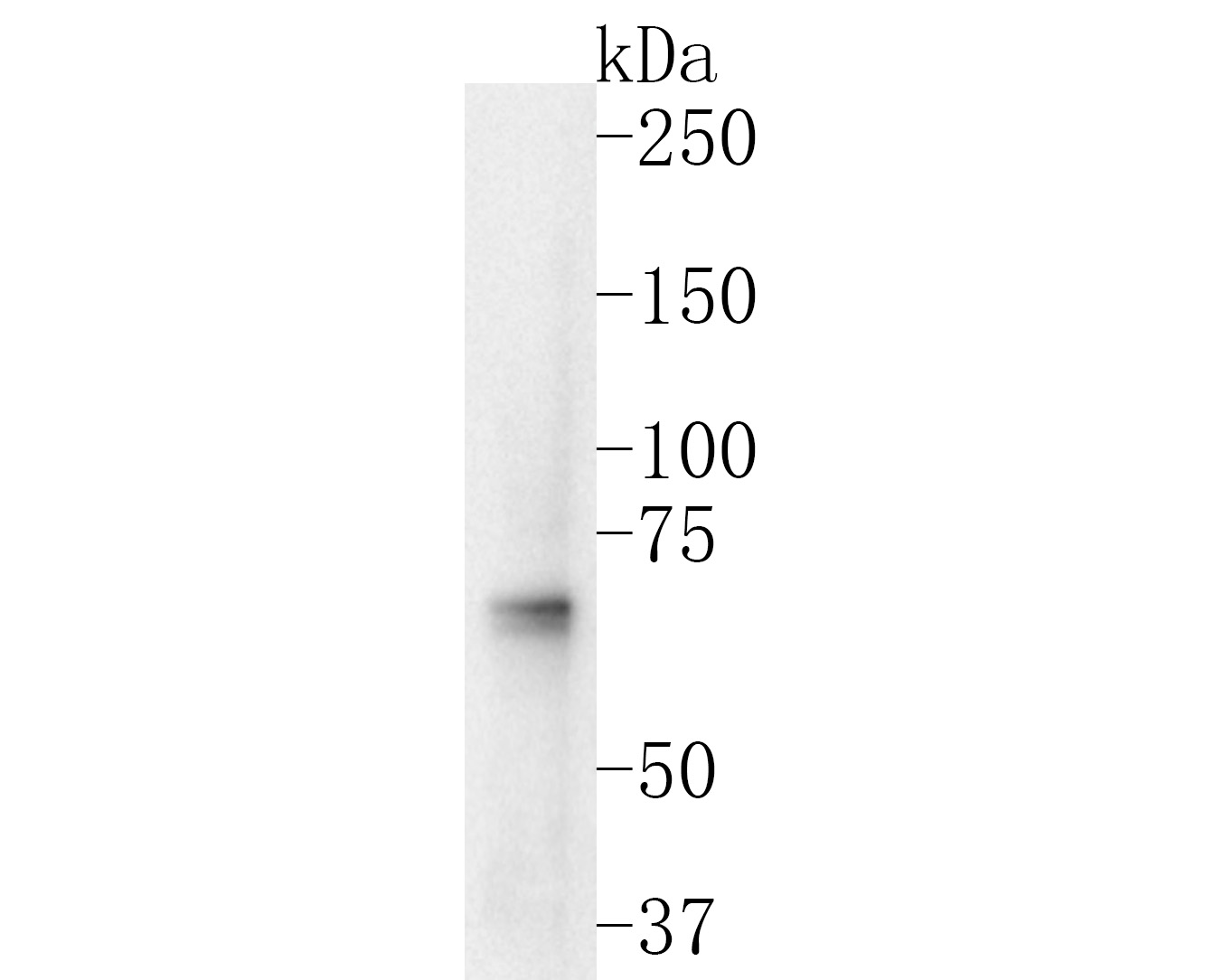 Sample:
Lane 1: Hela cell lysate
Primary: Anti-NRF1 (SLM-54140R) at 1:1000 dilution
Secondary: Goat Anti-Rabbit IgG - HRP at 1:5000 dilution
Predicted band size: 55 kD
Observed band size: 65 kD
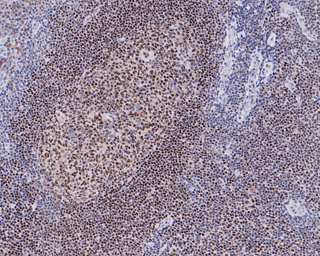 Paraformaldehyde-fixed, paraffin embedded (human tonsil); Antigen retrieval by boiling in sodium citrate buffer (pH6.0) for 15min; Block endogenous peroxidase by 3% hydrogen peroxide for 20 minutes; Blocking buffer (normal goat serum) at 37°C for 30min; Antibody incubation with (NRF1) Monoclonal Antibody, Unconjugated (SLM-54140R) at 1:50 overnight at 4°C, followed by operating according to SP Kit(Rabbit) (sp-0023) instructionsand DAB staining.
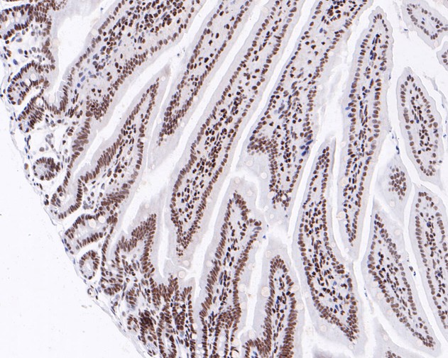 Paraformaldehyde-fixed, paraffin embedded (mouse colon); Antigen retrieval by boiling in sodium citrate buffer (pH6.0) for 15min; Block endogenous peroxidase by 3% hydrogen peroxide for 20 minutes; Blocking buffer (normal goat serum) at 37°C for 30min; Antibody incubation with (NRF1) Monoclonal Antibody, Unconjugated (SLM-54140R) at 1:50 overnight at 4°C, followed by operating according to SP Kit(Rabbit) (sp-0023) instructionsand DAB staining.
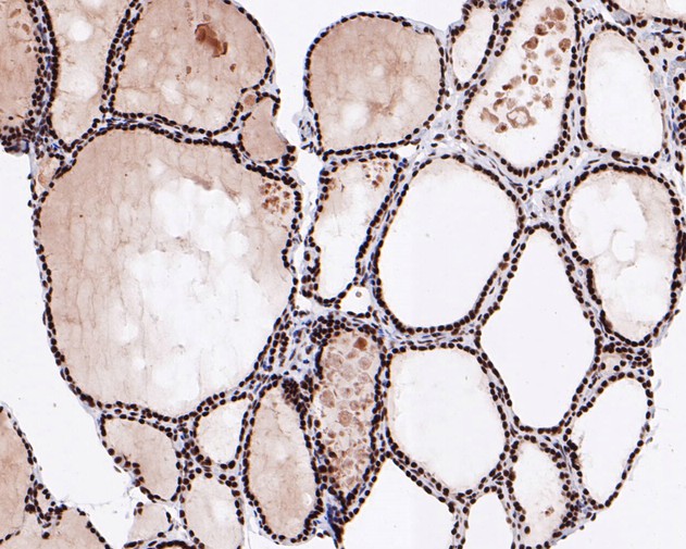 Paraformaldehyde-fixed, paraffin embedded (human thyroid); Antigen retrieval by boiling in sodium citrate buffer (pH6.0) for 15min; Block endogenous peroxidase by 3% hydrogen peroxide for 20 minutes; Blocking buffer (normal goat serum) at 37°C for 30min; Antibody incubation with (NRF1) Monoclonal Antibody, Unconjugated (SLM-54140R) at 1:50 overnight at 4°C, followed by operating according to SP Kit(Rabbit) (sp-0023) instructionsand DAB staining.
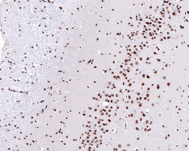 Paraformaldehyde-fixed, paraffin embedded (rat brain); Antigen retrieval by boiling in sodium citrate buffer (pH6.0) for 15min; Block endogenous peroxidase by 3% hydrogen peroxide for 20 minutes; Blocking buffer (normal goat serum) at 37°C for 30min; Antibody incubation with (NRF1) Monoclonal Antibody, Unconjugated (SLM-54140R) at 1:50 overnight at 4°C, followed by operating according to SP Kit(Rabbit) (sp-0023) instructionsand DAB staining.
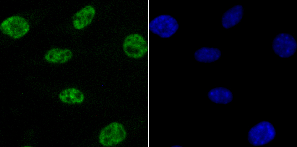 SH-SY5Y cell; 4% Paraformaldehyde-fixed; Triton X-100 at room temperature for 20 min; Blocking buffer (normal goat serum, C-0005) at 37°C for 20 min; Antibody incubation with (NRF1) monoclonal Antibody, Unconjugated (SLM-54140R) 1:50, 90 minutes at 37°C; followed by a conjugated Goat Anti-Rabbit IgG antibody at 37°C for 90 minutes, DAPI (blue, C02-04002) was used to stain the cell nuclei.
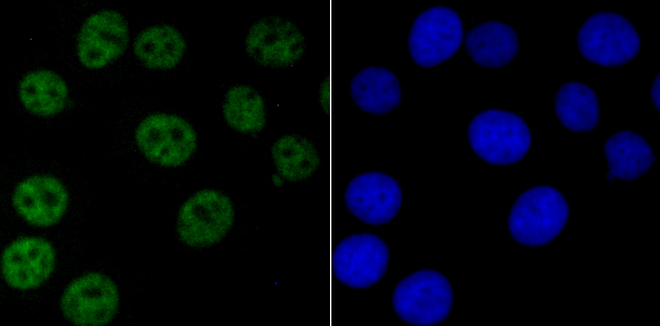 MCF-7 cell; 4% Paraformaldehyde-fixed; Triton X-100 at room temperature for 20 min; Blocking buffer (normal goat serum, C-0005) at 37°C for 20 min; Antibody incubation with (NRF1) monoclonal Antibody, Unconjugated (SLM-54140R) 1:50, 90 minutes at 37°C; followed by a conjugated Goat Anti-Rabbit IgG antibody at 37°C for 90 minutes, DAPI (blue, C02-04002) was used to stain the cell nuclei.
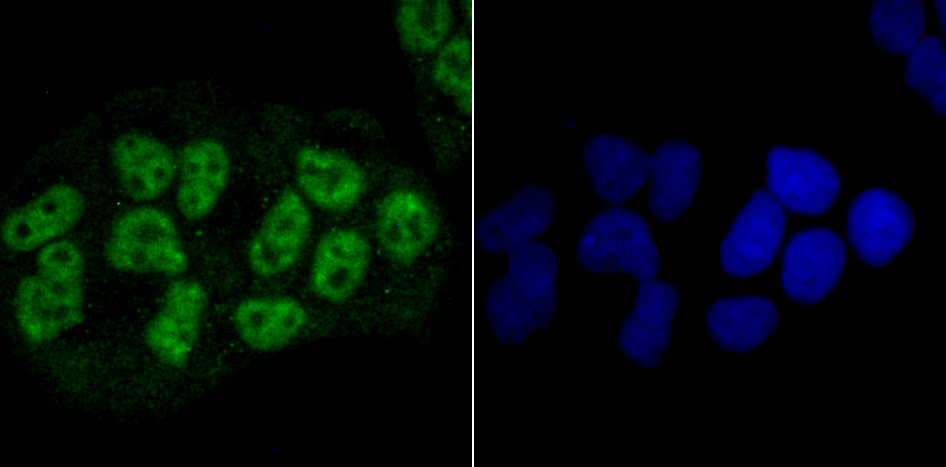 Hela cell; 4% Paraformaldehyde-fixed; Triton X-100 at room temperature for 20 min; Blocking buffer (normal goat serum, C-0005) at 37°C for 20 min; Antibody incubation with (NRF1) monoclonal Antibody, Unconjugated (SLM-54140R) 1:50, 90 minutes at 37°C; followed by a conjugated Goat Anti-Rabbit IgG antibody at 37°C for 90 minutes, DAPI (blue, C02-04002) was used to stain the cell nuclei.
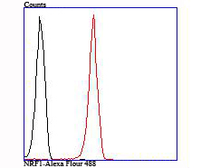 Blank control:293T.
Primary Antibody (green line): Rabbit Anti-NRF1 antibody (SLM-54140R)
Dilution: 1:50;
Isotype Control Antibody (orange line): Rabbit IgG .
Secondary Antibody : Goat anti-rabbit IgG-AF488
Dilution: 1:1000.
Protocol
The cells were fixed with 4% PFA (10min at room temperature)and then permeabilized with 90% ice-cold methanol for 20 min at -20℃. The cells were then incubated in 5%BSA to block non-specific protein-protein interactions for 30 min at room temperature .Cells stained with Primary Antibody for 30 min at room temperature. The secondary antibody used for 40 min at room temperature. Acquisition of 20,000 events was performed.
Cartpieces
Totalgoods,subtotals:¥Checkout
References (0)
No References
Bought notes(bought amounts latest0)
No one bought this product
User Comment(Total0User Comment Num)
- No comment


 +86 571 56623320
 +86 18668110335

