
Rabbit Anti-phospho-MLKL (Ser345)antibody
phospho-MLKL(S345); p-MLKL(S345); MLKL(p-S345); hMLKL; Mixed lineage kinase domain like; Mixed lineage kinase domain like protein; Mixed lineage kinase domain-like protein; mixed lineage kinase domain like pseudokinase; MLKL_HUMAN.
View History [Clear]
Details
Product Name phospho-MLKL (Ser345) Chinese Name 磷酸化MLKLRecombinant rabbit monoclonal anti Alias phospho-MLKL(S345); p-MLKL(S345); MLKL(p-S345); hMLKL; Mixed lineage kinase domain like; Mixed lineage kinase domain like protein; Mixed lineage kinase domain-like protein; mixed lineage kinase domain like pseudokinase; MLKL_HUMAN. literatures Research Area Cell biology Kinases and Phosphatases Immunogen Species Rabbit Clonality Monoclonal Clone NO. 7G4 React Species (predicted: Mouse, ) Applications WB=1:500-1000 IHC-P=1:100-500 IHC-F=1:50-100 (Paraffin sections need antigen repair)
not yet tested in other applications.
optimal dilutions/concentrations should be determined by the end user.Theoretical molecular weight 54kDa Cellular localization cytoplasmic The cell membrane Form Liquid Concentration 1mg/ml immunogen KLH conjugated Synthesised phosphopeptide derived from mouse MLKL around the phosphorylation site of Ser345 Lsotype IgG Purification affinity purified by Protein A Buffer Solution 0.01M TBS(pH7.4) with 1% BSA, 0.03% Proclin300 and 50% Glycerol. Storage Shipped at 4℃. Store at -20 °C for one year. Avoid repeated freeze/thaw cycles. Attention This product as supplied is intended for research use only, not for use in human, therapeutic or diagnostic applications. PubMed PubMed Product Detail This gene belongs to the protein kinase superfamily. The encoded protein contains a protein kinase-like domain; however, is thought to be inactive because it lacks several residues required for activity. This protein plays a critical role in tumor necrosis factor (TNF)-induced necroptosis, a programmed cell death process, via interaction with receptor-interacting protein 3 (RIP3), which is a key signaling molecule in necroptosis pathway. Inhibitor studies and knockdown of this gene inhibited TNF-induced necrosis. High levels of this protein and RIP3 are associated with inflammatory bowel disease in children. Alternatively spliced transcript variants have been described for this gene. [provided by RefSeq, Sep 2015].
Function:
Pseudokinase that plays a key role in TNF-induced necroptosis, a programmed cell death process. Activated following phosphorylation by RIPK3, leading to homotrimerization, localization to the plasma membrane and execution of programmed necrosis characterized by calcium influx and plasma membrane damage. Does not have protein kinase activity.
Subunit:
Homotrimer; forms homotrimers on necroptosis induction. Interacts with RIPK3; the interaction is direct. Upon TNF-induced necrosis, forms in complex with PGAM5, RIPK1 and RIPK3. Within this complex, may play a role in the proper targeting of RIPK1/RIPK3 to its downstream effector PGAM5.
Subcellular Location:
Cytoplasm. Cell membrane. Note=Localizes to the cytoplasm and translocates to the plasma membrane on necroptosis induction.
Post-translational modifications:
Phosphorylation by RIPK3 induces a conformational switch that is required for necroptosis. It also induces homotrimerization and localization to the plasma membrane.
Similarity:
Belongs to the protein kinase superfamily.
Contains 1 protein kinase domain.
SWISS:
Q9D2Y4
Gene ID:
74568
Database links:Entrez Gene: 197259 Human
Entrez Gene: 74568 Mouse
Omim: 615153 Human
SwissProt: Q8NB16 Human
SwissProt: Q9D2Y4 Mouse
Unigene: 119878 Human
Unigene: 207971 Mouse
Unigene: 105677 Rat
Product Picture 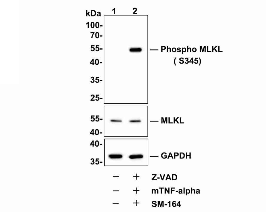 Western blot analysis of Phospho-MLKL (S345) on L929 cell lysates. Lane 1 : L929 cells, whole cell lysate, 10 μg /lane. Lane 2 : L929 cells were treated with 20 uM Z-VAD for 30 minutes, then added 20 ng/ml mTNF-alpha and 100 nM SM-164 for 4 hours, whole cell lysates, 10 μg/lane.Western blot analysis of Phospho-MLKL (S345) on L929 cell lysates.
Lane 1 : L929 cells, whole cell lysate, 10 μg /lane.
Lane 2 : L929 cells were treated with 20 uM Z-VAD for 30 minutes, then added 20 ng/ml mTNF-alpha and 100 nM SM-164 for 4 hours, whole cell lysates, 10 μg/lane.
Proteins were transferred to a PVDF membrane and blocked with 5% BSA in PBS for 1 hour at room temperature. The primary antibody Anti-Phospho-MLKL (S345) (SLM-54104R, 1/500) , Secondary Antibody at 1:200,000 dilution was used for 1 hour at room temperature.
Predicted band size: 54 kDa
Observed band size: 54 kDa
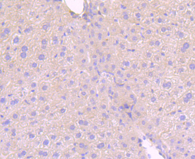 Immunohistochemical analysis of paraffin-embedded mouse liver tissue using anti-MLKL (phospho S345) antibody. The section was pre-treated using heat mediated antigen retrieval with Tris-EDTA buffer (pH 9.0) for 20 minutes.The tissues were blocked in 5% BSA for 30 minutes at room temperature, washed with ddH2O and PBS, and then probed with the primary antibody (SLM-54104R, 1/50) for 30 minutes at room temperature. The detection was performed using an HRP conjugated compact polymer system. DAB was used as the chromogen. Tissues were counterstained with hematoxylin and mounted with DPX.
 Immunohistochemical analysis of paraffin-embedded mouse colon tissue using anti-MLKL (phospho S345) antibody. The section was pre-treated using heat mediated antigen retrieval with Tris-EDTA buffer (pH 9.0) for 20 minutes.The tissues were blocked in 5% BSA for 30 minutes at room temperature, washed with ddH2O and PBS, and then probed with the primary antibody (SLM-54104R, 1/50) for 30 minutes at room temperature. The detection was performed using an HRP conjugated compact polymer system. DAB was used as the chromogen. Tissues were counterstained with hematoxylin and mounted with DPX.
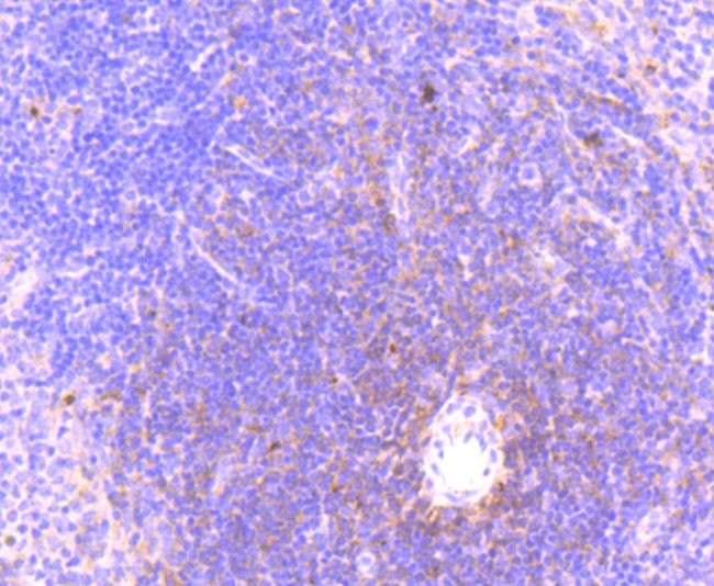 Immunohistochemical analysis of paraffin-embedded mouse spleen tissue using anti-MLKL (phospho S345) antibody. The section was pre-treated using heat mediated antigen retrieval with Tris-EDTA buffer (pH 9.0) for 20 minutes.The tissues were blocked in 5% BSA for 30 minutes at room temperature, washed with ddH2O and PBS, and then probed with the primary antibody (SLM-54104R, 1/50) for 30 minutes at room temperature. The detection was performed using an HRP conjugated compact polymer system. DAB was used as the chromogen. Tissues were counterstained with hematoxylin and mounted with DPX.
 Immunohistochemical analysis of paraffin-embedded mouse lung tissue using anti-MLKL (phospho S345) antibody. The section was pre-treated using heat mediated antigen retrieval with Tris-EDTA buffer (pH 9.0) for 20 minutes.The tissues were blocked in 5% BSA for 30 minutes at room temperature, washed with ddH2O and PBS, and then probed with the primary antibody (SLM-54104R, 1/50) for 30 minutes at room temperature. The detection was performed using an HRP conjugated compact polymer system. DAB was used as the chromogen. Tissues were counterstained with hematoxylin and mounted with DPX.
 Immunohistochemical analysis of paraffin-embedded mouse liver tissue using anti-Phospho-MLKL (S345) antibody. The section was pre-treated using heat mediated antigen retrieval with Tris-EDTA buffer (pH 9.0) for 20 minutes.The tissues were blocked in 5% BSA for 30 minutes at room temperature, washed with ddH2O and PBS, and then probed with the primary antibody (SLM-54104R, 1/50) for 30 minutes at room temperature. The detection was performed using an HRP conjugated compact polymer system. DAB was used as the chromogen. Tissues were counterstained with hematoxylin and mounted with DPX.
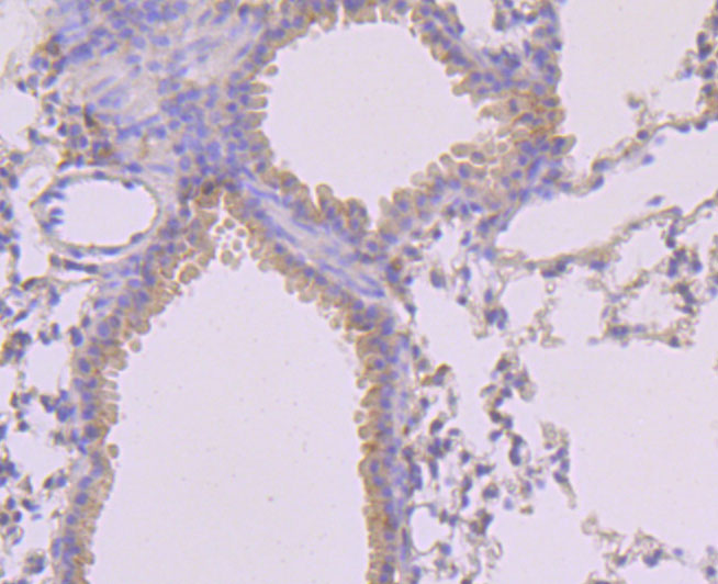 Immunohistochemical analysis of paraffin-embedded mouse lung tissue using anti-Phospho-MLKL (S345) antibody. The section was pre-treated using heat mediated antigen retrieval with Tris-EDTA buffer (pH 9.0) for 20 minutes.The tissues were blocked in 5% BSA for 30 minutes at room temperature, washed with ddH2O and PBS, and then probed with the primary antibody (SLM-54104R, 1/50) for 30 minutes at room temperature. The detection was performed using an HRP conjugated compact polymer system. DAB was used as the chromogen. Tissues were counterstained with hematoxylin and mounted with DPX.
 Immunohistochemical analysis of paraffin-embedded mouse spleen tissue using anti-Phospho-MLKL (S345) antibody. The section was pre-treated using heat mediated antigen retrieval with Tris-EDTA buffer (pH 9.0) for 20 minutes.The tissues were blocked in 5% BSA for 30 minutes at room temperature, washed with ddH2O and PBS, and then probed with the primary antibody (SLM-54104R, 1/50) for 30 minutes at room temperature. The detection was performed using an HRP conjugated compact polymer system. DAB was used as the chromogen. Tissues were counterstained with hematoxylin and mounted with DPX.
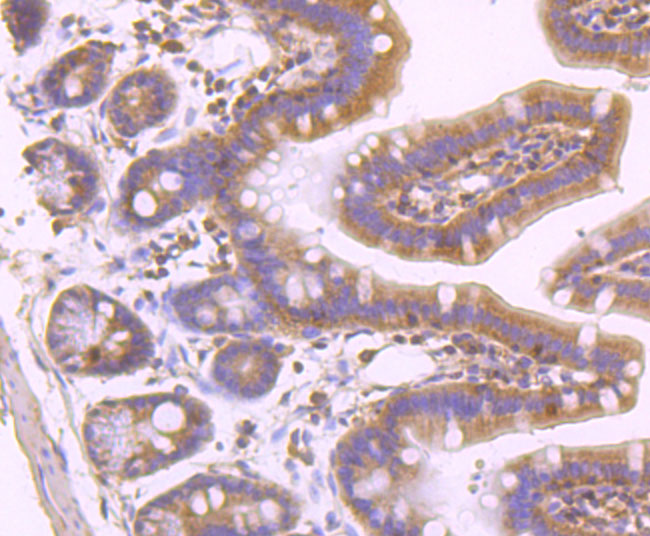 Immunohistochemical analysis of paraffin-embedded mouse colon tissue using anti-Phospho-MLKL (S345) antibody. The section was pre-treated using heat mediated antigen retrieval with Tris-EDTA buffer (pH 9.0) for 20 minutes.The tissues were blocked in 5% BSA for 30 minutes at room temperature, washed with ddH2O and PBS, and then probed with the primary antibody (SLM-54104R, 1/50) for 30 minutes at room temperature. The detection was performed using an HRP conjugated compact polymer system. DAB was used as the chromogen. Tissues were counterstained with hematoxylin and mounted with DPX.
Cartpieces
Totalgoods,subtotals:¥Checkout
Bought notes(bought amounts latest0)
No one bought this product
User Comment(Total0User Comment Num)
- No comment


 +86 571 56623320
 +86 18668110335

