
Rabbit Anti-CXCR3 antibody
CXCR3_HUMAN; Interferon-inducible protein 10 receptor; IP-10 receptor; C-X-C chemokine receptor type 3; CD 183; CD183; CD183 antigen; G protein-coupled receptor 9; G Protein Coupled Receptor 9; Chemokine (C X C motif) receptor 3; Chemokine (C X C) recepto
View History [Clear]
Details
Product Name CXCR3 Chinese Name 细胞表面Chemokine受体3(CD183)Recombinant rabbit monoclonal anti Alias CXCR3_HUMAN; Interferon-inducible protein 10 receptor; IP-10 receptor; C-X-C chemokine receptor type 3; CD 183; CD183; CD183 antigen; G protein-coupled receptor 9; G Protein Coupled Receptor 9; Chemokine (C X C motif) receptor 3; Chemokine (C X C) receptor 3; C-X-C Chemokine receptor; CXC Motif Receptor 3; Chemokine CXC Motif Receptor 3; CKRL2; CKR L2; CKR-L2; GPR9; CXC-R3; CXCR-3; IP10 receptor. Research Area Cardiovascular Cell biology immunology Signal transduction Cyclin The cell membrane受体 G protein-coupled receptor Cell differentiation G protein signal Immunogen Species Rabbit Clonality Monoclonal Clone NO. 5F2 React Species Human, Applications WB=1:500-2000 IP=1:20-50 ICC=1:50 IF=1:50-200
not yet tested in other applications.
optimal dilutions/concentrations should be determined by the end user.Theoretical molecular weight 41kDa Cellular localization The cell membrane Form Liquid Concentration 1mg/ml immunogen Recombinant human CXCR3 protein Lsotype IgG Purification affinity purified by Protein A Buffer Solution 0.01M TBS(pH7.4) with 1% BSA, 0.03% Proclin300 and 50% Glycerol. Storage Shipped at 4℃. Store at -20 °C for one year. Avoid repeated freeze/thaw cycles. Attention This product as supplied is intended for research use only, not for use in human, therapeutic or diagnostic applications. PubMed PubMed Product Detail This gene encodes a G protein-coupled receptor with selectivity for three chemokines, termed CXCL9/Mig (monokine induced by interferon-g), CXCL10/IP10 (interferon-g-inducible 10 kDa protein) and CXCL11/I-TAC (interferon-inducible T cell a-chemoattractant). Binding of chemokines to this protein induces cellular responses that are involved in leukocyte traffic, most notably integrin activation, cytoskeletal changes and chemotactic migration. Alternatively spliced transcript variants encoding different isoforms have been found for this gene. One of the isoforms (CXCR3-B) shows high affinity binding to chemokine, CXCL4/PF4 (PMID:12782716). [provided by RefSeq, Jun 2011].
Function:
Receptor for CXCL9, CXCL10 and CXCL11 and mediates the proliferation of human mesangial cells (HMC).
Isoform 2 is a receptor for CXCL4 and also mediates the inhibitory activities of CXCL9, CXCL10 and CXCL11 on the growth of human microvascular endothelial cells (HMVEC). Interaction with CXCL4 or CXCL10 leads to activation of the p38MAPK pathway and contributes to inhibition of angiogenesis. Overexpression in renal cancer cells down-regulates expression of the anti-apoptotic protein HMOX1 and promotes apoptosis.
Isoform 3 mediates the activity of CXCL11.
Subcellular Location:
Cell membrane; Multi-pass membrane protein.
Tissue Specificity:
Isoform 1 and isoform 2 are mainly expressed in heart, kidney, liver and skeletal muscle. Isoform 1 is also expressed in placenta.
Post-translational modifications:
Sulfation on Tyr-27 and Tyr-29 is essential for CXCL10 binding and subsequent signal transduction induction.
N-glycosylated.
Similarity:
Belongs to the G-protein coupled receptor 1 family.
SWISS:
P49682
Gene ID:
2833
Database links:Entrez Gene: 2833 Human
Omim: 300574 Human
SwissProt: P49682 Human
Unigene: 198252 Human
Product Picture 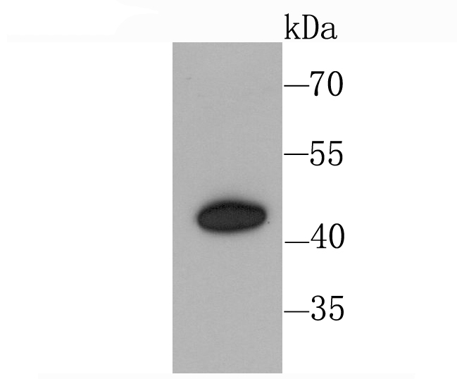 Sample:
Lane 1: Hela cell lysate
Primary: Anti-CXCR3 (SLM-54064R) at 1:1000 dilution
Secondary: Goat Anti-Rabbit IgG - HRP at 1:5000 dilution
Predicted band size: 41 kD
Observed band size: 43 kD
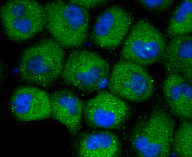 HUVEC cell; 4% Paraformaldehyde-fixed; Triton X-100 at room temperature for 20 min; Blocking buffer (normal goat serum, C-0005) at 37°C for 20 min; Antibody incubation with (CXCR3) monoclonal Antibody, Unconjugated (SLM-54064R) 1:50, 90 minutes at 37°C; followed by a conjugated Goat Anti-Rabbit IgG antibody at 37°C for 90 minutes, DAPI (blue, C02-04002) was used to stain the cell nuclei.
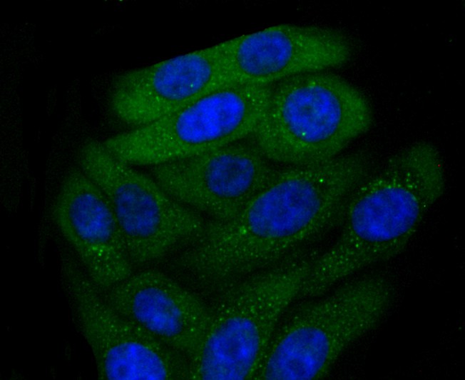 HepG2 cell; 4% Paraformaldehyde-fixed; Triton X-100 at room temperature for 20 min; Blocking buffer (normal goat serum, C-0005) at 37°C for 20 min; Antibody incubation with (CXCR3) monoclonal Antibody, Unconjugated (SLM-54064R) 1:50, 90 minutes at 37°C; followed by a conjugated Goat Anti-Rabbit IgG antibody at 37°C for 90 minutes, DAPI (blue, C02-04002) was used to stain the cell nuclei.
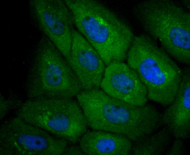 A431 cell; 4% Paraformaldehyde-fixed; Triton X-100 at room temperature for 20 min; Blocking buffer (normal goat serum, C-0005) at 37°C for 20 min; Antibody incubation with (CXCR3) monoclonal Antibody, Unconjugated (SLM-54064R) 1:50, 90 minutes at 37°C; followed by a conjugated Goat Anti-Rabbit IgG antibody at 37°C for 90 minutes, DAPI (blue, C02-04002) was used to stain the cell nuclei.
Cartpieces
Totalgoods,subtotals:¥Checkout
References (0)
No References
Bought notes(bought amounts latest0)
No one bought this product
User Comment(Total0User Comment Num)
- No comment


 +86 571 56623320
 +86 18668110335

