
Rabbit Anti-NG2 antibody
MELCSPG; AN2; AN2 proteoglycan; Chondroitin sulfate proteoglycan 4 (melanoma-associated); Chondroitin sulfate proteoglycan 4; CSPG4; Cspg4 chondroitin sulfate proteoglycan 4; HMW-MAA; HSN tumor-specific antigen; MCSP; MCSPG; MEL-CSPG; Melanoma chondroitin
View History [Clear]
Details
Product Name NG2 Chinese Name 黑色素瘤硫酸软骨素蛋白多糖Recombinant rabbit monoclonal anti Alias MELCSPG; AN2; AN2 proteoglycan; Chondroitin sulfate proteoglycan 4 (melanoma-associated); Chondroitin sulfate proteoglycan 4; CSPG4; Cspg4 chondroitin sulfate proteoglycan 4; HMW-MAA; HSN tumor-specific antigen; MCSP; MCSPG; MEL-CSPG; Melanoma chondroitin sulfate proteoglycan; Melanoma-associated chondroitin sulfate proteoglycan; MSK16; CSPG4_HUMAN. Research Area Tumour Cell biology immunology Neurobiology Cell adhesion molecule Immunogen Species Rabbit Clonality Monoclonal Clone NO. 3A8 React Species Human, Mouse, Rat, Applications WB=1:500-2000 IHC-P=1:50-200 Flow-Cyt=1:50 (Paraffin sections need antigen repair)
not yet tested in other applications.
optimal dilutions/concentrations should be determined by the end user.Theoretical molecular weight 248kDa Cellular localization cytoplasmic The cell membrane Form Liquid Concentration 1mg/ml immunogen KLH conjugated synthetic peptide derived from human NG2 Lsotype IgG Purification affinity purified by Protein A Buffer Solution 0.01M TBS(pH7.4) with 1% BSA, 0.03% Proclin300 and 50% Glycerol. Storage Shipped at 4℃. Store at -20 °C for one year. Avoid repeated freeze/thaw cycles. Attention This product as supplied is intended for research use only, not for use in human, therapeutic or diagnostic applications. PubMed PubMed Product Detail Proteoglycans (PGs) are a family of proteins composed of different polypeptide chains containing glycosaminoglycan (GAG) modifications. They vary in their cellular locations and have diverse structure and functions. NG2 is a proteoglycan found in the nervous system, comprising a large integral membrane PG with a core protein of 300 kDa and at least one covalently attached chondroitin sulfate (CS) GAG chain. In the CNS this protein is expressed mainly on the surfaces of developing and adult oligodendrocyte precursor cells, but is also associated with developing chondrocytes, cardiomyocytes, pericytes and several human tumours. NG2 also stimulates alpha-4, beta-1 integrin-mediated adhesion and spreading by recruiting and activating a signaling cascade through CDC42, ACK1 and BCAR1. May activate FAK and ERK1/ERK2 signaling cascades.
Function:
Proteoglycan playing a role in cell proliferation and migration which stimulates endothelial cells motility during microvascular morphogenesis. May also inhibit neurite outgrowth and growth cone collapse during axon regeneration. Cell surface receptor for collagen alpha 2(VI) which may confer cells ability to migrate on that substrate. Binds through its extracellular N-terminus growth factors, extracellular matrix proteases modulating their activity. May regulate MPP16-dependent degradation and invasion of type I collagen participating in melanoma cells invasion properties. May modulate the plasminogen system by enhancing plasminogen activation and inhibiting angiostatin. Functions also as a signal transducing protein by binding through its cytoplasmic C-terminus scaffolding and signaling proteins. May promote retraction fiber formation and cell polarization through Rho GTPase activation. May stimulate alpha-4, beta-1 integrin-mediated adhesion and spreading by recruiting and activating a signaling cascade through CDC42, ACK1 and BCAR1. May activate FAK and ERK1/ERK2 signaling cascades.
Subunit:
Interacts with the first PDZ domain of MPDZ. Interacts with PRKCA. Binds TNC, laminin-1, COL5A1 and COL6A2. Interacts with PLG and angiostatin. Binds FGF2 and PDGFA. Interacts with GRIP1, GRIP2 and GRIA2. Forms a ternary complex with GRIP1 and GRIA2 (By similarity). Interacts with LGALS3 and the integrin composed of ITGB1 and ITGA3. Interacts with ITGA4 through its chondroitin sulfate glycosaminoglycan. Interacts with BCAR1, CDC42 and ACK1. Interacts with MMP16.
Subcellular Location:
Apical cell membrane. Cell projection > lamellipodium membrane. Localized at the apical plasma membrane it relocalizes to the lamellipodia of astrocytoma upon phosphorylation by PRKCA. Localizes to the retraction fibers. Localizes to the plasma membrane of oligodendrocytes.
Tissue Specificity:
Detected only in malignant melanoma cells.
Post-translational modifications:
O-glycosylated; contains glycosaminoglycan chondroitin sulfate which are required for proper localization and function in stress fiber formation (By similarity). Involved in interaction with MMP16 and ITGA4.
Phosphorylation by PRKCA regulates its subcellular location and function in cell motility (By similarity).
Similarity:
Contains 15 CSPG (NG2) repeats.
Contains 2 laminin G-like domains.
SWISS:
Q6UVK1
Gene ID:
1464
Database links:Entrez Gene: 1464 Human
Entrez Gene: 121021 Mouse
Omim: 601172 Human
SwissProt: Q6UVK1 Human
SwissProt: Q8VHY0 Mouse
Unigene: 513044 Human
Unigene: 41329 Mouse
Product Picture 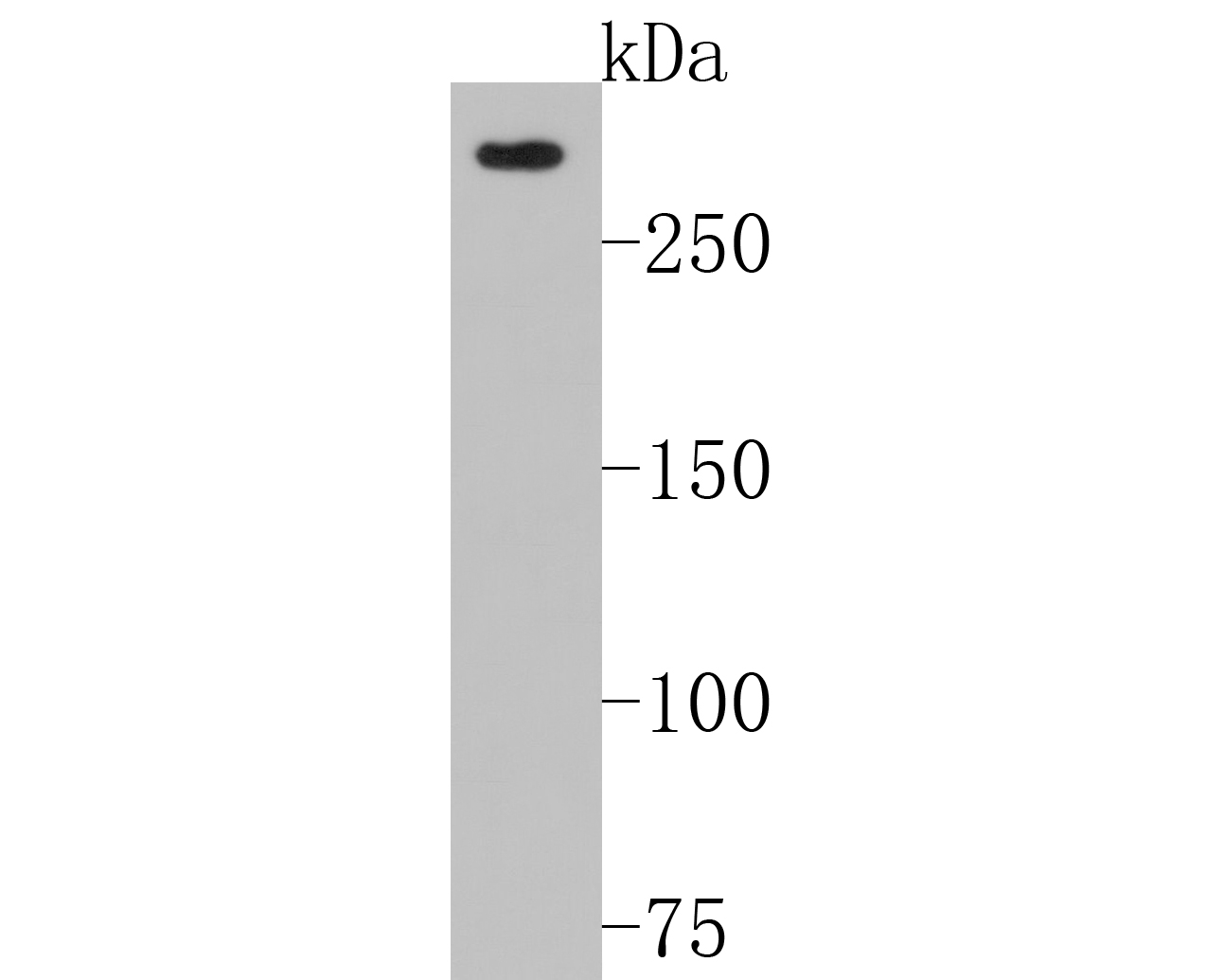 Sample:
Lane 1: SiHa cell lysates
Primary: Anti-NG2 (SLM-52891R) at 1:500 dilution
Secondary: Goat Anti-Rabbit IgG - HRP at 1:5000 dilution
Predicted band size: 248 kD
Observed band size: 280 kD
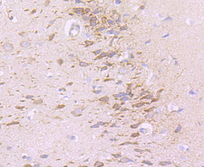 Paraformaldehyde-fixed, paraffin embedded (rat brain tissue); Antigen retrieval by boiling in sodium citrate buffer (pH6.0) for 15min; Block endogenous peroxidase by 3% hydrogen peroxide for 20 minutes; Blocking buffer (normal goat serum) at 37°C for 30min; Antibody incubation with (NG2) Monoclonal Antibody, Unconjugated (SLM-52891R) at 1:50 overnight at 4°C, followed by operating according to SP Kit(Rabbit) (sp-0023) instructionsand DAB staining.
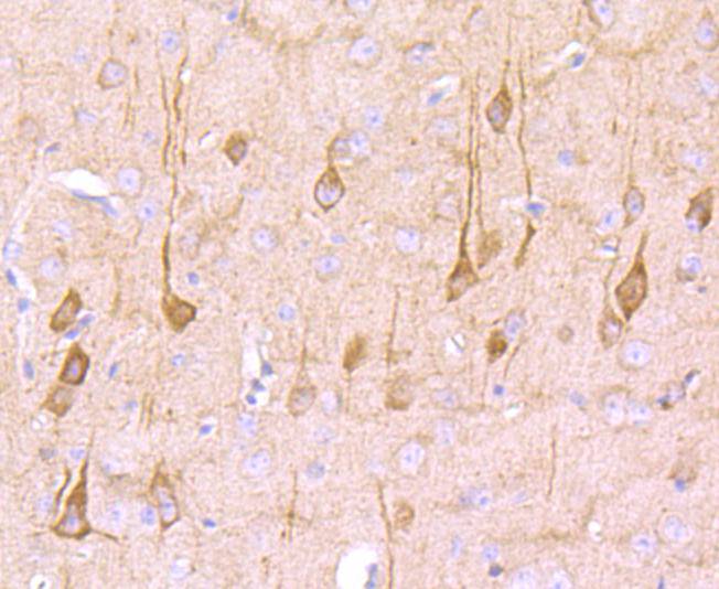 Paraformaldehyde-fixed, paraffin embedded (mouse brain tissue); Antigen retrieval by boiling in sodium citrate buffer (pH6.0) for 15min; Block endogenous peroxidase by 3% hydrogen peroxide for 20 minutes; Blocking buffer (normal goat serum) at 37°C for 30min; Antibody incubation with (NG2) Monoclonal Antibody, Unconjugated (SLM-52891R) at 1:50 overnight at 4°C, followed by operating according to SP Kit(Rabbit) (sp-0023) instructionsand DAB staining.
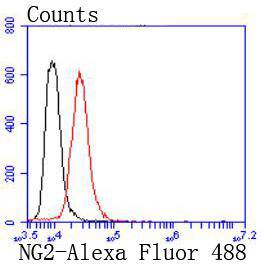 Blank control:SHG-44.
Primary Antibody (green line): Rabbit Anti-NG2 antibody (SLM-52819)
Dilution: 1:50;
Isotype Control Antibody (orange line): Rabbit IgG .
Secondary Antibody : Goat anti-rabbit IgG-AF488
Dilution: 1:1000.
Protocol
The cells were fixed with 4% PFA (10min at room temperature)and then permeabilized with 0.1% PBST for 20 min at room temperature. The cells were then incubated in 5%BSA to block non-specific protein-protein interactions for 30 min at room temperature .Cells stained with Primary Antibody for 30 min at room temperature. The secondary antibody used for 40 min at room temperature. Acquisition of 20,000 events was performed.
Cartpieces
Totalgoods,subtotals:¥Checkout
References (0)
No References
Bought notes(bought amounts latest0)
No one bought this product
User Comment(Total0User Comment Num)
- No comment


 +86 571 56623320
 +86 18668110335

