
Rabbit Anti-Bcl-2 antibody
Apoptosis regulator Bcl 2; Apoptosis regulator Bcl2; AW986256; B cell CLL/lymphoma 2; B cell leukemia/lymphoma 2; B cell lymphoma 2; Bcl 2; Bcl-2; Bcl2; BCL2 protein; C430015F12Rik; D630044D05Rik; D830018M01Rik; Leukemia/lymphoma, B-cell, 2; Oncogene B-ce
View History [Clear]
Details
Product Name Bcl-2 Chinese Name Bcl-2Recombinant rabbit monoclonal anti Alias Apoptosis regulator Bcl 2; Apoptosis regulator Bcl2; AW986256; B cell CLL/lymphoma 2; B cell leukemia/lymphoma 2; B cell lymphoma 2; Bcl 2; Bcl-2; Bcl2; BCL2 protein; C430015F12Rik; D630044D05Rik; D830018M01Rik; Leukemia/lymphoma, B-cell, 2; Oncogene B-cell leukemia 2; BCL2_HUMAN. literatures Research Area Cell biology Signal transduction Apoptosis The new supersedes the old Mitochondrion Immunogen Species Rabbit Clonality Monoclonal Clone NO. 6C4 React Species Human, Mouse, Applications WB=1:1000-2000 IP=1:10-50 IHC-P=1:50-200 IHC-F=1:50-200 Flow-Cyt=1:50 ICC=1:50 IF=1:50-200 (Paraffin sections need antigen repair)
not yet tested in other applications.
optimal dilutions/concentrations should be determined by the end user.Theoretical molecular weight 26kDa Cellular localization The nucleus cytoplasmic The cell membrane Mitochondrion Form Liquid Concentration 1mg/ml immunogen KLH conjugated synthetic peptide derived from human Bcl-2 Lsotype IgG Purification affinity purified by Protein A Buffer Solution 0.01M TBS(pH7.4) with 1% BSA, 0.03% Proclin300 and 50% Glycerol. Storage Shipped at 4℃. Store at -20 °C for one year. Avoid repeated freeze/thaw cycles. Attention This product as supplied is intended for research use only, not for use in human, therapeutic or diagnostic applications. PubMed PubMed Product Detail The Bcl-2 gene was isolated at the chromosomal breakpoint of t(14;18)-bearing follicular B cell lymphomas(1,2).Bcl-2 blocks cell death following a variety of stimuli and confers a death-sparing effect to certain hematopoietic cell lines following growth factor withdrawal (3,5).Bcl-2 appears to function in several subcellular locations yet lacks any known motifs that would confer insight into its mechanism of action (6,7).A more recently identified protein,designated Bax p21(i.e., Bcl-associated X protein ),has extensive amino acid homology with Bcl-2 and both homodimerizes and forms heterodimers with Bcl-2(8). Overexpression of Bax accelerates apoptotic death induced by cytokine deprivation in an IL-3 dependent cell line and Bax also counters the death repressor activty of Bcl-2(8).
Function:
Suppresses apoptosis in a variety of cell systems including factor-dependent lymphohematopoietic and neural cells. Regulates cell death by controlling the mitochondrial membrane permeability. Appears to function in a feedback loop system with caspases. Inhibits caspase activity either by preventing the release of cytochrome c from the mitochondria and/or by binding to the apoptosis-activating factor (APAF-1).
Subunit:
Forms homodimers, and heterodimers with BAX, BAD, BAK and Bcl-X(L). Heterodimerization with BAX requires intact BH1 and BH2 motifs, and is necessary for anti-apoptotic activity. Interacts with EI24 (By similarity). Also interacts with APAF1, BBC3, BCL2L1, BNIPL, MRPL41 and TP53BP2. Binding to FKBP8 seems to target BCL2 to the mitochondria and probably interferes with the binding of BCL2 to its targets. Interacts with BAG1 in an ATP-dependent manner. Interacts with RAF1 (the 'Ser-338' and 'Ser-339' phosphorylated form). Interacts (via the BH4 domain) with EGLN3; the interaction prevents the formation of the BAX-BCL2 complex and inhibits the anti-apoptotic activity of BCL2. Interacts with G0S2; this interaction also prevents the formation of the anti-apoptotic BAX-BCL2 complex.
Subcellular Location:
Mitochondrion outer membrane; Single-pass membrane protein. Nucleus membrane; Single-pass membrane protein. Endoplasmic reticulum membrane; Single-pass membrane protein.
Tissue Specificity:
Expressed in a variety of tissues.
Post-translational modifications:
Phosphorylation/dephosphorylation on Ser-70 regulates anti-apoptotic activity. Growth factor-stimulated phosphorylation on Ser-70 by PKC is required for the anti-apoptosis activity and occurs during the G2/M phase of the cell cycle. In the absence of growth factors, BCL2 appears to be phosphorylated by other protein kinases such as ERKs and stress-activated kinases. Phosphorylated by MAPK8/JNK1 at Thr-69, Ser-70 and Ser-87, wich stimulates starvation-induced autophagy. Dephosphorylated by protein phosphatase 2A (PP2A).
Proteolytically cleaved by caspases during apoptosis. The cleaved protein, lacking the BH4 motif, has pro-apoptotic activity, causes the release of cytochrome c into the cytosol promoting further caspase activity.
Monoubiquitinated by PARK2, leading to increase its stability.
DISEASE:
Note=A chromosomal aberration involving BCL2 has been found in chronic lymphatic leukemia. Translocation t(14;18)(q32;q21) with immunoglobulin gene regions. BCL2 mutations found in non-Hodgkin lymphomas carrying the chromosomal translocation could be attributed to the Ig somatic hypermutation mechanism resulting in nucleotide transitions.
Similarity:
Belongs to the Bcl-2 family.
SWISS:
P49950
Gene ID:
596
Database links:
Entrez Gene: 596 Human
Entrez Gene: 12043 Mouse
Omim: 151430 Human
SwissProt: P10415 Human
SwissProt: P10417 Mouse
Unigene: 150749 Human
Unigene: 257460 Mouse
Unigene: 9996 Rat
Product Picture 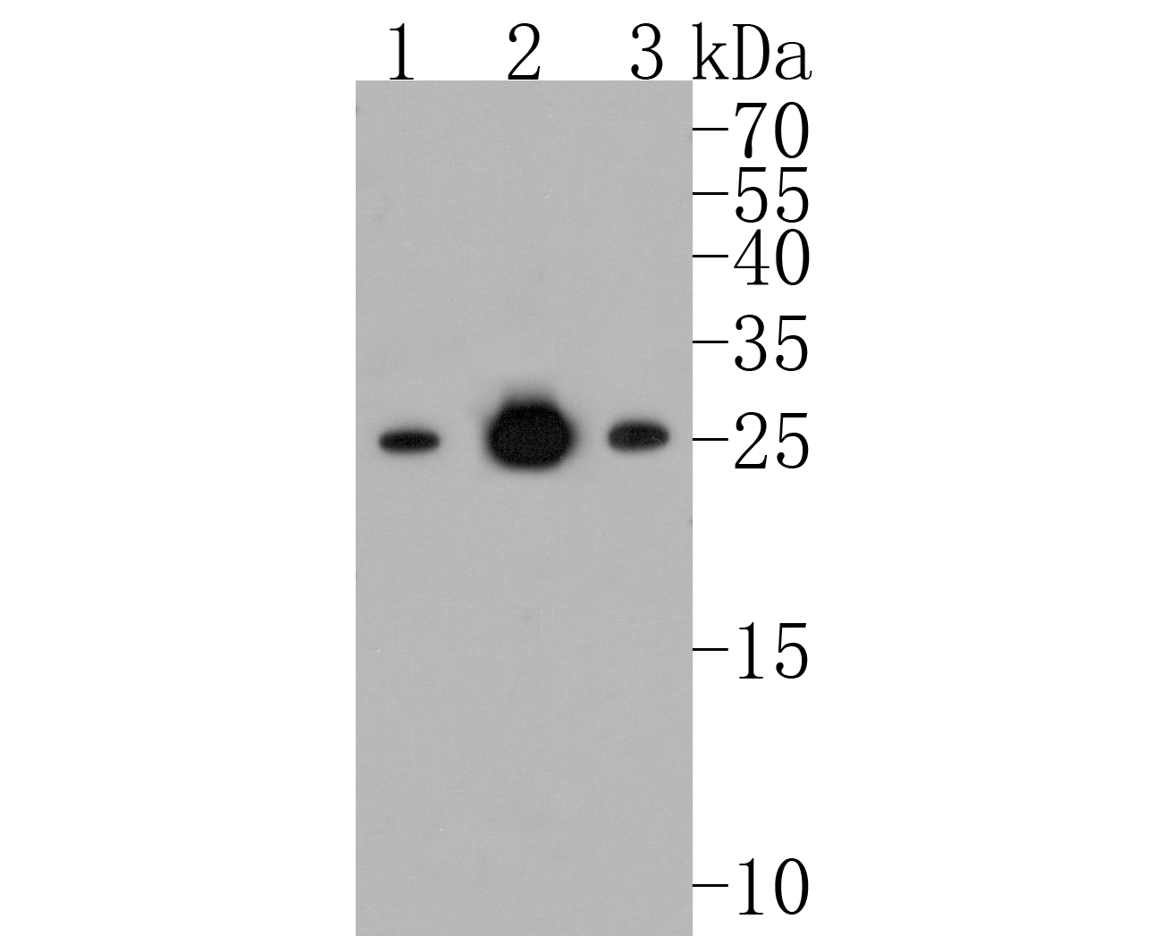 Sample:
Lane 1: Hela cell lysate
Lane 2: Jurkat cell lysate
Lane 3: MCF-7 cell lysate
Primary: Anti-Bcl-2 (SLM-52833R) at 1:500 dilution
Secondary: Goat Anti-Rabbit IgG - HRP at 1:5000 dilution
Predicted band size: 26 kD
Observed band size: 26 kD
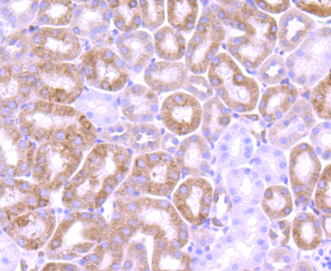 Paraformaldehyde-fixed, paraffin embedded (mouse kidney tissue); Antigen retrieval by boiling in sodium citrate buffer (pH6.0) for 15min; Block endogenous peroxidase by 3% hydrogen peroxide for 20 minutes; Blocking buffer (normal goat serum) at 37°C for 30min; Antibody incubation with (Bcl-2) Monoclonal Antibody, Unconjugated (SLM-52833R) at 1:50 overnight at 4°C, followed by operating according to SP Kit(Rabbit) (sp-0023) instructionsand DAB staining.
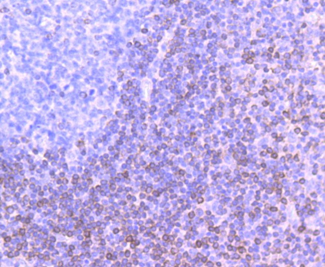 Paraformaldehyde-fixed, paraffin embedded (human tonsil); Antigen retrieval by boiling in sodium citrate buffer (pH6.0) for 15min; Block endogenous peroxidase by 3% hydrogen peroxide for 20 minutes; Blocking buffer (normal goat serum) at 37°C for 30min; Antibody incubation with (Bcl-2) Monoclonal Antibody, Unconjugated (SLM-52833R) at 1:50 overnight at 4°C, followed by operating according to SP Kit(Rabbit) (sp-0023) instructionsand DAB staining.
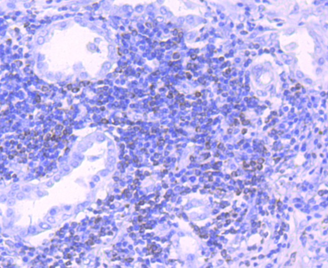 Paraformaldehyde-fixed, paraffin embedded (human lung carcinoma); Antigen retrieval by boiling in sodium citrate buffer (pH6.0) for 15min; Block endogenous peroxidase by 3% hydrogen peroxide for 20 minutes; Blocking buffer (normal goat serum) at 37°C for 30min; Antibody incubation with (Bcl-2) Monoclonal Antibody, Unconjugated (SLM-52833R) at 1:50 overnight at 4°C, followed by operating according to SP Kit(Rabbit) (sp-0023) instructionsand DAB staining.
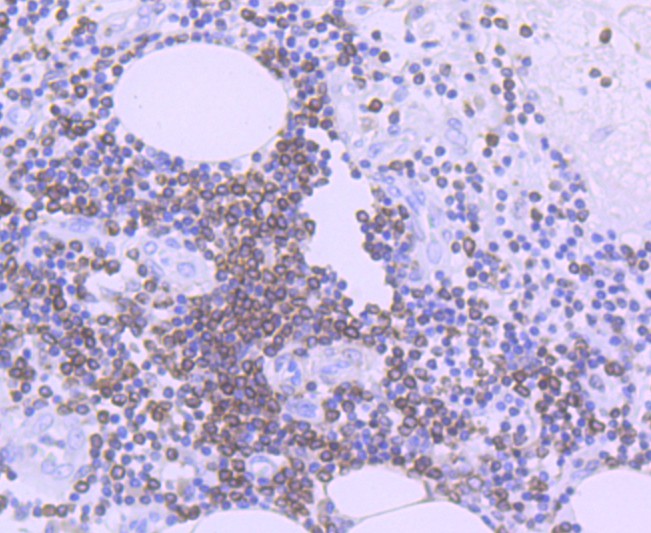 Paraformaldehyde-fixed, paraffin embedded (human colon carcinoma); Antigen retrieval by boiling in sodium citrate buffer (pH6.0) for 15min; Block endogenous peroxidase by 3% hydrogen peroxide for 20 minutes; Blocking buffer (normal goat serum) at 37°C for 30min; Antibody incubation with (Bcl-2) Monoclonal Antibody, Unconjugated (SLM-52833R) at 1:50 overnight at 4°C, followed by operating according to SP Kit(Rabbit) (sp-0023) instructionsand DAB staining.
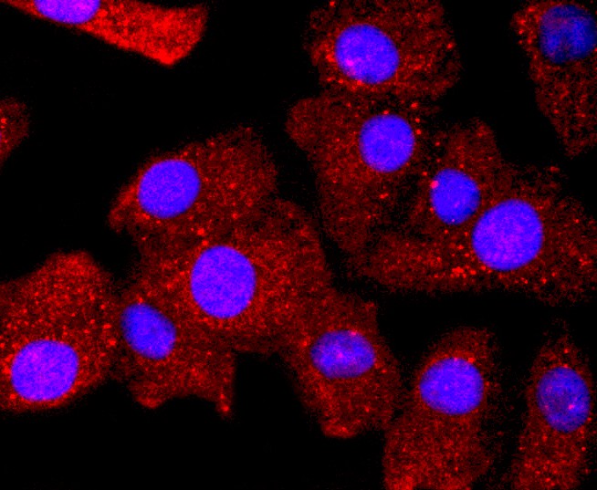 A549 cell; 4% Paraformaldehyde-fixed; Triton X-100 at room temperature for 20 min; Blocking buffer (normal goat serum,C-0005) at 37°C for 20 min; Antibody incubation with (Bcl-2) monoclonal Antibody, Unconjugated (SLM-52833R) 1:50, 90 minutes at 37°C; followed by a conjugated Goat Anti-Rabbit IgG antibody at 37°C for 90 minutes, DAPI (blue, C02-04002) was used to stain the cell nuclei.
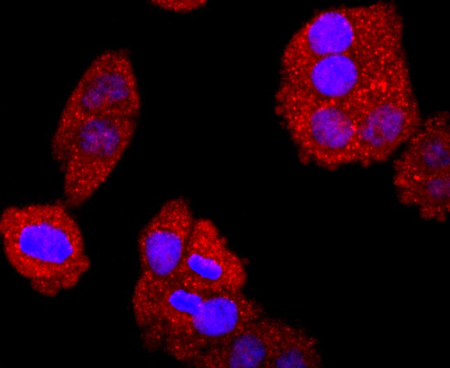 Hela cell; 4% Paraformaldehyde-fixed; Triton X-100 at room temperature for 20 min; Blocking buffer (normal goat serum,C-0005) at 37°C for 20 min; Antibody incubation with (Bcl-2) monoclonal Antibody, Unconjugated (SLM-52833R) 1:50, 90 minutes at 37°C; followed by a conjugated Goat Anti-Rabbit IgG antibody at 37°C for 90 minutes, DAPI (blue, C02-04002) was used to stain the cell nuclei.
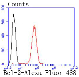 Blank control:Jurkat.
Primary Antibody (green line): Rabbit Anti-Bcl-2antibody (SLM-52833R)
Dilution: 1:50;
Isotype Control Antibody (orange line): Rabbit IgG .
Secondary Antibody : Goat anti-rabbit IgG-AF488
Dilution: 1:1000.
Protocol
The cells were fixed with 4% PFA (10min at room temperature)and then permeabilized with 90% ice-cold methanol for 20 min at-20℃.The cells were then incubated in 5%BSA to block non-specific protein-protein interactions for 30 min at room temperature .Cells stained with Primary Antibody for 30 min at room temperature. The secondary antibody used for 40 min at room temperature. Acquisition of 20,000 events was performed.
Cartpieces
Totalgoods,subtotals:¥Checkout
References (0)
No References
Bought notes(bought amounts latest0)
No one bought this product
User Comment(Total0User Comment Num)
- No comment


 +86 571 56623320
 +86 18668110335

