
Rabbit Anti-TFRC antibody
Transferrin receptor protein 1; Transferrin receptor; CD71; CD 71; CD71 antigen; p90; T9; TFR 1; TFR; TFR1; TR; sTfR; Transferrin receptor (p90 CD71); TRFR; TFR1_HUMAN.
View History [Clear]
Details
Product Name TFRC Chinese Name 转铁蛋白受体(CD71)Recombinant rabbit monoclonal anti Alias Transferrin receptor protein 1; Transferrin receptor; CD71; CD 71; CD71 antigen; p90; T9; TFR 1; TFR; TFR1; TR; sTfR; Transferrin receptor (p90 CD71); TRFR; TFR1_HUMAN. Research Area Tumour Cardiovascular Cell biology immunology Neurobiology Stem cells Diabetes Cell Surface Molecule lymphocyte Immunogen Species Rabbit Clonality Monoclonal Clone NO. 8G6 React Species Human, Mouse, Rat, Applications WB=1:500-2000 IP=1:10-50 IHC-P=1:50-200 IHC-F=1:50-200 ICC=1:100 (Paraffin sections need antigen repair)
not yet tested in other applications.
optimal dilutions/concentrations should be determined by the end user.Theoretical molecular weight 95kDa Cellular localization The cell membrane Form Liquid Concentration 1mg/ml immunogen KLH conjugated synthetic peptide derived from human Transferrin receptor Lsotype IgG Purification affinity purified by Protein A Buffer Solution 0.01M TBS(pH7.4) with 1% BSA, 0.03% Proclin300 and 50% Glycerol. Storage Shipped at 4℃. Store at -20 °C for one year. Avoid repeated freeze/thaw cycles. Attention This product as supplied is intended for research use only, not for use in human, therapeutic or diagnostic applications. PubMed PubMed Product Detail This gene encodes a cell surface receptor necessary for cellular iron uptake by the process of receptor-mediated endocytosis. This receptor is required for erythropoiesis and neurologic development. Multiple alternatively spliced variants have been identified. [provided by RefSeq, Sep 2015]
Function:
Cellular uptake of iron occurs via receptor-mediated endocytosis of ligand-occupied transferrin receptor into specialized endosomes. Endosomal acidification leads to iron release. The apotransferrin-receptor complex is then recycled to the cell surface with a return to neutral pH and the concomitant loss of affinity of apotransferrin for its receptor. Transferrin receptor is necessary for development of erythrocytes and the nervous system. A second ligand, the heditary hemochromatosis protein HFE, competes for binding with transferring for an overlapping C-terminal binding site.
Subunit:
Homodimer; disulfide-linked. Binds one transferrin or HFE molecule per subunit. Binds the HLA class II histocompatibility antigen, DR1. Interacts with SH3BP3. Interacts with Machupo arenavirus GPC.
Subcellular Location:
Cell membrane; Single-pass type II membrane protein. Melanosome. Note=Identified by mass spectrometry in melanosome fractions from stage I to stage IV.
Transferrin receptor protein 1, serum form: Secreted
Post-translational modifications:
N- and O-glycosylated, phosphorylated and palmitoylated. The serum form is only glycosylated.
Proteolytically cleaved on Arg-100 to produce the soluble serum form (sTfR).
Palmitoylated on both Cys-62 and Cys-67. Cys-62 seems to be the major site of palmitoylation.
Similarity:
Belongs to the peptidase M28 family. M28B subfamily.
Contains 1 PA (protease associated) domain.
SWISS:
P02786
Gene ID:
7037
Database links:Entrez Gene: 7037 Human
Entrez Gene: 22042 Mouse
Omim: 190010 Human
SwissProt: P02786 Human
SwissProt: Q62351 Mouse
Unigene: 529618 Human
Unigene: 28683 Mouse
Unigene: 98672 Rat
Product Picture  Sample:
Lane 1: Placenta (Mouse) Lysate at 40 ug
Lane 2: Hela (Human) Cell Lysate at 30 ug
Lane 3: HT1080 (Human) Cell Lysate at 30 ug
Lane 4: K562 (Human) Cell Lysate at 30 ug
Primary: Anti-CD71/Transferrin receptor (SLM-52793R) at 1/1000 dilution
Secondary: IRDye800CW Goat Anti-Rabbit IgG at 1/20000 dilution
Predicted band size: 95 kD
Observed band size: 100 kD
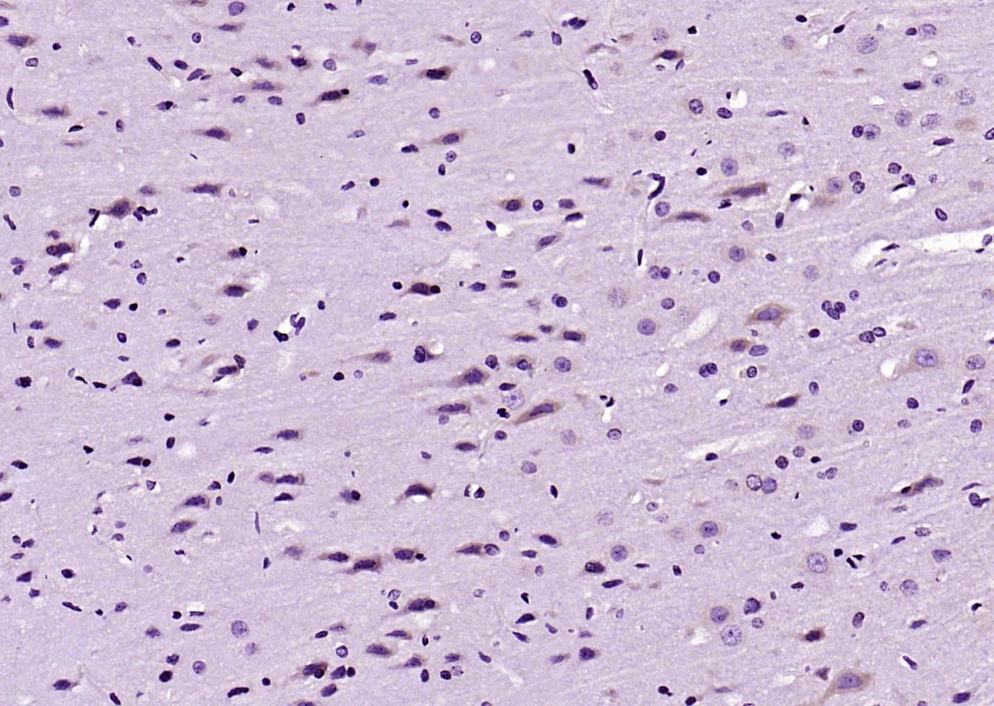 Paraformaldehyde-fixed, paraffin embedded (rat brain); Antigen retrieval by boiling in sodium citrate buffer (pH6.0) for 15min; Block endogenous peroxidase by 3% hydrogen peroxide for 20 minutes; Blocking buffer (normal goat serum) at 37°C for 30min; Antibody incubation with (CD71 Transferrin receptor) Monoclonal Antibody, Unconjugated (SLM-52793R) at 1:200 overnight at 4°C, followed by operating according to SP Kit(Rabbit) (sp-0023) instructionsand DAB staining.
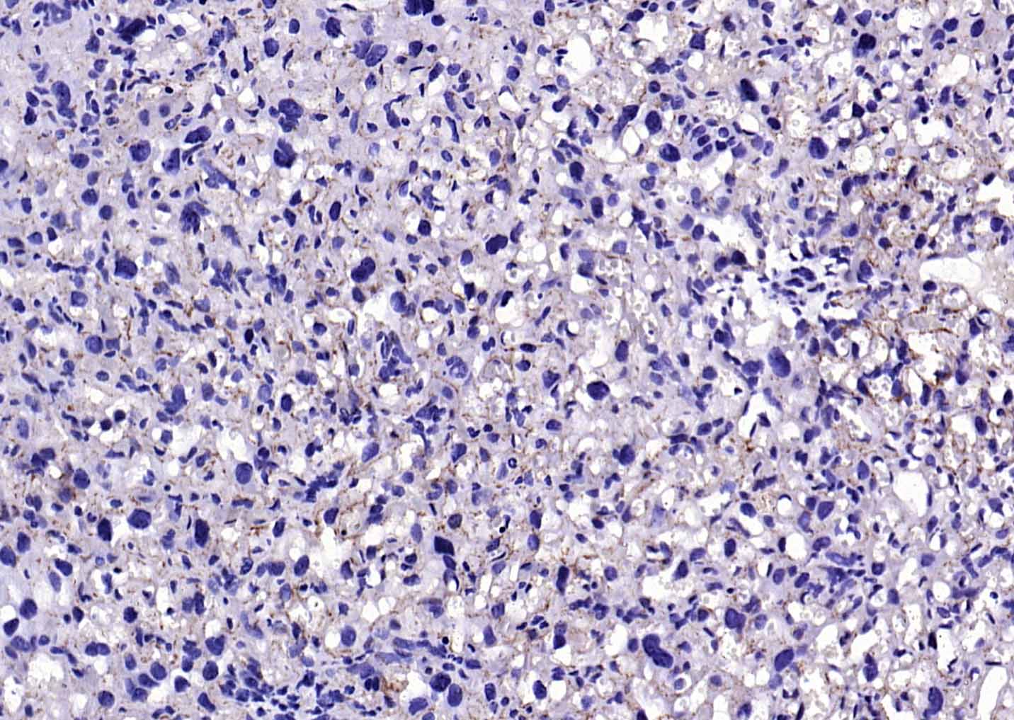 Paraformaldehyde-fixed, paraffin embedded (mouse placenta); Antigen retrieval by boiling in sodium citrate buffer (pH6.0) for 15min; Block endogenous peroxidase by 3% hydrogen peroxide for 20 minutes; Blocking buffer (normal goat serum) at 37°C for 30min; Antibody incubation with (CD71 Transferrin receptor) Monoclonal Antibody, Unconjugated (SLM-52793R) at 1:200 overnight at 4°C, followed by operating according to SP Kit(Rabbit) (sp-0023) instructionsand DAB staining.
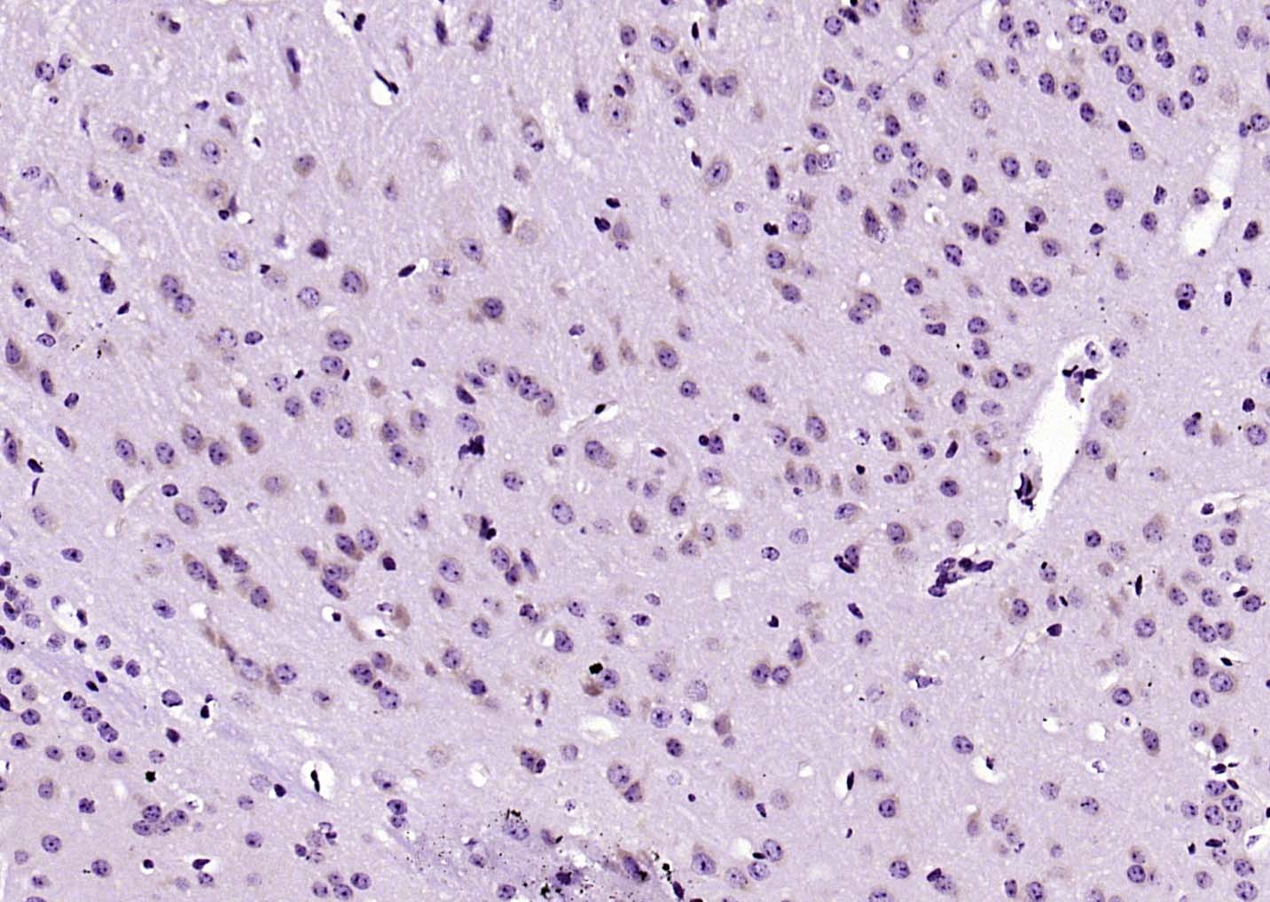 Paraformaldehyde-fixed, paraffin embedded (mouse brain); Antigen retrieval by boiling in sodium citrate buffer (pH6.0) for 15min; Block endogenous peroxidase by 3% hydrogen peroxide for 20 minutes; Blocking buffer (normal goat serum) at 37°C for 30min; Antibody incubation with (CD71 Transferrin receptor) Monoclonal Antibody, Unconjugated (SLM-52793R) at 1:200 overnight at 4°C, followed by operating according to SP Kit(Rabbit) (sp-0023) instructionsand DAB staining.
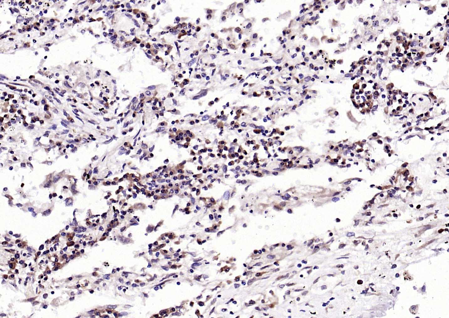 Paraformaldehyde-fixed, paraffin embedded (human lung carcinoma); Antigen retrieval by boiling in sodium citrate buffer (pH6.0) for 15min; Block endogenous peroxidase by 3% hydrogen peroxide for 20 minutes; Blocking buffer (normal goat serum) at 37°C for 30min; Antibody incubation with (CD71 Transferrin receptor) Monoclonal Antibody, Unconjugated (SLM-52793R) at 1:200 overnight at 4°C, followed by operating according to SP Kit(Rabbit) (sp-0023) instructionsand DAB staining.
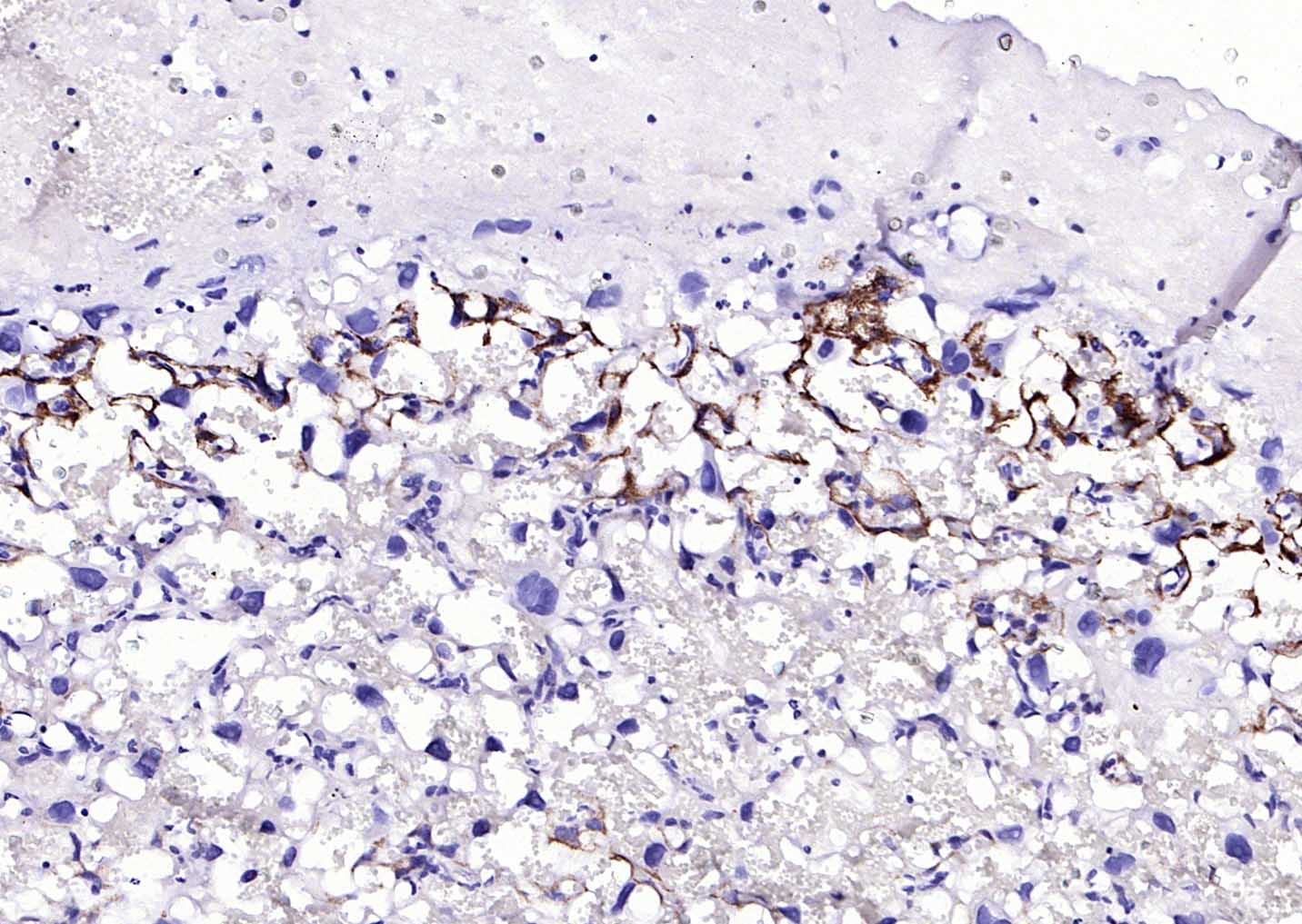 Paraformaldehyde-fixed, paraffin embedded (rat placenta); Antigen retrieval by boiling in sodium citrate buffer (pH6.0) for 15min; Block endogenous peroxidase by 3% hydrogen peroxide for 20 minutes; Blocking buffer (normal goat serum) at 37°C for 30min; Antibody incubation with (CD71 Transferrin receptor) Monoclonal Antibody, Unconjugated (SLM-52793R) at 1:200 overnight at 4°C, followed by operating according to SP Kit(Rabbit) (sp-0023) instructionsand DAB staining.
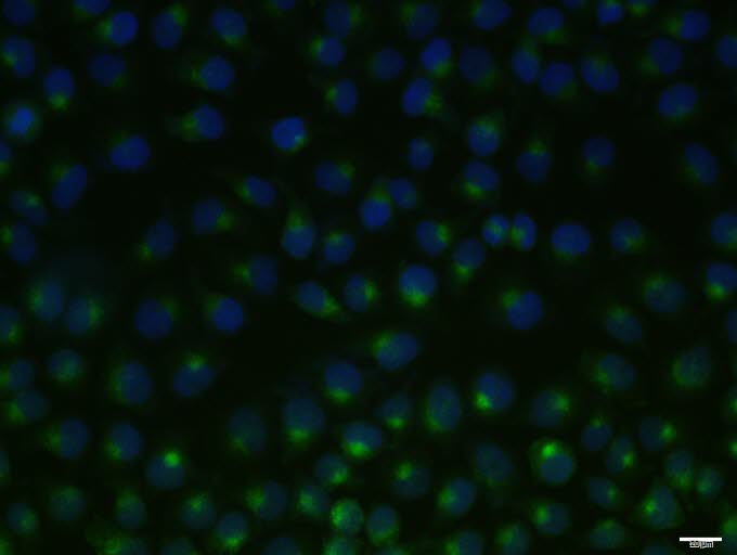 Hela cell; 4% Paraformaldehyde-fixed; Triton X-100 at room temperature for 20 min; Blocking buffer (normal goat serum, C-0005) at 37°C for 20 min; Antibody incubation with (TFRC) monoclonal Antibody, Unconjugated (SLM-52793R) 1:100, 90 minutes at 37°C; followed by a conjugated Goat Anti-Rabbit IgG antibody at 37°C for 90 minutes, DAPI (blue, C02-04002) was used to stain the cell nuclei.
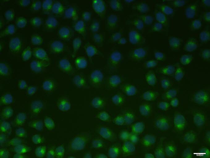 Hela cell; 4% Paraformaldehyde-fixed; Triton X-100 at room temperature for 20 min; Blocking buffer (normal goat serum, C-0005) at 37°C for 20 min; Antibody incubation with (TFRC) monoclonal Antibody, Unconjugated (SLM-52793R) 1:100, 90 minutes at 37°C; followed by a conjugated Goat Anti-Rabbit IgG antibody at 37°C for 90 minutes, DAPI (blue, C02-04002) was used to stain the cell nuclei.
Cartpieces
Totalgoods,subtotals:¥Checkout
References (0)
No References
Bought notes(bought amounts latest0)
No one bought this product
User Comment(Total0User Comment Num)
- No comment


 +86 571 56623320
 +86 18668110335

