
Rabbit Anti-ATF4 antibody
Cyclic AMP response element binding protein 2; DNA binding protein TAXREB67; Activating Transcription Factor 4; ATF 4; ATF4 protein; CREB 2; CREB2; CREB-2; Cyclic AMP dependent transcription factor ATF 4; Tax Responsive Enhancer Element B67; TAXREB67; TXR
View History [Clear]
Details
Product Name ATF4 Chinese Name 活化转录因子4Recombinant rabbit monoclonal anti Alias Cyclic AMP response element binding protein 2; DNA binding protein TAXREB67; Activating Transcription Factor 4; ATF 4; ATF4 protein; CREB 2; CREB2; CREB-2; Cyclic AMP dependent transcription factor ATF 4; Tax Responsive Enhancer Element B67; TAXREB67; TXREB; ATF4_HUMAN. Research Area Tumour Signal transduction Apoptosis transcriptional regulatory factor Immunogen Species Rabbit Clonality Monoclonal Clone NO. 9C3 React Species Human, Rat, (predicted: Mouse, ) Applications WB=1:500-1000 IHC-P=1:50-200 IHC-F=1:50-200 Flow-Cyt=1:50 ICC=1:50 IF=1:50-200 (Paraffin sections need antigen repair)
not yet tested in other applications.
optimal dilutions/concentrations should be determined by the end user.Theoretical molecular weight 38kDa Cellular localization The nucleus cytoplasmic The cell membrane Form Liquid Concentration 1mg/ml immunogen KLH conjugated synthetic peptide derived from human ATF4 Lsotype IgG Purification affinity purified by Protein A Buffer Solution 0.01M TBS(pH7.4) with 1% BSA, 0.03% Proclin300 and 50% Glycerol. Storage Shipped at 4℃. Store at -20 °C for one year. Avoid repeated freeze/thaw cycles. Attention This product as supplied is intended for research use only, not for use in human, therapeutic or diagnostic applications. PubMed PubMed Product Detail This gene encodes a transcription factor that was originally identified as a widely expressed mammalian DNA binding protein that could bind a tax-responsive enhancer element in the LTR of HTLV-1. The encoded protein was also isolated and characterized as the cAMP-response element binding protein 2 (CREB-2). The protein encoded by this gene belongs to a family of DNA-binding proteins that includes the AP-1 family of transcription factors, cAMP-response element binding proteins (CREBs) and CREB-like proteins. These transcription factors share a leucine zipper region that is involved in protein-protein interactions, located C-terminal to a stretch of basic amino acids that functions as a DNA binding domain. Two alternative transcripts encoding the same protein have been described. Two pseudogenes are located on the X chromosome at q28 in a region containing a large inverted duplication. [provided by RefSeq, Sep 2011]
Function:
Transcriptional activator. Binds the cAMP response element (CRE) (consensus: 5'-GTGACGT[AC][AG]-3'), a sequence present in many viral and cellular promoters. Cooperates with FOXO1 in osteoblasts to regulate glucose homeostasis through suppression of beta-cell production and decrease in insulin production. It binds to a Tax-responsive enhancer element in the long terminal repeat of HTLV-I. Regulates the induction of DDIT3/CHOP and asparagine synthetase (ASNS) in response to ER stress. In concert with DDIT3/CHOP, activates the transcription of TRIB3 and promotes ER stress-induced neuronal apoptosis by regulating the transcriptional induction of BBC3/PUMA. Activates transcription of SIRT4.
Subunit:
Binds DNA as a homo- or heterodimer. Interacts (via its leucine zipper domain) with GABBR1 and GABBR2 (via their C-termini). Interacts (via its DNA binding domain) with FOXO1 (C-terminal half); the interaction occurs in osteoblasts and regulates glucose homeostasis through suppression of beta-cell proliferation and a decrease in insulin production. Interacts with SATB2; the interaction results in enhanced DNA binding and transactivation by these transcription factors. Interacts with CEP290 (via an N-terminal region). Interacts with NEK6, DAPK2 (isoform 2) and ZIPK/DAPK3. Forms a heterodimer with TXLNG in osteoblasts. Interacts with DDIT3/CHOP.
Subcellular Location:
Cytoplasm. Cell membrane. Nucleus. Cytoplasm, cytoskeleton, microtubule organizing center, centrosome. Note=Colocalizes with GABBR1 in hippocampal neuron dendritic membranes. Co- localizes with NEK6 in the centrosome.
Post-translational modifications:
Ubiquitinated by SCF(BTRC) in response to mTORC1 signal, followed by proteasomal degradation and leading to down-regulate expression of SIRT4.
Phosphorylated by NEK6. Phosphorylated on the betaTrCP degron motif at Ser-219, followed by phosphorylation at Thr-213, Ser-224, Ser-231, Ser-235 and Ser-248, promoting interaction with BTRC and ubiquitination. Phosphorylation is promoted by mTORC1.
Phosphorylated by NEK6.
Similarity:
Belongs to the bZIP family.
Contains 1 bZIP (basic-leucine zipper) domain.
SWISS:
P18848
Gene ID:
468
Database links:Entrez Gene: 468 Human
Entrez Gene: 11911 Mouse
NCBI: 33469976 Human
Omim: 604064 Human
SwissProt: P18848 Human
SwissProt: Q96AQ3 Human
SwissProt: Q06507 Mouse
SwissProt: Q5U4B2 Mouse
SwissProt: Q8CF69 Mouse
Unigene: 496487 Human
Unigene: 641 Mouse
Unigene: 2423 Rat
Product Picture 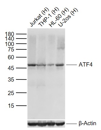 Sample:
Lane 1: Human Jurkat cell lysates
Lane 2: Human THP-1 cell lysates
Lane 3: Human HL-60 cell lysates
Lane 4: Human U2os cell lysates
Primary: Anti-ATF4 (SLM-52667R) at 1/1000 dilution
Secondary: IRDye800CW Goat Anti-Rabbit IgG at 1/20000 dilution
Predicted band size: 45 kDa
Observed band size: 45 kDa
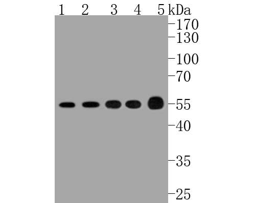 Sample:
Lane 1: Hela cell lysate
Lane 2: PC-12 cell lysate
Lane 3: HL-60 cell lysate
Lane 4: K562 cell lysate
Lane 5: human lung carcinoma tissue lysate
Primary: Anti-ATF4 (SLM-52667R) at 1:500 dilution
Secondary: Goat Anti-Rabbit IgG - HRP at 1:5000 dilution
Predicted band size: 38 kD
Observed band size: 55 kD
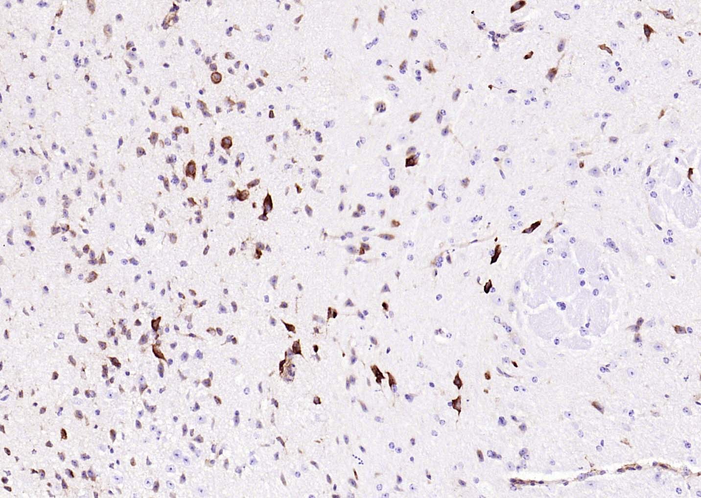 Paraformaldehyde-fixed, paraffin embedded (mouse brain); Antigen retrieval by boiling in sodium citrate buffer (pH6.0) for 15min; Block endogenous peroxidase by 3% hydrogen peroxide for 20 minutes; Blocking buffer (normal goat serum) at 37°C for 30min; Antibody incubation with (ATF4) Monoclonal Antibody, Unconjugated (SLM-52667R) at 1:200 overnight at 4°C, followed by operating according to SP Kit(Rabbit) (sp-0023) instructionsand DAB staining.
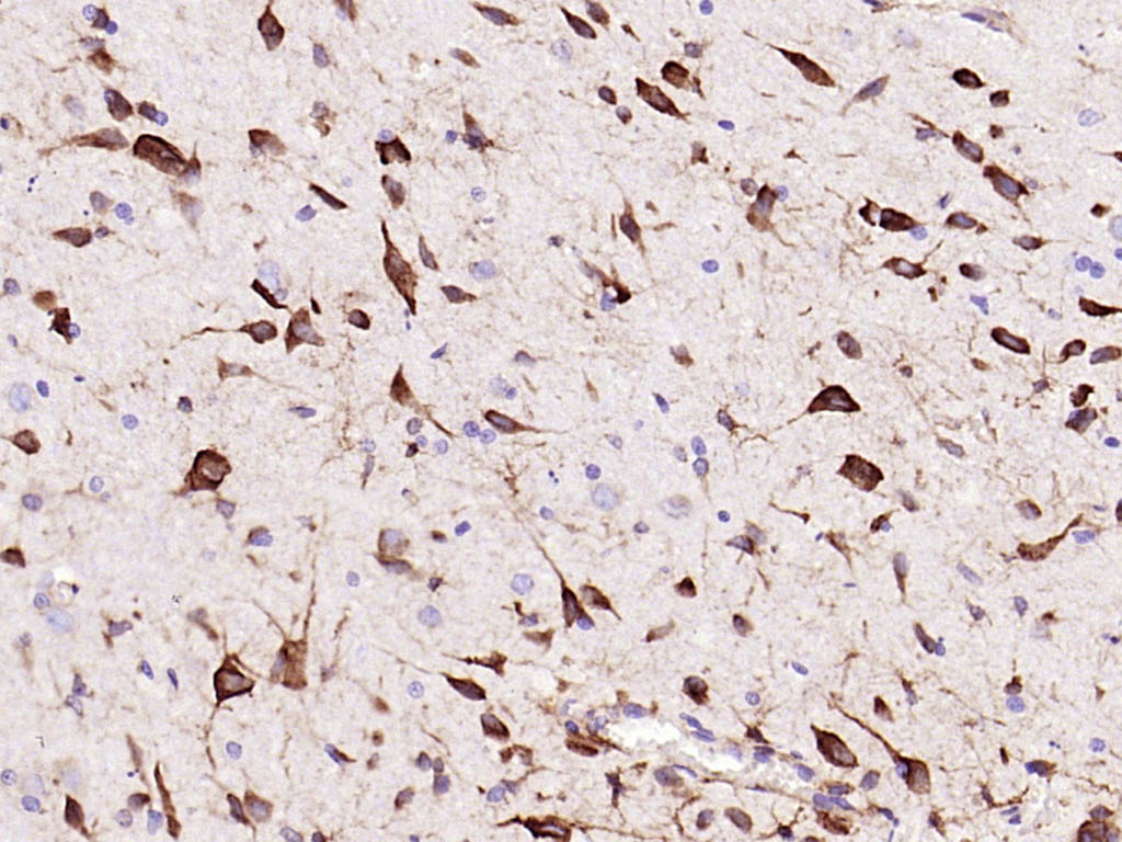 Paraformaldehyde-fixed, paraffin embedded (rat brain); Antigen retrieval by boiling in sodium citrate buffer (pH6.0) for 15min; Block endogenous peroxidase by 3% hydrogen peroxide for 20 minutes; Blocking buffer (normal goat serum) at 37°C for 30min; Antibody incubation with (ATF4) Monoclonal Antibody, Unconjugated (SLM-52667R) at 1:200 overnight at 4°C, followed by operating according to SP Kit(Rabbit) (sp-0023) instructionsand DAB staining.
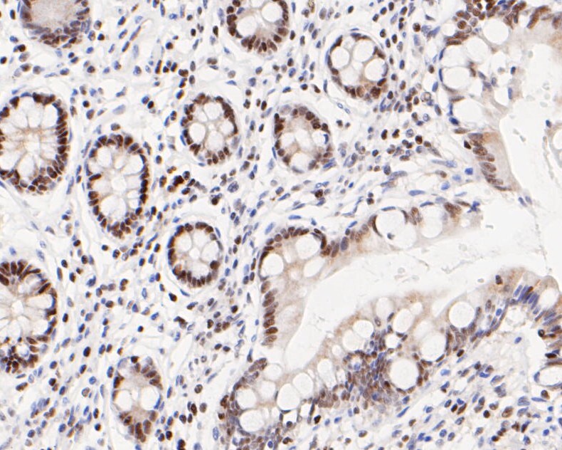 Paraformaldehyde-fixed, paraffin embedded (human small intestine); Antigen retrieval by boiling in sodium citrate buffer (pH6.0) for 15min; Block endogenous peroxidase by 3% hydrogen peroxide for 20 minutes; Blocking buffer (normal goat serum) at 37°C for 30min; Antibody incubation with (ATF4) Monoclonal Antibody, Unconjugated (SLM-52667R) at 1:50 overnight at 4°C, followed by operating according to SP Kit(Rabbit) (sp-0023) instructionsand DAB staining.
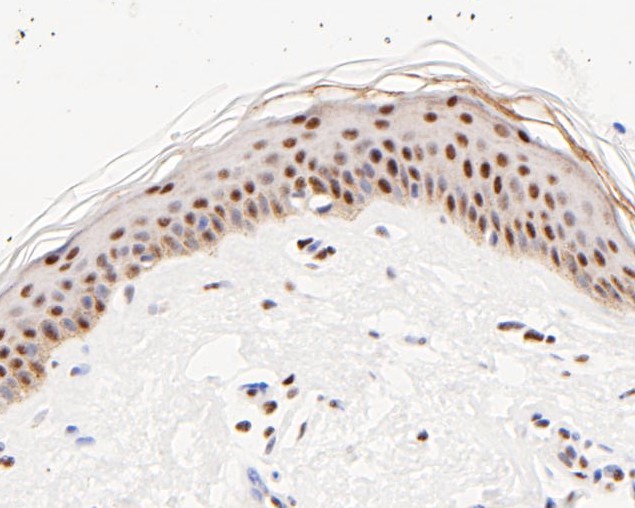 Paraformaldehyde-fixed, paraffin embedded (human skin tissue); Antigen retrieval by boiling in sodium citrate buffer (pH6.0) for 15min; Block endogenous peroxidase by 3% hydrogen peroxide for 20 minutes; Blocking buffer (normal goat serum) at 37°C for 30min; Antibody incubation with (ATF4) Monoclonal Antibody, Unconjugated (SLM-52667R) at 1:50 overnight at 4°C, followed by operating according to SP Kit(Rabbit) (sp-0023) instructionsand DAB staining.
 Paraformaldehyde-fixed, paraffin embedded (human prostate carcinoma); Antigen retrieval by boiling in sodium citrate buffer (pH6.0) for 15min; Block endogenous peroxidase by 3% hydrogen peroxide for 20 minutes; Blocking buffer (normal goat serum) at 37°C for 30min; Antibody incubation with (ATF4) Monoclonal Antibody, Unconjugated (SLM-52667R) at 1:50 overnight at 4°C, followed by operating according to SP Kit(Rabbit) (sp-0023) instructionsand DAB staining.
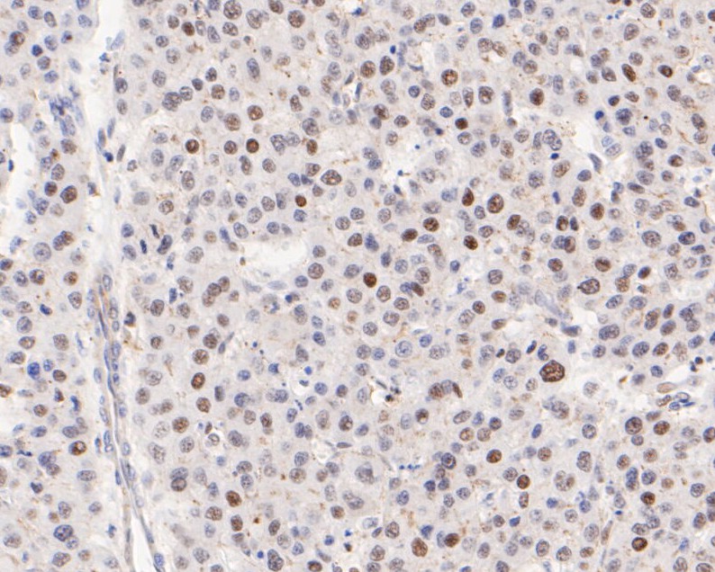 Paraformaldehyde-fixed, paraffin embedded (human liver carcinoma); Antigen retrieval by boiling in sodium citrate buffer (pH6.0) for 15min; Block endogenous peroxidase by 3% hydrogen peroxide for 20 minutes; Blocking buffer (normal goat serum) at 37°C for 30min; Antibody incubation with (ATF4) Monoclonal Antibody, Unconjugated (SLM-52667R) at 1:50 overnight at 4°C, followed by operating according to SP Kit(Rabbit) (sp-0023) instructionsand DAB staining.
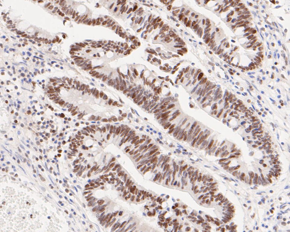 Paraformaldehyde-fixed, paraffin embedded (human colon carcinoma); Antigen retrieval by boiling in sodium citrate buffer (pH6.0) for 15min; Block endogenous peroxidase by 3% hydrogen peroxide for 20 minutes; Blocking buffer (normal goat serum) at 37°C for 30min; Antibody incubation with (ATF4) Monoclonal Antibody, Unconjugated (SLM-52667R) at 1:50 overnight at 4°C, followed by operating according to SP Kit(Rabbit) (sp-0023) instructionsand DAB staining.
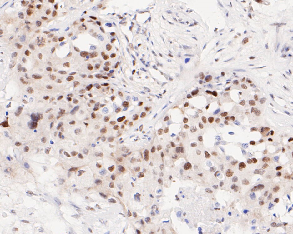 Paraformaldehyde-fixed, paraffin embedded (human breast carcinoma); Antigen retrieval by boiling in sodium citrate buffer (pH6.0) for 15min; Block endogenous peroxidase by 3% hydrogen peroxide for 20 minutes; Blocking buffer (normal goat serum) at 37°C for 30min; Antibody incubation with (ATF4) Monoclonal Antibody, Unconjugated (SLM-52667R) at 1:50 overnight at 4°C, followed by operating according to SP Kit(Rabbit) (sp-0023) instructionsand DAB staining.
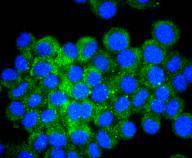 N2A cell; 4% Paraformaldehyde-fixed; Triton X-100 at room temperature for 20 min; Blocking buffer (normal goat serum,C-0005) at 37°C for 20 min; Antibody incubation with (ATF4) monoclonal Antibody, Unconjugated (SLM-52667R) 1:50, 90 minutes at 37°C; followed by a conjugated Goat Anti-Rabbit IgG antibody at 37°C for 90 minutes, DAPI (blue, C02-04002) was used to stain the cell nuclei.
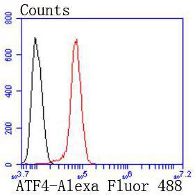 Blank control:Hela.
Primary Antibody (green line): Rabbit Anti-ATF4 antibody (SLM-52667R)
Dilution: 1:50;
Isotype Control Antibody (orange line): Rabbit IgG .
Secondary Antibody : Goat anti-rabbit IgG-AF488
Dilution: 1:1000.
Protocol
The cells were fixed with 4% PFA (10min at room temperature)and then permeabilized with 0.1% PBST for 20 min at room temperature.The cells were then incubated in 5%BSA to block non-specific protein-protein interactions for 30 min at room temperature .Cells stained with Primary Antibody for 30 min at room temperature. The secondary antibody used for 40 min at room temperature. Acquisition of 20,000 events was performed.
Cartpieces
Totalgoods,subtotals:¥Checkout
References (0)
No References
Bought notes(bought amounts latest0)
No one bought this product
User Comment(Total0User Comment Num)
- No comment


 +86 571 56623320
 +86 18668110335

