
Rabbit Anti-S100 beta antibody
NEF; Protein S100 B; Protein S100-B; S 100 calcium binding protein beta chain; S 100 protein beta chain; S 100 protein beta subunit; S-100 protein beta chain; S-100 protein subunit beta; S100; S100 calcium binding protein B; S100 calcium binding protein b
View History [Clear]
Details
Product Name S100 beta Chinese Name S100B蛋白Recombinant rabbit monoclonal anti Alias NEF; Protein S100 B; Protein S100-B; S 100 calcium binding protein beta chain; S 100 protein beta chain; S 100 protein beta subunit; S-100 protein beta chain; S-100 protein subunit beta; S100; S100 calcium binding protein B; S100 calcium binding protein beta (neural); S100 calcium binding protein beta chain; S100 calcium-binding protein B; S100 protein beta chain; S100B_HUMAN; S100beta. Research Area Tumour Cell biology Neurobiology Cyclin Cell differentiation Epigenetics Immunogen Species Rabbit Clonality Monoclonal Clone NO. 26C3 React Species Human, Mouse, Rat, Zebrafish, Applications WB=1:500-2000 IHC-P=1:50-200 (Paraffin sections need antigen repair)
not yet tested in other applications.
optimal dilutions/concentrations should be determined by the end user.Theoretical molecular weight 10kDa Cellular localization The nucleus cytoplasmic Form Liquid Concentration 1mg/ml immunogen KLH conjugated synthetic peptide derived from human S100B Lsotype IgG Purification affinity purified by Protein A Buffer Solution 0.01M TBS(pH7.4) with 1% BSA, 0.03% Proclin300 and 50% Glycerol. Storage Shipped at 4℃. Store at -20 °C for one year. Avoid repeated freeze/thaw cycles. Attention This product as supplied is intended for research use only, not for use in human, therapeutic or diagnostic applications. PubMed PubMed Product Detail S100 beta is a member of the S100 family of proteins containing 2 EF-hand calcium binding motifs. S100 proteins are localized in the cytoplasm and/or nucleus of a wide range of cells, and involved in the regulation of a number of cellular processes such as cell cycle progression and differentiation. S100 genes include at least 13 members which are located as a cluster on chromosome 1q21; however, this gene is located at 21q22.3. This protein may function in neurite extension, proliferation of melanoma cells, stimulation of Ca2+ fluxes, inhibition of PKC mediated phosphorylation, astrocytosis and axonal proliferation, and inhibition of microtubule assembly. Chromosomal rearrangements and altered expression of this gene have been implicated in several neurological, neoplastic, and other types of diseases, including Alzheimer's disease, Down's syndrome, epilepsy, amyotrophic lateral sclerosis, melanoma, and type I diabetes.
Function:
Weakly binds calcium but binds zinc very tightly-distinct binding sites with different affinities exist for both ions on each monomer. Physiological concentrations of potassium ion antagonize the binding of both divalent cations, especially affecting high-affinity calcium-binding sites. Binds to and initiates the activation of STK38 by releasing autoinhibitory intramolecular interactions within the kinase. Interaction with AGER after myocardial infarction may play a role in myocyte apoptosis by activating ERK1/2 and p53/TP53 signaling.
Subunit:
Dimer of either two alpha chains, or two beta chains, or one alpha and one beta chain. The S100B dimer binds two molecules of STK38 (By similarity). The S100B dimer interacts with two molecules of CAPZA1.
Subcellular Location:
Cytoplasm. Nucleus.
Tissue Specificity:
Although predominant among the water-soluble brain proteins, S100 is also found in a variety of other tissues.
Similarity:
Belongs to the S-101 family.
Contains 2 EF-hand domains.
SWISS:
P04271
Gene ID:
6285
Database links:Entrez Gene: 6285 Human
Entrez Gene: 20203 Mouse
Omim: 176990 Human
SwissProt: P04271 Human
SwissProt: P50114 Mouse
Unigene: 422181 Human
Unigene: 235998 Mouse
Unigene: 8937 Rat
Product Picture 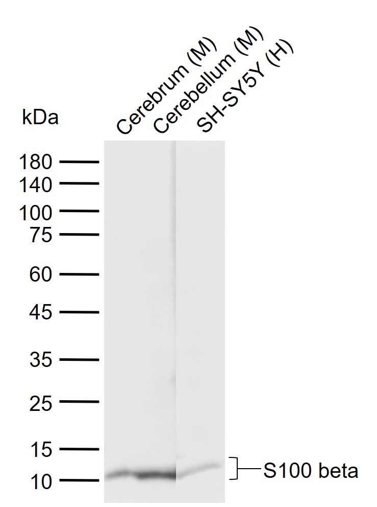 Sample:
Lane 1: Mouse Cerebrum tissue lysates
Lane 2: Mouse Cerebellum tissue lysates
Lane 3: Human SH-SY5Y cell lysates
Primary: Anti-S100 beta (SLM-52506R) at 1/1000 dilution
Secondary: IRDye800CW Goat Anti-Rabbit IgG at 1/20000 dilution
Predicted band size: 10 kDa
Observed band size: 10 kDa
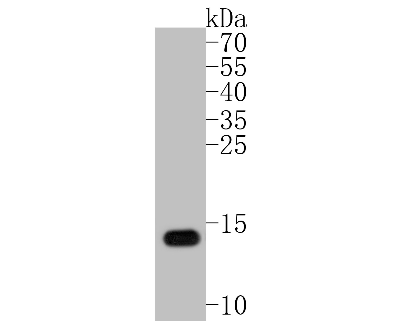 Sample:
Lane 1: mouse liver tissue lysates
Primary: Anti-S100 beta (SLM-52506R) at 1:500 dilution
Secondary: Goat Anti-Rabbit IgG - HRP at 1:5000 dilution
Predicted band size: 10 kD
Observed band size: 13 kD
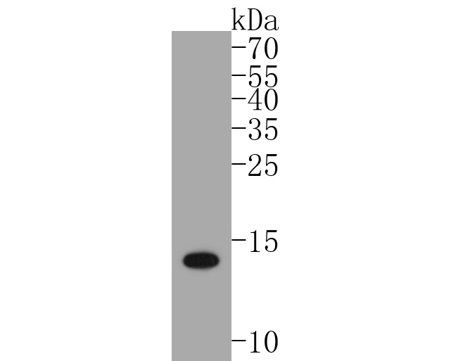 Sample:
Lane 1: zebrafish tissue tissue lysates
Primary: Anti-S100 beta (SLM-52506R) at 1:500 dilution
Secondary: Goat Anti-Rabbit IgG - HRP at 1:5000 dilution
Predicted band size: 10 kD
Observed band size: 13 kD
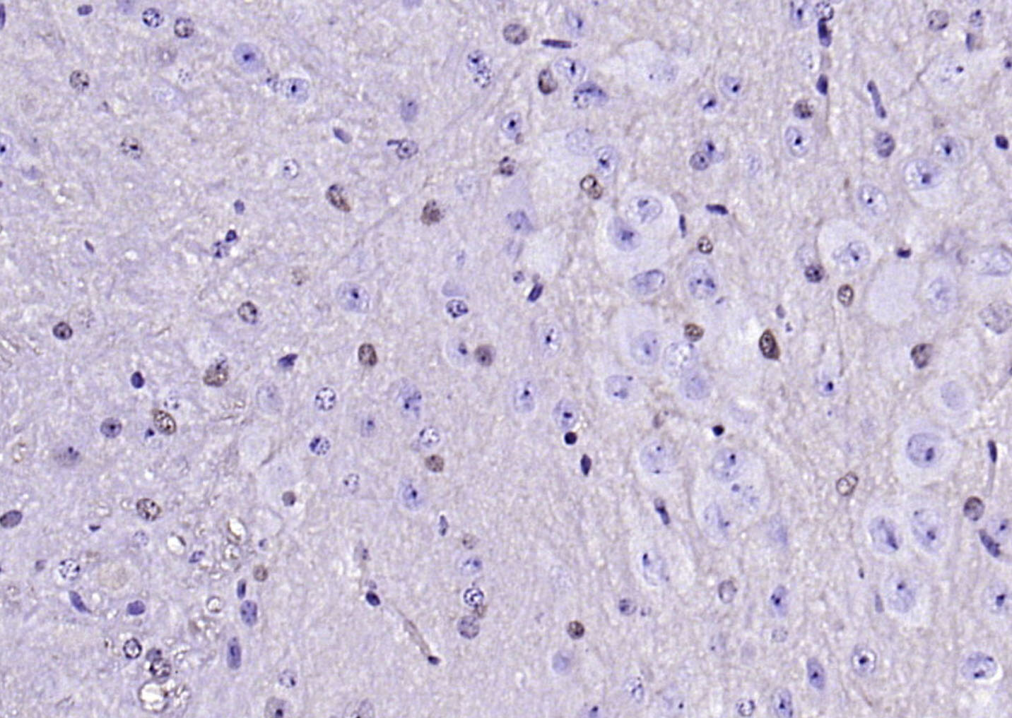 Paraformaldehyde-fixed, paraffin embedded (mouse cerebellum); Antigen retrieval by boiling in sodium citrate buffer (pH6.0) for 15min; Block endogenous peroxidase by 3% hydrogen peroxide for 20 minutes; Blocking buffer (normal goat serum) at 37°C for 30min; Antibody incubation with (S100 beta ) Monoclonal Antibody, Unconjugated (SLM-52506R) at 1:200 overnight at 4°C, followed by operating according to SP Kit(Rabbit) (sp-0023) instructionsand DAB staining.
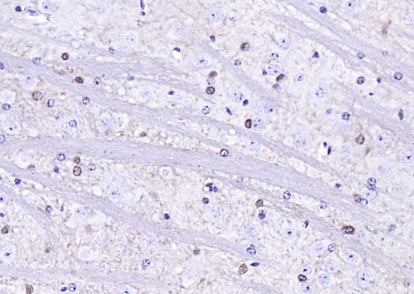 Paraformaldehyde-fixed, paraffin embedded (mouse brain); Antigen retrieval by boiling in sodium citrate buffer (pH6.0) for 15min; Block endogenous peroxidase by 3% hydrogen peroxide for 20 minutes; Blocking buffer (normal goat serum) at 37°C for 30min; Antibody incubation with (S100 beta ) Monoclonal Antibody, Unconjugated (SLM-52506R) at 1:200 overnight at 4°C, followed by operating according to SP Kit(Rabbit) (sp-0023) instructionsand DAB staining.
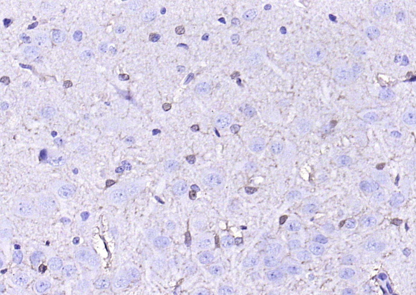 Paraformaldehyde-fixed, paraffin embedded (rat brain); Antigen retrieval by boiling in sodium citrate buffer (pH6.0) for 15min; Block endogenous peroxidase by 3% hydrogen peroxide for 20 minutes; Blocking buffer (normal goat serum) at 37°C for 30min; Antibody incubation with (S100 beta ) Monoclonal Antibody, Unconjugated (SLM-52506R) at 1:200 overnight at 4°C, followed by operating according to SP Kit(Rabbit) (sp-0023) instructionsand DAB staining.
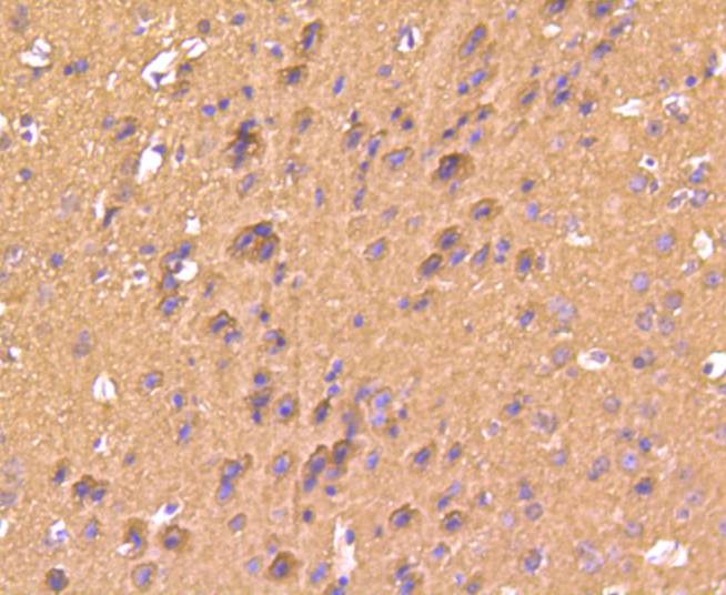 Paraformaldehyde-fixed, paraffin embedded (mouse brain); Antigen retrieval by boiling in sodium citrate buffer (pH6.0) for 15min; Block endogenous peroxidase by 3% hydrogen peroxide for 20 minutes; Blocking buffer (normal goat serum) at 37°C for 30min; Antibody incubation with (S100 beta) Monoclonal Antibody, Unconjugated (SLM-52506R) at 1:50 overnight at 4°C, followed by operating according to SP Kit(Rabbit) (sp-0023) instructionsand DAB staining.
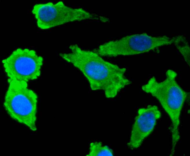 SH-SY5Y cell; 4% Paraformaldehyde-fixed; Triton X-100 at room temperature for 20 min; Blocking buffer (normal goat serum,C-0005) at 37°C for 20 min; Antibody incubation with (S100 beta) monoclonal Antibody, Unconjugated (SLM-52506R) 1:50, 90 minutes at 37°C; followed by a conjugated Goat Anti-Rabbit IgG antibody at 37°C for 90 minutes, DAPI (blue, C02-04002) was used to stain the cell nuclei.
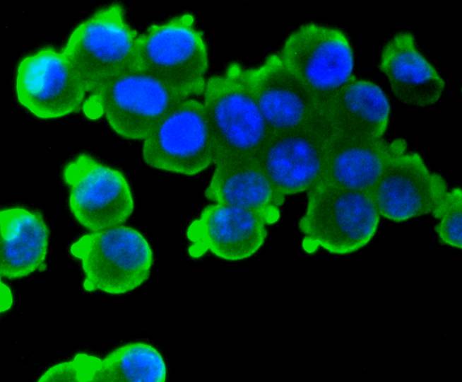 N2A cell; 4% Paraformaldehyde-fixed; Triton X-100 at room temperature for 20 min; Blocking buffer (normal goat serum,C-0005) at 37°C for 20 min; Antibody incubation with (S100 beta) monoclonal Antibody, Unconjugated (SLM-52506R) 1:50, 90 minutes at 37°C; followed by a conjugated Goat Anti-Rabbit IgG antibody at 37°C for 90 minutes, DAPI (blue, C02-04002) was used to stain the cell nuclei.
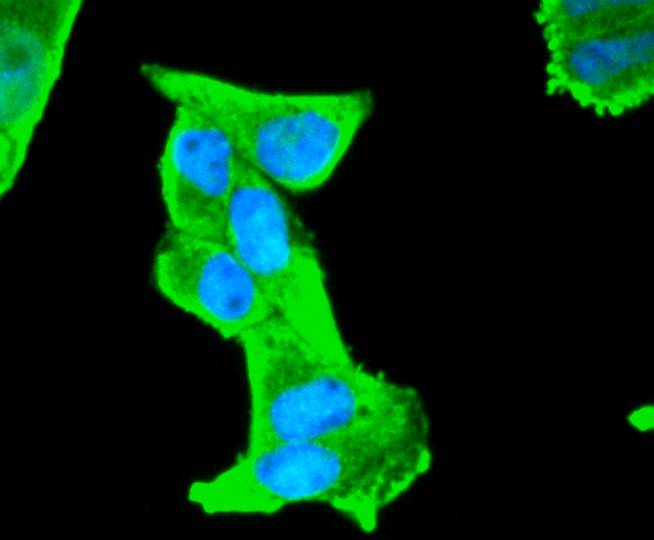 Hela cell; 4% Paraformaldehyde-fixed; Triton X-100 at room temperature for 20 min; Blocking buffer (normal goat serum,C-0005) at 37°C for 20 min; Antibody incubation with (S100 beta) monoclonal Antibody, Unconjugated (SLM-52506R) 1:50, 90 minutes at 37°C; followed by a conjugated Goat Anti-Rabbit IgG antibody at 37°C for 90 minutes, DAPI (blue, C02-04002) was used to stain the cell nuclei.
Cartpieces
Totalgoods,subtotals:¥Checkout
References (0)
No References
Bought notes(bought amounts latest0)
No one bought this product
User Comment(Total0User Comment Num)
- No comment


 +86 571 56623320
 +86 18668110335

