
Rabbit Anti-GLUT1 antibody
Glucose Transporter GLUT1; GT-1; GLUT-1; GLUT 1; Solute carrier family 2; facilitated glucose transporter member 1; Glucose transporter type 1; erythrocyte/brain; DYT17; DYT18; Erythrocyte/brain HepG2 glucose transporter; Erythrocyte/hepatoma glucose tran
View History [Clear]
Details
Product Name GLUT1 Chinese Name 葡萄糖Transporter1Recombinant rabbit monoclonal anti Alias Glucose Transporter GLUT1; GT-1; GLUT-1; GLUT 1; Solute carrier family 2; facilitated glucose transporter member 1; Glucose transporter type 1; erythrocyte/brain; DYT17; DYT18; Erythrocyte/brain HepG2 glucose transporter; Erythrocyte/hepatoma glucose transporter; Glucose transporter 1; Glucose transporter type 1; Glucose transporter type 1 erythrocyte/brain; Glucose transporter type 1, erythrocyte/brain; GLUT; GLUT1; GLUT1DS; GLUTB; GT1; GTG1; Gtg3; GTR1_HUMAN; HepG2 glucose transporter; MGC141895; MGC141896; PED; RATGTG1; SLC2A 1; SLC2A1; Solute carrier family 2 (facilitated glucose transporter), member 1; Solute carrier family 2 facilitated glucose transporter member 1. literatures Research Area Tumour immunology Growth factors and hormones Transporter Immunogen Species Rabbit Clonality Monoclonal Clone NO. 2D5 React Species Human, Mouse, Rat, Applications WB=1:300-500 IHC-P=1:50-200 IHC-F=1:50-200 ICC=1:50-200 (Paraffin sections need antigen repair)
not yet tested in other applications.
optimal dilutions/concentrations should be determined by the end user.Theoretical molecular weight 54kDa Cellular localization The cell membrane Extracellular matrix Form Liquid Concentration 1mg/ml immunogen KLH conjugated synthetic peptide derived from human GLUT1 Lsotype IgG Purification affinity purified by Protein A Buffer Solution 0.01M TBS(pH7.4) with 1% BSA, 0.03% Proclin300 and 50% Glycerol. Storage Shipped at 4℃. Store at -20 °C for one year. Avoid repeated freeze/thaw cycles. Attention This product as supplied is intended for research use only, not for use in human, therapeutic or diagnostic applications. PubMed PubMed Product Detail This gene encodes a major glucose transporter in the mammalian blood-brain barrier. Mutations in this gene have been found in a family with paroxysmal exertion-induced dyskinesia. [provided by RefSeq, Jul 2008].
Function:
Facilitative glucose transporter. This isoform may be responsible for constitutive or basal glucose uptake. Has a very broad substrate specificity; can transport a wide range of aldoses including both pentoses and hexoses.
Subcellular Location:
Cell membrane; Multi-pass membrane protein. Melanosome. Note=Localizes primarily at the cell surface. Identified by mass spectrometry in melanosome fractions from stage I to stage IV.
Tissue Specificity:
Expressed at variable levels in many human tissues.
Post-translational modifications:
Phosphorylated upon DNA damage, probably by ATM or ATR.
DISEASE:
Defects in SLC2A1 are the cause of GLUT1 deficiency syndrome type 1 (GLUT1DS1) [MIM:606777]; also known as blood-brain barrier glucose transport defect. A neurologic disorder showing wide phenotypic variability. The most severe 'classic' phenotype comprises infantile-onset epileptic encephalopathy associated with delayed development, acquired microcephaly, motor incoordination, and spasticity. Onset of seizures, usually characterized by apneic episodes, staring spells, and episodic eye movements, occurs within the first 4 months of life. Other paroxysmal findings include intermittent ataxia, confusion, lethargy, sleep disturbance, and headache. Varying degrees of cognitive impairment can occur, ranging from learning disabilities to severe mental retardation.
Defects in SLC2A1 are the cause of GLUT1 deficiency syndrome type 2 (GLUT1DS2) [MIM:612126]. A clinically variable disorder characterized primarily by onset in childhood of paroxysmal exercise-induced dyskinesia. The dyskinesia involves transient abnormal involuntary movements, such as dystonia and choreoathetosis, induced by exercise or exertion, and affecting the exercised limbs. Some patients may also have epilepsy, most commonly childhood absence epilepsy. Mild mental retardation may also occur. In some patients involuntary exertion-induced dystonic, choreoathetotic, and ballistic movements may be associated with macrocytic hemolytic anemia.
Similarity:
Belongs to the major facilitator superfamily. Sugar transporter (TC 2.A.1.1) family. Glucose transporter subfamily.
SWISS:
P11166
Gene ID:
6513
Database links:Entrez Gene: 6513 Human
Entrez Gene: 20525 Mouse
Omim: 138140 Human
SwissProt: P11166 Human
SwissProt: P17809 Mouse
Unigene: 473721 Human
Unigene: 721551 Human
Unigene: 21002 Mouse
Unigene: 3205 Rat
Product Picture 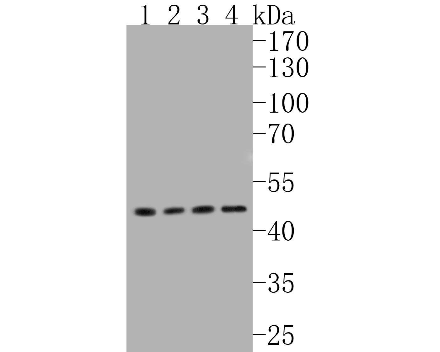 Western blot analysis of Glucose Transporter GLUT1 on different lysates. Proteins were transferred to a PVDF membrane and blocked with 5% BSA in PBS for 1 hour at room temperature. The primary antibody (SLM-52240R, 1/500) was used in 5% BSA at room temperature for 2 hours. Goat Anti-Rabbit IgG - HRP Secondary Antibody at 1:5,000 dilution was used for 1 hour at room temperature.
Positive control:
Lane 1: Hela cell lysate
Lane 2: SK-Br-3 cell lysate
Lane 3: NIH/3T3 cell lysate
Lane 4: HepG2 cell lysate
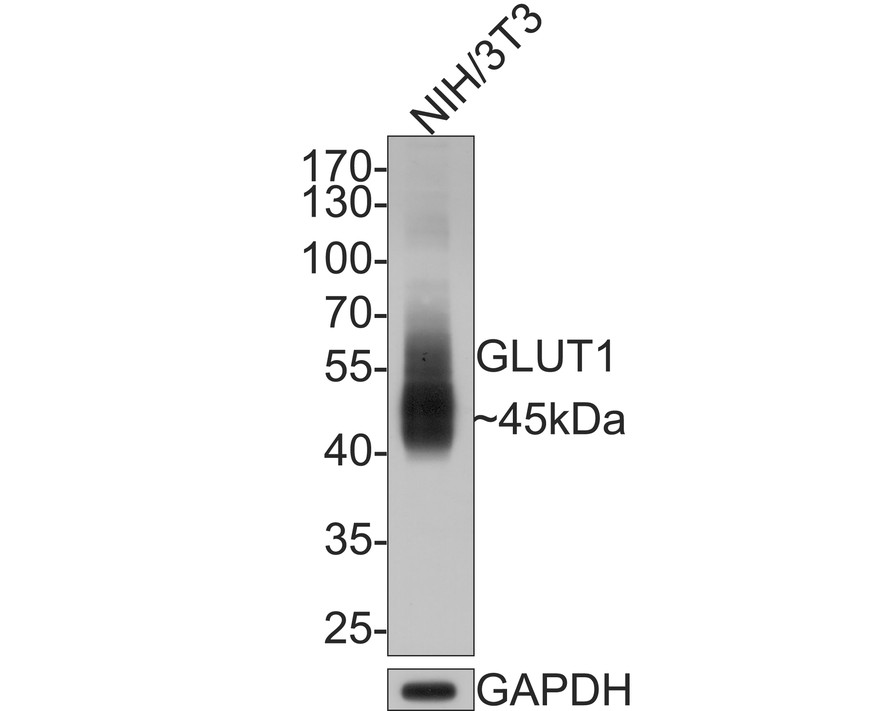 Western blot analysis of Glucose Transporter GLUT1 on NIH/3T3 cell lysates with Rabbit anti-Glucose Transporter GLUT1 antibody (SLM-52240R) at 1/500 dilution.
Lysates/proteins at 10 ug/Lane.
Predicted band size: 54 kDa
Observed band size: 45 kDa
Exposure time: 2 minutes;
10% SDS-PAGE gel.
 Paraformaldehyde-fixed, paraffin embedded (human liver); Antigen retrieval by boiling in sodium citrate buffer (pH6.0) for 15min; Block endogenous peroxidase by 3% hydrogen peroxide for 20 minutes; Blocking buffer (normal goat serum) at 37°C for 30min; Antibody incubation with (GLUT1) Monoclonal Antibody, Unconjugated (SLM-52240R) at 1:200 overnight at 4°C, followed by operating according to SP Kit(Rabbit) (sp-0023) instructionsand DAB staining.
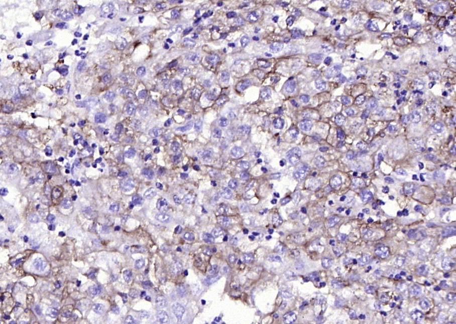 Paraformaldehyde-fixed, paraffin embedded (human endometrial carcinoma); Antigen retrieval by boiling in sodium citrate buffer (pH6.0) for 15min; Block endogenous peroxidase by 3% hydrogen peroxide for 20 minutes; Blocking buffer (normal goat serum) at 37°C for 30min; Antibody incubation with (GLUT1) Monoclonal Antibody, Unconjugated (SLM-52240R) at 1:200 overnight at 4°C, followed by operating according to SP Kit(Rabbit) (sp-0023) instructionsand DAB staining.
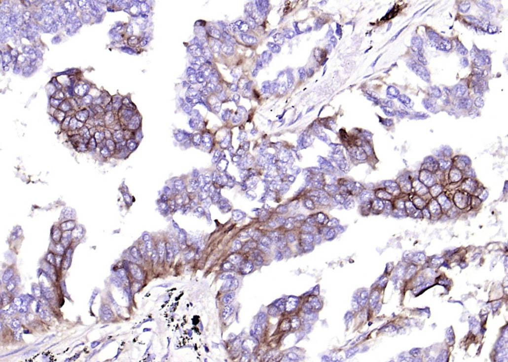 Paraformaldehyde-fixed, paraffin embedded (human lung carcinoma); Antigen retrieval by boiling in sodium citrate buffer (pH6.0) for 15min; Block endogenous peroxidase by 3% hydrogen peroxide for 20 minutes; Blocking buffer (normal goat serum) at 37°C for 30min; Antibody incubation with (GLUT1) Monoclonal Antibody, Unconjugated (SLM-52240R) at 1:200 overnight at 4°C, followed by operating according to SP Kit(Rabbit) (sp-0023) instructionsand DAB staining.
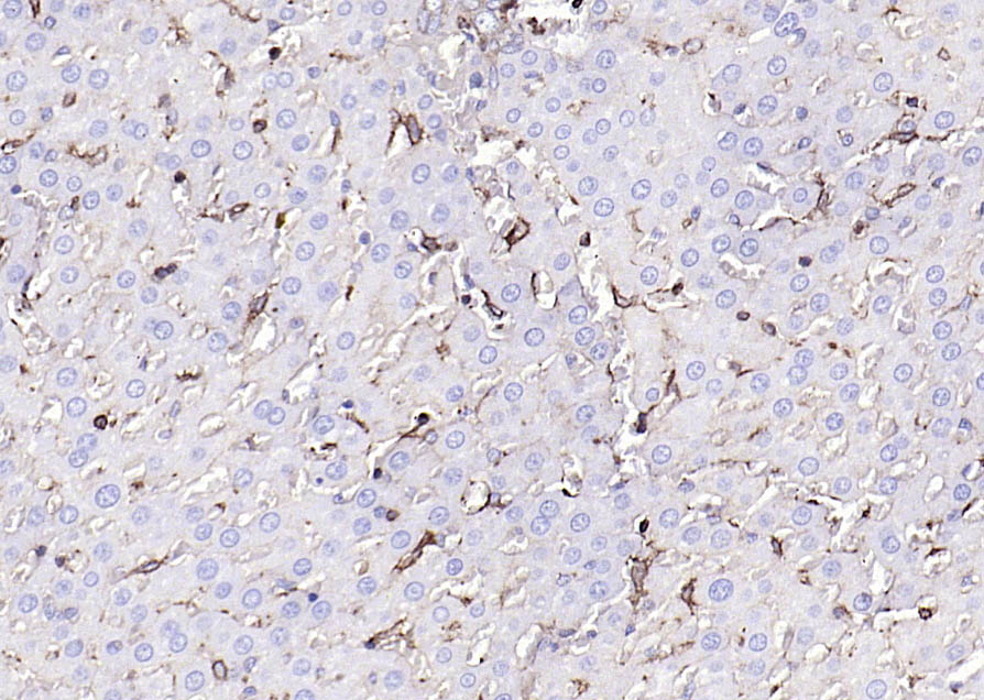 Paraformaldehyde-fixed, paraffin embedded (rat liver); Antigen retrieval by boiling in sodium citrate buffer (pH6.0) for 15min; Block endogenous peroxidase by 3% hydrogen peroxide for 20 minutes; Blocking buffer (normal goat serum) at 37°C for 30min; Antibody incubation with (GLUT1) Monoclonal Antibody, Unconjugated (SLM-52240R) at 1:200 overnight at 4°C, followed by operating according to SP Kit(Rabbit) (sp-0023) instructionsand DAB staining.
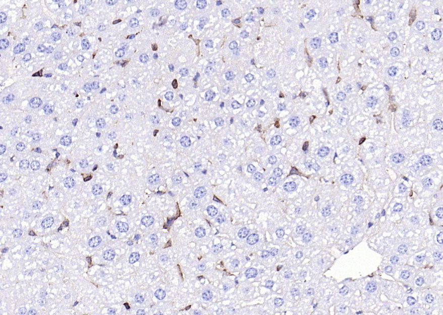 Paraformaldehyde-fixed, paraffin embedded (mouse liver); Antigen retrieval by boiling in sodium citrate buffer (pH6.0) for 15min; Block endogenous peroxidase by 3% hydrogen peroxide for 20 minutes; Blocking buffer (normal goat serum) at 37°C for 30min; Antibody incubation with (GLUT1) Monoclonal Antibody, Unconjugated (SLM-52240R) at 1:200 overnight at 4°C, followed by operating according to SP Kit(Rabbit) (sp-0023) instructionsand DAB staining.
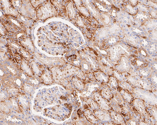 Immunohistochemical analysis of paraffin-embedded human kidney tissue using anti-Glucose Transporter GLUT1 antibody. The section was pre-treated using heat mediated antigen retrieval with Tris-EDTA buffer (pH 8.0-8.4) for 20 minutes.The tissues were blocked in 5% BSA for 30 minutes at room temperature, washed with ddH2O and PBS, and then probed with the primary antibody (SLM-52240R, 1/200) for 30 minutes at room temperature. The detection was performed using an HRP conjugated compact polymer system. DAB was used as the chromogen. Tissues were counterstained with hematoxylin and mounted with DPX.
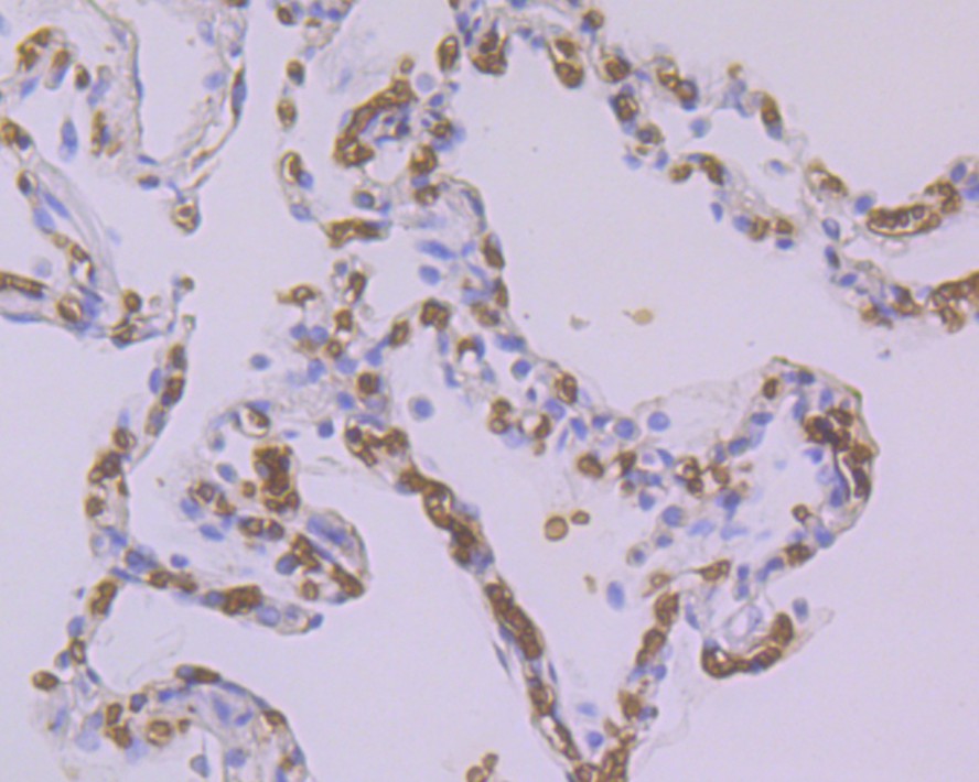 Immunohistochemical analysis of paraffin-embedded human lung cancer tissue using anti-Glucose Transporter GLUT1 antibody. The section was pre-treated using heat mediated antigen retrieval with Tris-EDTA buffer (pH 9.0) for 20 minutes.The tissues were blocked in 5% BSA for 30 minutes at room temperature, washed with ddH2O and PBS, and then probed with the primary antibody (SLM-52240R, 1/200) for 30 minutes at room temperature. The detection was performed using an HRP conjugated compact polymer system. DAB was used as the chromogen. Tissues were counterstained with hematoxylin and mounted with DPX.
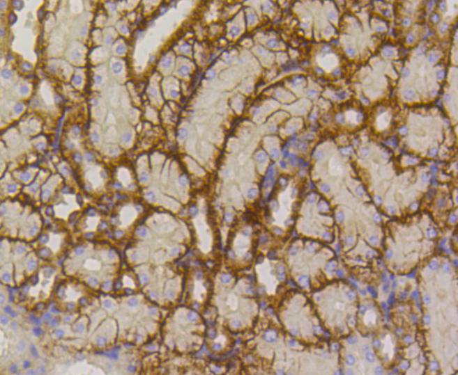 Immunohistochemical analysis of paraffin-embedded mouse kidney tissue using anti-Glucose Transporter GLUT1 antibody. The section was pre-treated using heat mediated antigen retrieval with Tris-EDTA buffer (pH 8.0-8.4) for 20 minutes.The tissues were blocked in 5% BSA for 30 minutes at room temperature, washed with ddH2O and PBS, and then probed with the primary antibody (SLM-52240R, 1/200) for 30 minutes at room temperature. The detection was performed using an HRP conjugated compact polymer system. DAB was used as the chromogen. Tissues were counterstained with hematoxylin and mounted with DPX.
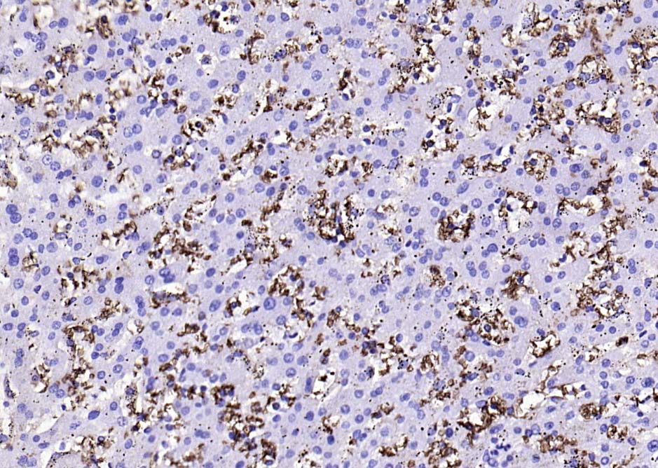
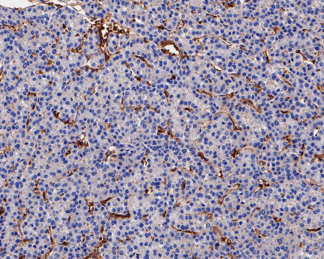 Immunohistochemical analysis of paraffin-embedded human liver carcinoma tissue using anti-Glucose Transporter GLUT1 antibody. The section was pre-treated using heat mediated antigen retrieval with Tris-EDTA buffer (pH 8.0-8.4) for 20 minutes.The tissues were blocked in 5% BSA for 30 minutes at room temperature, washed with ddH2O and PBS, and then probed with the primary antibody (SLM-52240R, 1/50) for 30 minutes at room temperature. The detection was performed using an HRP conjugated compact polymer system. DAB was used as the chromogen. Tissues were counterstained with hematoxylin and mounted with DPX.
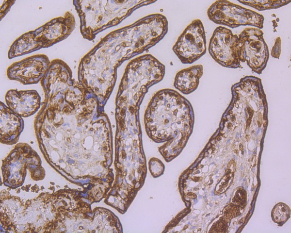 Immunohistochemical analysis of paraffin-embedded human placenta tissue using anti-Glucose Transporter GLUT1 antibody. The section was pre-treated using heat mediated antigen retrieval with Tris-EDTA buffer (pH 8.0-8.4) for 20 minutes.The tissues were blocked in 5% BSA for 30 minutes at room temperature, washed with ddH2O and PBS, and then probed with the primary antibody (SLM-52240R, 1/50) for 30 minutes at room temperature. The detection was performed using an HRP conjugated compact polymer system. DAB was used as the chromogen. Tissues were counterstained with hematoxylin and mounted with DPX.
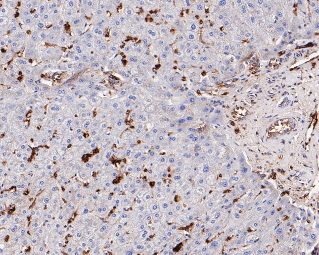 Immunohistochemical analysis of paraffin-embedded human liver tissue using anti-Glucose Transporter GLUT1 antibody. The section was pre-treated using heat mediated antigen retrieval with Tris-EDTA buffer (pH 8.0-8.4) for 20 minutes.The tissues were blocked in 5% BSA for 30 minutes at room temperature, washed with ddH2O and PBS, and then probed with the primary antibody (SLM-52240R, 1/200) for 30 minutes at room temperature. The detection was performed using an HRP conjugated compact polymer system. DAB was used as the chromogen. Tissues were counterstained with hematoxylin and mounted with DPX.
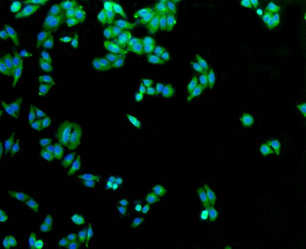 ICC staining of Glucose Transporter GLUT1 in HepG2 cells (green). Formalin fixed cells were permeabilized with 0.1% Triton X-100 in TBS for 10 minutes at room temperature and blocked with 1% Blocker BSA for 15 minutes at room temperature. Cells were probed with the primary antibody (SLM-52240R, 1/50) for 1 hour at room temperature, washed with PBS. Alexa Fluor®488 Goat anti-Rabbit IgG was used as the secondary antibody at 1/1,000 dilution. The nuclear counter stain is DAPI (blue).
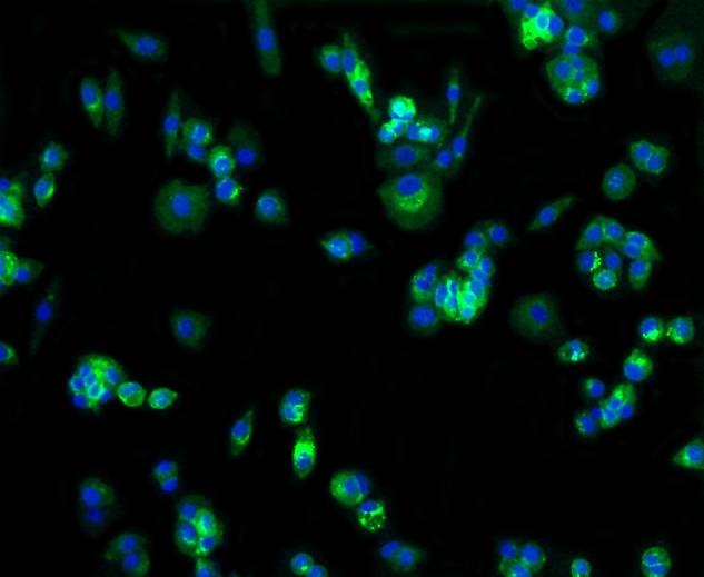 ICC staining of Glucose Transporter GLUT1 in MCF-7 cells (green). Formalin fixed cells were permeabilized with 0.1% Triton X-100 in TBS for 10 minutes at room temperature and blocked with 1% Blocker BSA for 15 minutes at room temperature. Cells were probed with the primary antibody (SLM-52240R, 1/50) for 1 hour at room temperature, washed with PBS. Alexa Fluor®488 Goat anti-Rabbit IgG was used as the secondary antibody at 1/1,000 dilution. The nuclear counter stain is DAPI (blue).
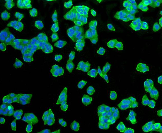 ICC staining of Glucose Transporter GLUT1 in Hela cells (green). Formalin fixed cells were permeabilized with 0.1% Triton X-100 in TBS for 10 minutes at room temperature and blocked with 1% Blocker BSA for 15 minutes at room temperature. Cells were probed with the primary antibody (SLM-52240R, 1/50) for 1 hour at room temperature, washed with PBS. Alexa Fluor®488 Goat anti-Rabbit IgG was used as the secondary antibody at 1/1,000 dilution. The nuclear counter stain is DAPI (blue).
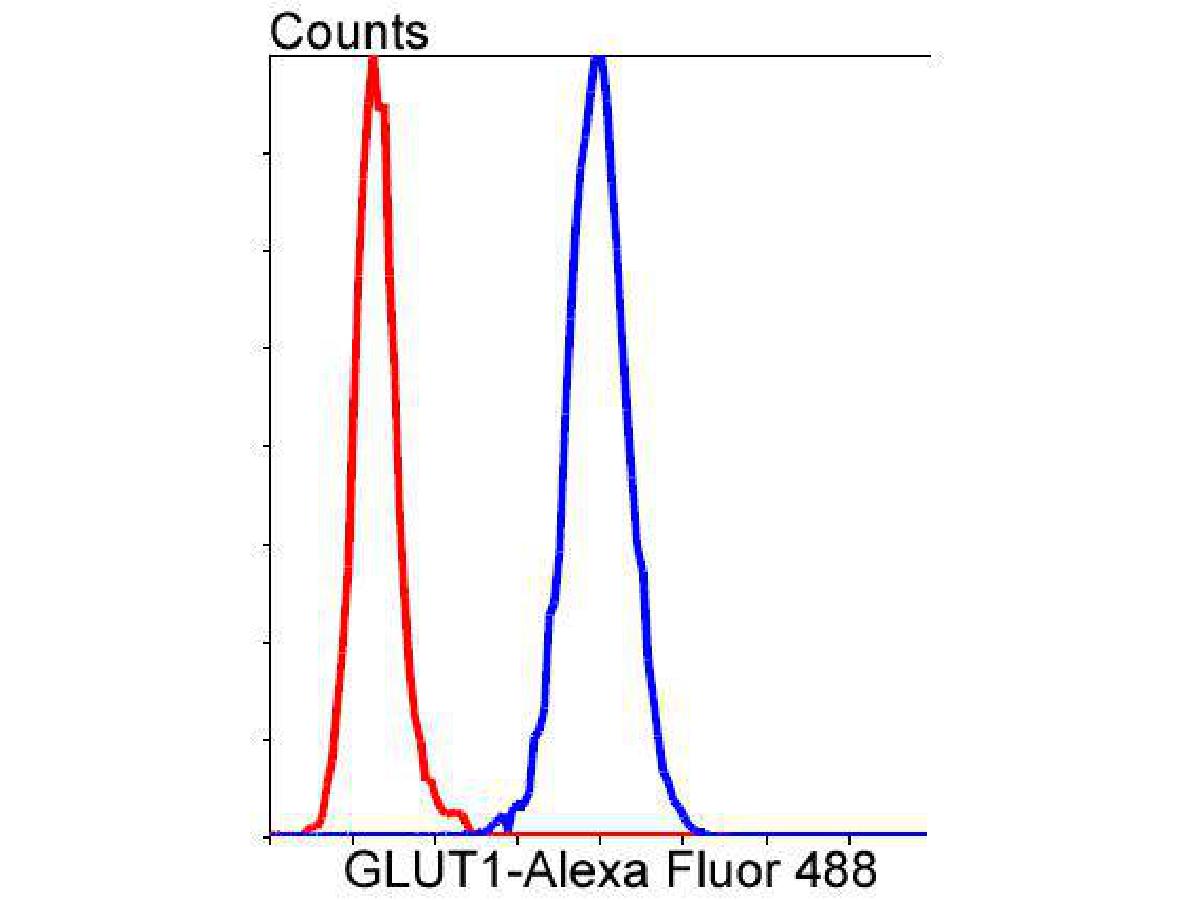 Flow cytometric analysis of Glucose Transporter GLUT1 was done on Hela cells. The cells were fixed, permeabilized and stained with the primary antibody (SLM-52240R, 1/50) (blue). After incubation of the primary antibody at room temperature for an hour, the cells were stained with a Alexa Fluor 488-conjugated Goat anti-Rabbit IgG Secondary antibody at 1/1,000 dilution for 30 minutes.Unlabelled sample was used as a control (cells without incubation with primary antibody; red).
Cartpieces
Totalgoods,subtotals:¥Checkout
References (0)
No References
Bought notes(bought amounts latest0)
No one bought this product
User Comment(Total0User Comment Num)
- No comment


 +86 571 56623320
 +86 18668110335

