
Rabbit Anti-STAT2 antibody
signal transducers and activators of transduction2; Homo sapiens interferon alpha induced transcriptional activator; interferon alpha induced transcriptional activator; ISGF3; P113; signal transducer and activator of transcription 2 113kD; STAT113; STAT2_
View History [Clear]
Details
Product Name STAT2 Chinese Name Signal transduction和转录激活因子2Recombinant rabbit monoclonal anti Alias signal transducers and activators of transduction2; Homo sapiens interferon alpha induced transcriptional activator; interferon alpha induced transcriptional activator; ISGF3; P113; signal transducer and activator of transcription 2 113kD; STAT113; STAT2_HUMAN; p113. Research Area Cell biology Neurobiology Immunogen Species Rabbit Clonality Monoclonal Clone NO. 4B1 React Species (predicted: Human, ) Applications WB=1:500-1000 IHC-P=1:100-500 IHC-F=1:400-800 ICC=1:50-200 IF=1:50-200 (Paraffin sections need antigen repair)
not yet tested in other applications.
optimal dilutions/concentrations should be determined by the end user.Theoretical molecular weight 97kDa Cellular localization The nucleus cytoplasmic Form Liquid Concentration 1mg/ml immunogen Recombinant human STAT2 protein (1-100aa) Lsotype IgG Purification affinity purified by Protein A Buffer Solution 0.01M TBS(pH7.4) with 1% BSA, 0.03% Proclin300 and 50% Glycerol. Storage Shipped at 4℃. Store at -20 °C for one year. Avoid repeated freeze/thaw cycles. Attention This product as supplied is intended for research use only, not for use in human, therapeutic or diagnostic applications. PubMed PubMed Product Detail The protein encoded by this gene is a member of the STAT protein family. In response to cytokines and growth factors, STAT family members are phosphorylated by the receptor associated kinases, and then form homo- or heterodimers that translocate to the cell nucleus where they act as transcription activators. In response to interferon (IFN), this protein forms a complex with STAT1 and IFN regulatory factor family protein p48 (ISGF3G), in which this protein acts as a transactivator, but lacks the ability to bind DNA directly. Transcription adaptor P300/CBP (EP300/CREBBP) has been shown to interact specifically with this protein, which is thought to be involved in the process of blocking IFN-alpha response by adenovirus. Multiple transcript variants encoding different isoforms have been found for this gene. [provided by RefSeq, Mar 2010].
Function:
Signal transducer and activator of transcription that mediates signaling by type I IFNs (IFN-alpha and IFN-beta). Following type I IFN binding to cell surface receptors, Jak kinases (TYK2 and JAK1) are activated, leading to tyrosine phosphorylation of STAT1 and STAT2. The phosphorylated STATs dimerize, associate with ISGF3G/IRF-9 to form a complex termed ISGF3 transcription factor, that enters the nucleus. ISGF3 binds to the IFN stimulated response element (ISRE) to activate the transcription of interferon stimulated genes, which drive the cell in an antiviral state.
Subunit:
Interacts with ISGF3G/IRF-9 in the cytoplasm. Heterodimer with STAT1 upon IFN-alpha/beta induced phosphorylation. Interacts with CRSP2 and CRSP6. Interacts with Simian virus 5 protein V and rabies virus phosphoprotein (By similarity). Can form a homodimer upon IFN-alpha induced phosphorylation. Interacts with IFNAR1; the interaction requires the phosphorylation of IFNAR1 at 'Tyr-466'. Interacts with IFNAR2. Interacts with dengue virus NS5; this interaction inhibits the phosphorylation of STAT2, and, when all viral proteins are present (polyprotein), targets STAT2 for degradation. Interacts with human cytomegalovirus/HHV-5 protein UL123; this interaction promotes viral growth.
Subcellular Location:
Cytoplasm. Nucleus. Note=Translocated into the nucleus upon activation by IFN-alpha/beta.
Post-translational modifications:
Tyrosine phosphorylated in response to IFN-alpha.
Similarity:
Belongs to the transcription factor STAT family.
Contains 1 SH2 domain.
SWISS:
P52630
Gene ID:
6773
Database links:Entrez Gene: 6773 Human
Entrez Gene: 20847 Mouse
Omim: 600556 Human
SwissProt: P52630 Human
SwissProt: Q9WVL2 Mouse
Unigene: 530595 Human
Unigene: 293120 Mouse
Unigene: 471333 Mouse
Unigene: 24237 Rat
Product Picture 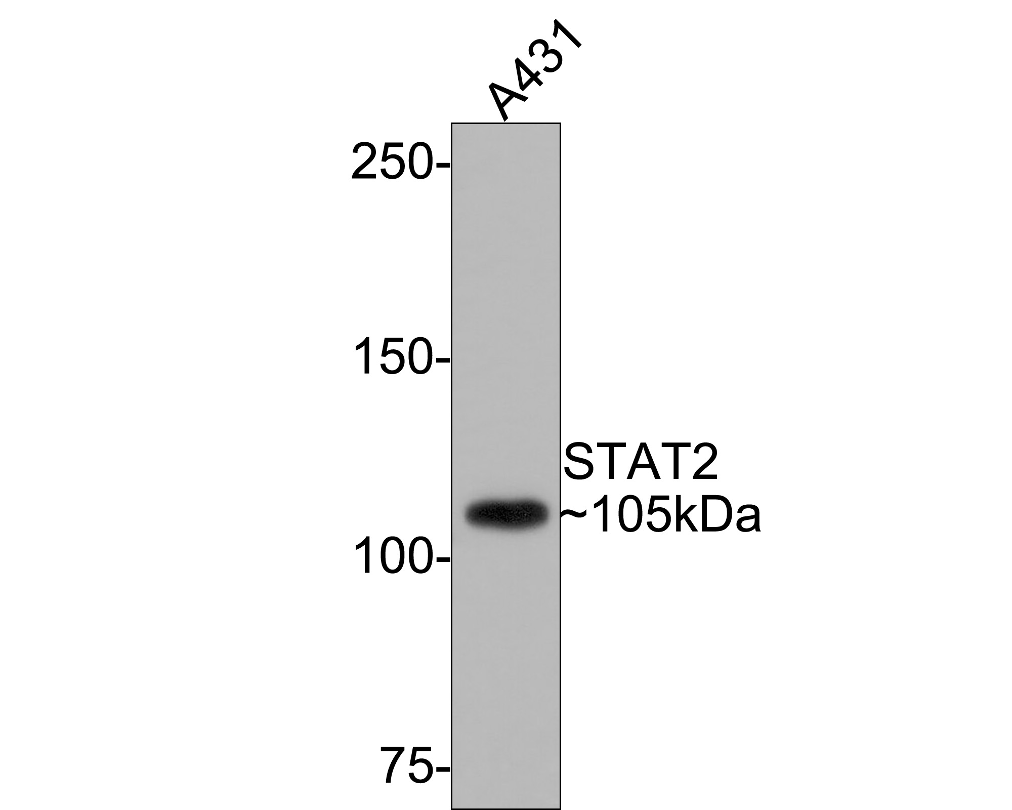 Western blot analysis of STAT2 on A431 cell lysates with Rabbit anti-STAT2 antibody (SLM-52234R) at 1/500 dilution.
Lysates/proteins at 10 µg/Lane.
Predicted band size: 98 kDa
Observed band size: 105 kDa
Exposure time: 1 minute;
6% SDS-PAGE gel.
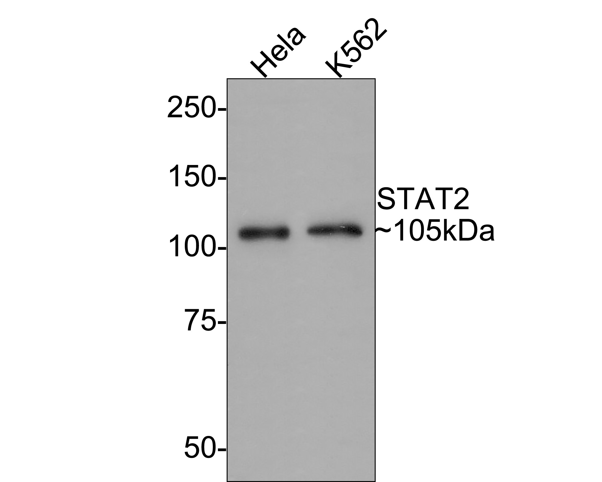 Western blot analysis of STAT2 on different lysates with Rabbit anti-STAT2 antibody (SLM-52234R) at 1/500 dilution.
Lane 1: Hela cell lysate
Lane 2: K562 cell lysate
Lysates/proteins at 10 µg/Lane.
Predicted band size: 98 kDa
Observed band size: 105 kDa
Exposure time: 2 minutes;
8% SDS-PAGE gel.
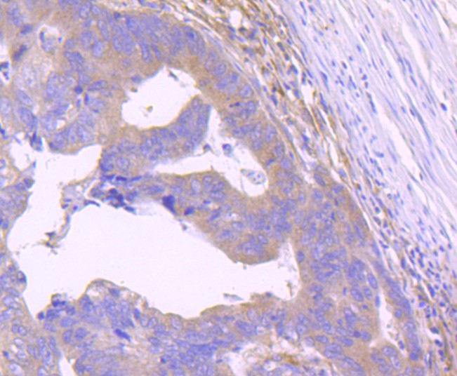 Immunohistochemical analysis of paraffin-embedded human colon cancer tissue with Rabbit anti-STAT2 antibody (SLM-52234R) at 1/50 dilution. The section was pre-treated using heat mediated antigen retrieval with Tris-EDTA buffer (pH 9.0) for 20 minutes. The tissues were blocked in 1% BSA for 20 minutes at room temperature, washed with ddH2O and PBS, and then probed with the primary antibody (SLM-52234R) at 1/400 dilution for 1 hour at room temperature. The detection was performed using an HRP conjugated compact polymer system. DAB was used as the chromogen. Tissues were counterstained with hematoxylin and mounted with DPX.
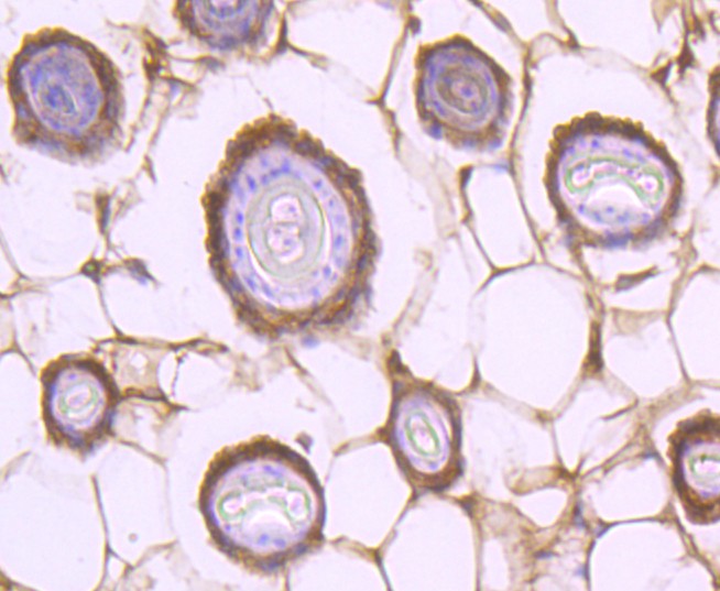 Immunohistochemical analysis of paraffin-embedded mouse skin cancer tissue with Rabbit anti-STAT2 antibody (SLM-52234R) at 1/50 dilution. The section was pre-treated using heat mediated antigen retrieval with Tris-EDTA buffer (pH 9.0) for 20 minutes. The tissues were blocked in 1% BSA for 20 minutes at room temperature, washed with ddH2O and PBS, and then probed with the primary antibody (SLM-52234R) at 1/400 dilution for 1 hour at room temperature. The detection was performed using an HRP conjugated compact polymer system. DAB was used as the chromogen. Tissues were counterstained with hematoxylin and mounted with DPX.
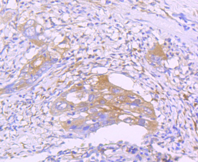 Immunohistochemical analysis of paraffin-embedded human lung cancer tissue with Rabbit anti-STAT2 antibody (SLM-52234R) at 1/50 dilution. The section was pre-treated using heat mediated antigen retrieval with Tris-EDTA buffer (pH 9.0) for 20 minutes. The tissues were blocked in 1% BSA for 20 minutes at room temperature, washed with ddH2O and PBS, and then probed with the primary antibody (SLM-52234R) at 1/400 dilution for 1 hour at room temperature. The detection was performed using an HRP conjugated compact polymer system. DAB was used as the chromogen. Tissues were counterstained with hematoxylin and mounted with DPX.
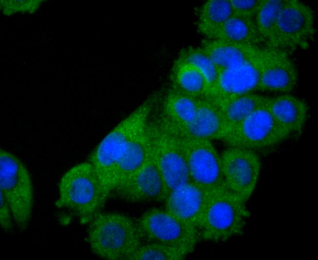 Immunocytochemistry analysis of SW480 cells labeling STAT2 with Rabbit anti-STAT2 antibody (SLM-52234R) at 1/50 dilution. Cells were fixed in 4% paraformaldehyde for 10 minutes at 37 ℃, permeabilized with 0.05% Triton X-100 in PBS for 20 minutes, and then blocked with 2% negative goat serum for 30 minutes at room temperature. Cells were then incubated with Rabbit anti-STAT2 antibody (SLM-52234R) at 1/200 dilution in 2% negative goat serum overnight at 4 ℃. Goat Anti-Rabbit IgG H&L (iFluor™ 488, HA1121) was used as the secondary antibody at 1/1,000 dilution. PBS instead of the primary antibody was used as the secondary antibody only control. Nuclear DNA was labelled in blue with DAPI.
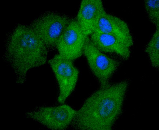 Immunocytochemistry analysis of A549 cells labeling STAT2 with Rabbit anti-STAT2 antibody (SLM-52234R) at 1/50 dilution. Cells were fixed in 4% paraformaldehyde for 10 minutes at 37 ℃, permeabilized with 0.05% Triton X-100 in PBS for 20 minutes, and then blocked with 2% negative goat serum for 30 minutes at room temperature. Cells were then incubated with Rabbit anti-STAT2 antibody (SLM-52234R) at 1/200 dilution in 2% negative goat serum overnight at 4 ℃. Goat Anti-Rabbit IgG H&L (iFluor™ 488, HA1121) was used as the secondary antibody at 1/1,000 dilution. PBS instead of the primary antibody was used as the secondary antibody only control. Nuclear DNA was labelled in blue with DAPI.
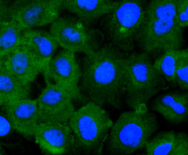 Immunocytochemistry analysis of A431 cells labeling STAT2 with Rabbit anti-STAT2 antibody (SLM-52234R) at 1/50 dilution. Cells were fixed in 4% paraformaldehyde for 10 minutes at 37 ℃, permeabilized with 0.05% Triton X-100 in PBS for 20 minutes, and then blocked with 2% negative goat serum for 30 minutes at room temperature. Cells were then incubated with Rabbit anti-STAT2 antibody (SLM-52234R) at 1/200 dilution in 2% negative goat serum overnight at 4 ℃. Goat Anti-Rabbit IgG H&L (iFluor™ 488) was used as the secondary antibody at 1/1,000 dilution. PBS instead of the primary antibody was used as the secondary antibody only control. Nuclear DNA was labelled in blue with DAPI.
Cartpieces
Totalgoods,subtotals:¥Checkout
References (0)
No References
Bought notes(bought amounts latest0)
No one bought this product
User Comment(Total0User Comment Num)
- No comment


 +86 571 56623320
 +86 18668110335

