
Rabbit Anti-phospho-STAT3 (Ser727)antibody
STAT3 (phospho S727); p-STAT3 (phospho S727); STAT3(Phospho Ser727); STAT3(Phospho S727); p-STAT3(Ser727); Acute Phase Response Factor; APRF; DNA binding protein APRF; Signal Transductor and Activator of Transcription 3; STAT 3; STAT3_HUMAN.
View History [Clear]
Details
Product Name phospho-STAT3 (Ser727) Chinese Name 磷酸化Signal transduction和转录激活因子3Recombinant rabbit monoclonal anti Alias STAT3 (phospho S727); p-STAT3 (phospho S727); STAT3(Phospho Ser727); STAT3(Phospho S727); p-STAT3(Ser727); Acute Phase Response Factor; APRF; DNA binding protein APRF; Signal Transductor and Activator of Transcription 3; STAT 3; STAT3_HUMAN. literatures Product Type Phosphorylated anti Recombinant rabbit monoclonal anti Research Area Tumour Cell biology immunology Signal transduction Apoptosis transcriptional regulatory factor Immunogen Species Rabbit Clonality Monoclonal Clone NO. 4G1 React Species Human, Rat, Applications WB=1:500-2000 IHC-P=1:100-200 IHC-F=1:100-200 Flow-Cyt=2ug/Test ICC=1:50-100 (Paraffin sections need antigen repair)
not yet tested in other applications.
optimal dilutions/concentrations should be determined by the end user.Theoretical molecular weight 88kDa Cellular localization The nucleus cytoplasmic Form Liquid Concentration 1mg/ml immunogen KLH conjugated Synthesised phosphopeptide derived from human STAT3 around the phosphorylation site of Ser727: PM(p-S)PR Lsotype IgG Purification affinity purified by Protein A Buffer Solution 0.01M TBS(pH7.4) with 1% BSA, 0.03% Proclin300 and 50% Glycerol. Storage Shipped at 4℃. Store at -20 °C for one year. Avoid repeated freeze/thaw cycles. Attention This product as supplied is intended for research use only, not for use in human, therapeutic or diagnostic applications. PubMed PubMed Product Detail STATs (signal transducers and activators of transcription) were originally discovered as two proteins (STAT1 and STAT2) which were involved in interferon alpha (IFN alpha) and IFN gamma signal transduction. Since then, several additional STAT proteins have been identified (STAT3, 4, 5a, 5b, and 6). STATs undergo tyrosine phosphorylation in response to growth factor or cytokine signaling. This phosphorylation results in dimerization and translocation of STAT proteins to the nucleus. In some cases this process is mediated by JAK Kinases (Janus Kinases 1, 2, and 3). For maximum activation of these proteins, phosphorylation at specific tyrosine and serine residues may be required in STAT1 alpha, 3, 4, and 5. Specific functions of the various members of the STAT family are poorly understood. STAT3 has been shown to be activated by IFN alpha but not IFN beta. The transcription factors associated with STAT3 are cJun and cyclic AMP responsive enhancer binding protein (CREB). Deletion of the STAT3 gene in knock out mice was lethal at the early embryonic stage.
Function:
Signal transducer and transcription activator that mediates cellular responses to interleukins, KITLG/SCF and other growth factors. May mediate cellular responses to activated FGFR1, FGFR2, FGFR3 and FGFR4. Binds to the interleukin-6 (IL-6)-responsive elements identified in the promoters of various acute-phase protein genes. Activated by IL31 through IL31RA. Cytoplasmic STAT3 represses macroautophagy by inhibiting EIF2AK2/PKR activity. Plays an important role in host defense in methicillin-resistant S.aureus lung infection by regulating the expression of the antimicrobial lectin REG3G (By similarity).
Subunit:
Forms a homodimer or a heterodimer with a related family member (at least STAT1). Interacts with IL31RA, NCOA1, PELP1, SIPAR, SOCS7, STATIP1 and TMF1. Interacts with HCV core protein. Interacts with IL23R in presence of IL23. Interacts (via SH2 domain) with NLK. Interacts with ARL2BP; the interaction is enhanced by LIF and JAK1 expression (By similarity). Interacts with KPNA4 and KPNA5; KPNA4 may be the primary mediator of nuclear import (By similarity). Interacts with CAV2; the interaction is increased on insulin-induced tyrosine phosphorylation of CAV2 and leads to STAT3 activation (By similarity). Interacts with ARL2BP; interaction is enhanced with ARL2. Interacts with NEK6 (By similarity). Binds to CDK9 when activated and nuclear. Interacts with BMX. Interacts with ZIPK/DAPK3. Interacts with PIAS3; the interaction occurs on stimulation by IL6, CNTF or OSM and inhibits the DNA binding activity of STAT3. In prostate cancer cells, interacts with STAT3 and promotes DNA binding activity of STAT3. Interacts with STMN3, antagonizing its microtubule-destabilizing activity. Interacts with the 'Lys-129' acetylated form of BIRC5/survivin. Interacts with FER. Interacts (via SH2 domain) with EIF2AK2/PKR (via the kinase catalytic domain).
Subcellular Location:
Cytoplasm. Nucleus. Note=Shuttles between the nucleus and the cytoplasm. Translocated into the nucleus upon tyrosine phosphorylation and dimerization, in response to signaling by activated FGFR1, FGFR2, FGFR3 or FGFR4. Constitutive nuclear presence is independent of tyrosine phosphorylation. Predominantly present in the cytoplasm without stimuli. Upon leukemia inhibitory factor (LIF) stimulation, accumulates in the nucleus. The complex composed of BART and ARL2 plays an important role in the nuclear translocation and retention of STAT3. Identified in a complex with LYN and PAG1.
Tissue Specificity:
Heart, brain, placenta, lung, liver, skeletal muscle, kidney and pancreas.
Post-translational modifications:
Tyrosine phosphorylated upon stimulation with EGF. Tyrosine phosphorylated in response to constitutively activated FGFR1, FGFR2, FGFR3 and FGFR4 (By similarity). Activated through tyrosine phosphorylation by BMX. Tyrosine phosphorylated in response to IL6, IL11, LIF, CNTF, KITLG/SCF, CSF1, EGF, PDGF, IFN-alpha and OSM. Activated KIT promotes phosphorylation on tyrosine residues and subsequent translocation to the nucleus. Phosphorylated on serine upon DNA damage, probably by ATM or ATR. Serine phosphorylation is important for the formation of stable DNA-binding STAT3 homodimers and maximal transcriptional activity. ARL2BP may participate in keeping the phosphorylated state of STAT3 within the nucleus. Upon LPS challenge, phosphorylated within the nucleus by IRAK1. Upon erythropoietin treatment, phosphorylated on Ser-727 by RPS6KA5. Phosphoryation at Tyr-705 by PTK6 or FER leads to an increase of its transcriptional activity.
DISEASE:
Hyperimmunoglobulin E recurrent infection syndrome, autosomal dominant (AD-HIES) [MIM:147060]: A rare disorder of immunity and connective tissue characterized by immunodeficiency, chronic eczema, recurrent Staphylococcal infections, increased serum IgE, eosinophilia, distinctive coarse facial appearance, abnormal dentition, hyperextensibility of the joints, and bone fractures. Note=The disease is caused by mutations affecting the gene represented in this entry.
Similarity:
Belongs to the transcription factor STAT family.
Contains 1 SH2 domain.
SWISS:
P40763
Gene ID:
6774
Database links:Entrez Gene: 6774 Human
Entrez Gene: 20848 Mouse
Omim: 102582 Human
SwissProt: P40763 Human
SwissProt: P42227 Mouse
Unigene: 463059 Human
Product Picture 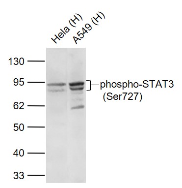 Sample:
Lane 1: Hela (Human) Cell Lysate at 30 ug
Lane 2: A549 (Human) Cell Lysate at 30 ug
Primary:
Anti- phospho-STAT3 (Ser727) (SLM-52210R) at 1/1000 dilution
Secondary: IRDye800CW Goat Anti-Rabbit IgG at 1/20000 dilution
Predicted band size: 92/84 kD
Observed band size: 92/84 kD
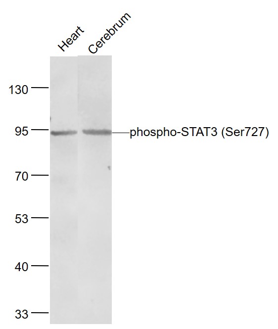 Sample:
Heart (Rat) Lysate at 40 ug
Cerebrum (Rat) Lysate at 40 ug
Primary: Anti- phospho-STAT3 (Ser727) (SLM-52210R) at 1/1000 dilution
Secondary: IRDye800CW Goat Anti-Rabbit IgG at 1/20000 dilution
Predicted band size: 98 kD
Observed band size: 94 kD
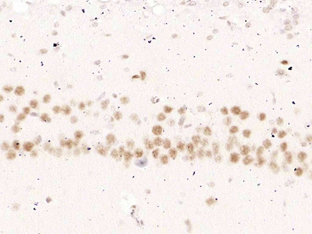 Paraformaldehyde-fixed, paraffin embedded (rat brain); Antigen retrieval by boiling in sodium citrate buffer (pH6.0) for 15min; Block endogenous peroxidase by 3% hydrogen peroxide for 20 minutes; Blocking buffer (normal goat serum) at 37°C for 30min; Antibody incubation with (phospho-STAT3 (Ser727)) Polyclonal Antibody, Unconjugated (SLM-52210R) at 1:200 overnight at 4°C, followed by operating according to SP Kit(Rabbit) (sp-0023) instructionsand DAB staining.
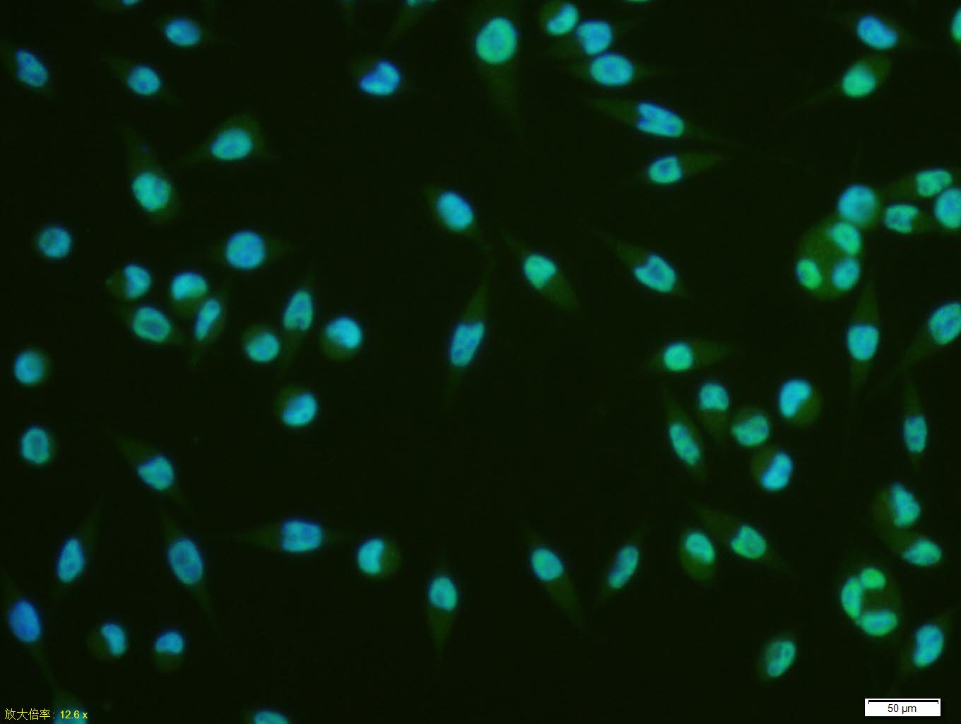 A431 cell; 4% Paraformaldehyde-fixed; Triton X-100 at room temperature for 20 min; Blocking buffer (normal goat serum, C-0005) at 37°C for 20 min; Antibody incubation with (phospho-STAT3 (Ser727)) monoclonal Antibody, Unconjugated (SL52210R) 1:100, 90 minutes at 37°C; followed by a conjugated Goat Anti-Rabbit IgG antibody at 37°C for 90 minutes, DAPI (blue, C02-04002) was used to stain the cell nuclei.A431 cell; 4% Paraformaldehyde-fixed; Triton X-100 at room temperature for 20 min; Blocking buffer (normal goat serum, C-0005) at 37°C for 20 min; Antibody incubation with (phospho-STAT3 (Ser727)) monoclonal Antibody, Unconjugated (SL52210R) 1:100, 90 minutes at 37°C; followed by a conjugated Goat Anti-Rabbit IgG antibody at 37°C for 90 minutes, DAPI (blue, C02-04002) was used to stain the cell nuclei.
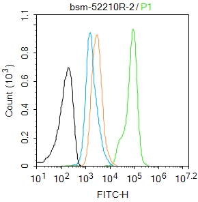 Blank control: A431.
Primary Antibody (green line): Rabbit Anti-phospho-STAT3 (Ser727) antibody (SLM-52210R)
Dilution: 1μg /10^6 cells;
Isotype Control Antibody (orange line): Rabbit IgG .
Secondary Antibody : Goat anti-rabbit IgG-AF488
Dilution: 1μg /test.
Protocol
The cells were fixed with 4% PFA (10min at room temperature)and then permeabilized with 90% ice-cold methanol for 20 min at-20℃. The cells were then incubated in 5%BSA to block non-specific protein-protein interactions for 30 min at at room temperature .Cells stained with Primary Antibody for 30 min at room temperature. The secondary antibody used for 40 min at room temperature. Acquisition of 20,000 events was performed.
Cartpieces
Totalgoods,subtotals:¥Checkout
Bought notes(bought amounts latest0)
No one bought this product
User Comment(Total0User Comment Num)
- No comment


 +86 571 56623320
 +86 18668110335

