
Rabbit Anti-phospho-SIRT1 (Thr530)antibody
SIR2L1; NAD-dependent protein deacetylase sirtuin-1; EC:2.3.1.286; hSIRT1; NAD-dependent protein deacylase sirtuin-1; Regulatory protein SIR2 homolog 1; SIR2-like protein 1 (hSIR2); SirtT1 75 kDa fragment; SIR1_HUMAN;
View History [Clear]
Details
Product Name phospho-SIRT1 (Thr530) Chinese Name 磷酸化沉默调节蛋白1Recombinant rabbit monoclonal anti Alias SIR2L1; NAD-dependent protein deacetylase sirtuin-1; EC:2.3.1.286; hSIRT1; NAD-dependent protein deacylase sirtuin-1; Regulatory protein SIR2 homolog 1; SIR2-like protein 1 (hSIR2); SirtT1 75 kDa fragment; SIR1_HUMAN; Product Type Phosphorylated anti Recombinant rabbit monoclonal anti Research Area Cell biology Apoptosis Epigenetics Immunogen Species Rabbit Clonality Monoclonal Clone NO. 6H5 React Species (predicted: Human, ) Applications WB=1:500-1000 IHC-P=1:100-500 IHC-F=1:50-200 ICC=1:50-200 IF=1:50-200 (Paraffin sections need antigen repair)
not yet tested in other applications.
optimal dilutions/concentrations should be determined by the end user.Theoretical molecular weight 81kDa Cellular localization cytoplasmic Form Liquid Concentration 1mg/ml immunogen KLH conjugated synthesised phosphopeptide derived from human SIRT1 around the phosphorylation site of Thr530: PP(p-T)PL Lsotype IgG Purification affinity purified by Protein A Buffer Solution 0.01M TBS(pH7.4) with 1% BSA, 0.03% Proclin300 and 50% Glycerol. Storage Shipped at 4℃. Store at -20 °C for one year. Avoid repeated freeze/thaw cycles. Attention This product as supplied is intended for research use only, not for use in human, therapeutic or diagnostic applications. PubMed PubMed Product Detail This gene encodes a member of the sirtuin family of proteins, homologs to the yeast Sir2 protein. Members of the sirtuin family are characterized by a sirtuin core domain and grouped into four classes. The functions of human sirtuins have not yet been determined; however, yeast sirtuin proteins are known to regulate epigenetic gene silencing and suppress recombination of rDNA. Studies suggest that the human sirtuins may function as intracellular regulatory proteins with mono-ADP-ribosyltransferase activity. The protein encoded by this gene is included in class I of the sirtuin family. Alternative splicing results in multiple transcript variants. [provided by RefSeq, Dec 2008]
Subcellular Location:
Cytoplasm. Mitochondrion and Nucleus > PML body. Cytoplasm. Recruited to the nuclear bodies via its interaction with PML. Colocalized with APEX1 in the nucleus. May be found in nucleolus, nuclear euchromatin, heterochromatin and inner membrane. Shuttles between nucleus and cytoplasm.
Tissue Specificity:
Widely expressed.
Post-translational modifications:
Methylated on multiple lysine residues; methylation is enhanced after DNA damage and is dispensable for deacetylase activity toward p53/TP53.
Phosphorylated. Phosphorylated by STK4/MST1, resulting in inhibition of SIRT1-mediated p53/TP53 deacetylation. Phosphorylation by MAPK8/JNK1 at Ser-27, Ser-47, and Thr-530 leads to increased nuclear localization and enzymatic activity. Phosphorylation at Thr-530 by DYRK1A and DYRK3 activates deacetylase activity and promotes cell survival. Phosphorylation by mammalian target of rapamycin complex 1 (mTORC1) at Ser-47 inhibits deacetylation activity. Phosphorylated by CaMK2, leading to increased p53/TP53 and NF-kappa-B p65/RELA deacetylation activity (By similarity). Phosphorylation at Ser-27 implicating MAPK9 is linked to protein stability. There is some ambiguity for some phosphosites: Ser-159/Ser-162 and Thr-544/Ser-545.
Proteolytically cleaved by cathepsin B upon TNF-alpha treatment to yield catalytic inactive but stable SirtT1 75 kDa fragment (75SirT1).
S-nitrosylated by GAPDH, leading to inhibit the NAD-dependent protein deacetylase activity.
Similarity:
Belongs to the sirtuin family. Class I subfamily.
Contains 1 deacetylase sirtuin-type domain.
SWISS:
Q96EB6
Gene ID:
23411
Database links:Entrez Gene: 23411 Human
Entrez Gene: 93759 Mouse
Omim: 604479 Human
SwissProt: Q96EB6 Human
SwissProt: Q923E4 Mouse
Unigene: 369779 Human
Unigene: 351459 Mouse
Product Picture  Immunohistochemical analysis of paraffin-embedded human kidney tissue using anti-Phospho-SIRT1(T530) antibody. The section was pre-treated using heat mediated antigen retrieval with Tris-EDTA buffer (pH 8.0-8.4) for 20 minutes.The tissues were blocked in 5% BSA for 30 minutes at room temperature, washed with ddH2O and PBS, and then probed with the primary antibody (SLM-52717R7, 1/50) for 30 minutes at room temperature. The detection was performed using an HRP conjugated compact polymer system. DAB was used as the chromogen. Tissues were counterstained with hematoxylin and mounted with DPX.
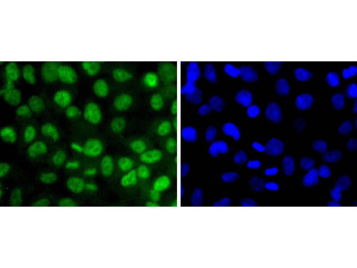 ICC staining of Phospho-SIRT1(T530) in 293 cells (green). Formalin fixed cells were permeabilized with 0.1% Triton X-100 in TBS for 10 minutes at room temperature and blocked with 1% Blocker BSA for 15 minutes at room temperature. Cells were probed with the primary antibody (SLM-52717R7, 1/50) for 1 hour at room temperature, washed with PBS. Alexa Fluor®488 Goat anti-Rabbit IgG was used as the secondary antibody at 1/1,000 dilution. The nuclear counter stain is DAPI (blue).
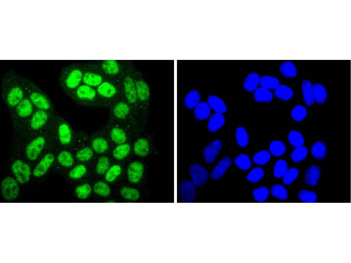 ICC staining of Phospho-SIRT1(T530) in Hela cells (green). Formalin fixed cells were permeabilized with 0.1% Triton X-100 in TBS for 10 minutes at room temperature and blocked with 1% Blocker BSA for 15 minutes at room temperature. Cells were probed with the primary antibody (SLM-52717R7, 1/50) for 1 hour at room temperature, washed with PBS. Alexa Fluor®488 Goat anti-Rabbit IgG was used as the secondary antibody at 1/1,000 dilution. The nuclear counter stain is DAPI (blue).
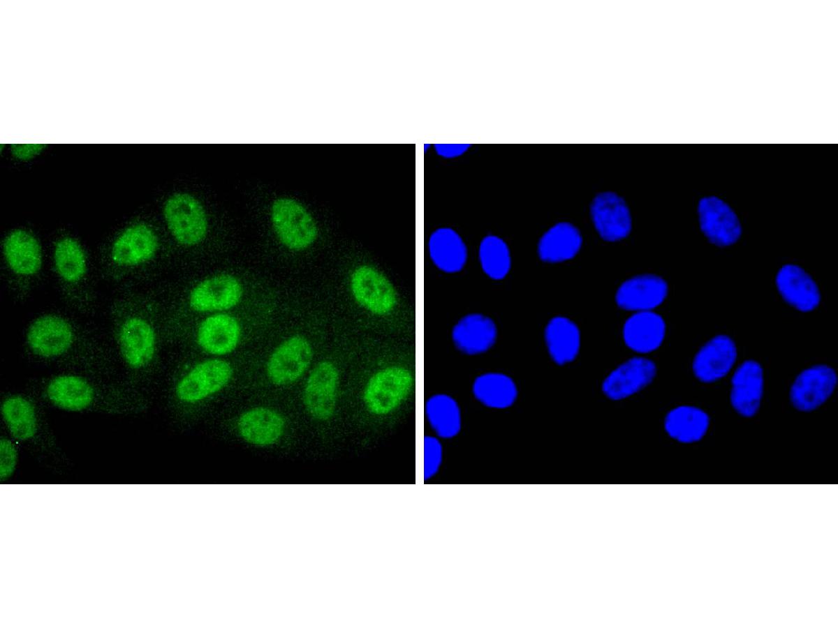 ICC staining of Phospho-SIRT1(T530) in HepG2 cells (green). Formalin fixed cells were permeabilized with 0.1% Triton X-100 in TBS for 10 minutes at room temperature and blocked with 1% Blocker BSA for 15 minutes at room temperature. Cells were probed with the primary antibody (SLM-52717R7, 1/50) for 1 hour at room temperature, washed with PBS. Alexa Fluor®488 Goat anti-Rabbit IgG was used as the secondary antibody at 1/1,000 dilution. The nuclear counter stain is DAPI (blue).
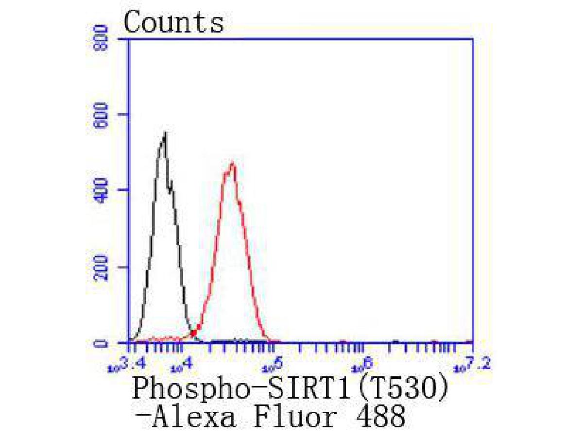 Flow cytometric analysis of Phospho-SIRT1(T530) was done on 293 cells. The cells were fixed, permeabilized and stained with the primary antibody (SLM-52717R7, 1/50) (red). After incubation of the primary antibody at room temperature for an hour, the cells were stained with a Alexa Fluor 488-conjugated Goat anti-Rabbit IgG Secondary antibody at 1/1,000 dilution for 30 minutes.Unlabelled sample was used as a control (cells without incubation with primary antibody; black).
Cartpieces
Totalgoods,subtotals:¥Checkout
References (0)
No References
Bought notes(bought amounts latest0)
No one bought this product
User Comment(Total0User Comment Num)
- No comment


 +86 571 56623320
 +86 18668110335

