
Rabbit Anti-Phospho-PKC alpha (Thr638)antibody
PKC alpha(T638); PKC(Phospho-Thr641); Phospho-PKC alpha/beta II (Thr638/641); Phospho-PKC alpha/beta II (Thr638 + 641); PKC alpha; AAG6; Aging associated gene 6; PICK 1; PKC A; PKC alpha; PKCA; PRKACA; PRKC A; PRKCA; Protein Kinase C alpha; Protein kinase
View History [Clear]
Details
Product Name Phospho-PKC alpha (Thr638) Chinese Name 磷酸化蛋白激酶C α/β2Recombinant rabbit monoclonal anti Alias PKC alpha(T638); PKC(Phospho-Thr641); Phospho-PKC alpha/beta II (Thr638/641); Phospho-PKC alpha/beta II (Thr638 + 641); PKC alpha; AAG6; Aging associated gene 6; PICK 1; PKC A; PKC alpha; PKCA; PRKACA; PRKC A; PRKCA; Protein Kinase C alpha; Protein kinase C alpha type; KPCB_HUMAN; PKC α. Product Type Phosphorylated anti Recombinant rabbit monoclonal anti Research Area Tumour immunology Signal transduction Apoptosis transcriptional regulatory factor Kinases and Phosphatases Immunogen Species Rabbit Clonality Monoclonal Clone NO. 4B3 React Species Human, Mouse, Rat, (predicted: Chicken, Pig, Cow, Horse, ) Applications WB=1:500-2000 IHC-P=1:50-1:500 IHC-F=1:50-1:500 Flow-Cyt=2ug/Test ICC=1:50 IF=1:50-1:500 (Paraffin sections need antigen repair)
not yet tested in other applications.
optimal dilutions/concentrations should be determined by the end user.Theoretical molecular weight 77kDa Cellular localization The nucleus cytoplasmic The cell membrane Form Liquid Concentration 1mg/ml immunogen KLH conjugated synthesised phosphopeptide derived from human PKC alpha around the phosphorylation site of Thr638: VL(p-T)PP Lsotype IgG Purification affinity purified by Protein A Buffer Solution 0.01M TBS(pH7.4) with 1% BSA, 0.03% Proclin300 and 50% Glycerol. Storage Shipped at 4℃. Store at -20 °C for one year. Avoid repeated freeze/thaw cycles. Attention This product as supplied is intended for research use only, not for use in human, therapeutic or diagnostic applications. PubMed PubMed Product Detail Protein Kinase c alpha (PKC alpha) is an 77 kDa member of the conventional group (cPKCs: sensitive to calcium, diacylglycerol, phosphatidylserine and phorbol esters) of the PKC family of serine/ threonine kinases that are involved in a wide range of physiological processes including mitogenesis, cell survival and transcriptional regulation. PKC alpha is an ubiquitously expressed PKC isozyme that has been implicated in the regulation of a broad range of cellular functions including proliferation, differentiation, development, migration, cell cell adhesion, cell extracellular matrix adhesion, and solute transport. The activation loop threonine (threonine 497 in PKC alpha) of conventional PKCs is phosphorylated by phosphoinositide dependent kinase 1 (PDK1). This phosphorylation is necessary for the autophosphorylation of threonine 638 in the carboxy terminus of PKC alpha, a step that is critical for regulating the rate of PKC alpha dephosphorylation and inactivation.
Function:
Calcium-activated, phospholipid- and diacylglycerol (DAG)-dependent serine/threonine-protein kinase involved in various cellular processes such as regulation of the B-cell receptor (BCR) signalosome, oxidative stress-induced apoptosis, androgen receptor-dependent transcription regulation, insulin signaling and endothelial cells proliferation. Plays a key role in B-cell activation by regulating BCR-induced NF-kappa-B activation. Mediates the activation of the canonical NF-kappa-B pathway (NFKB1) by direct phosphorylation of CARD11/CARMA1 at 'Ser-559', 'Ser-644' and 'Ser-652'. Phosphorylation induces CARD11/CARMA1 association with lipid rafts and recruitment of the BCL10-MALT1 complex as well as MAP3K7/TAK1, which then activates IKK complex, resulting in nuclear translocation and activation of NFKB1. Plays a direct role in the negative feedback regulation of the BCR signaling, by down-modulating BTK function via direct phosphorylation of BTK at 'Ser-180', which results in the alteration of BTK plasma membrane localization and in turn inhibition of BTK activity. Involved in apoptosis following oxidative damage: in case of oxidative conditions, specifically phosphorylates 'Ser-36' of isoform p66Shc of SHC1, leading to mitochondrial accumulation of p66Shc, where p66Shc acts as a reactive oxygen species producer. Acts as a coactivator of androgen receptor (ANDR)-dependent transcription, by being recruited to ANDR target genes and specifically mediating phosphorylation of 'Thr-6' of histone H3 (H3T6ph), a specific tag for epigenetic transcriptional activation that prevents demethylation of histone H3 'Lys-4' (H3K4me) by LSD1/KDM1A. In insulin signaling, may function downstream of IRS1 in muscle cells and mediate insulin-dependent DNA synthesis through the RAF1-MAPK/ERK signaling cascade. May participate in the regulation of glucose transport in adipocytes by negatively modulating the insulin-stimulated translocation of the glucose transporter SLC2A4/GLUT4. Under high glucose in pancreatic beta-cells, is probably involved in the inhibition of the insulin gene transcription, via regulation of MYC expression. In endothelial cells, activation of PRKCB induces increased phosphorylation of RB1, increased VEGFA-induced cell proliferation, and inhibits PI3K/AKT-dependent nitric oxide synthase (NOS3/eNOS) regulation by insulin, which causes endothelial dysfunction. Also involved in triglyceride homeostasis.
Subunit:
Recruited in a circadian manner into a nuclear complex which also includes BMAL1 and GNB2L1/RACK1 (By similarity). Interacts with ADAP1/CENTA1, CSPG4 and PRKCABP. Binds to SDPR in the presence of phosphatidylserine. Interacts with PICK1 (via PDZ domain). Interacts with TRIM41.
Subcellular Location:
Cytoplasm. Cell membrane; Peripheral membrane protein. Mitochondrion membrane; Peripheral membrane protein (Probable). Nucleus.
Post-translational modifications:
Phosphorylation on Thr-500 within the activation loop renders it competent to autophosphorylate. Subsequent autophosphorylation of Thr-642 maintains catalytic competence, and autophosphorylation on Ser-661 appears to release the kinase into the cytosol. Autophosphorylation on other sites i.e. in the N-terminal and hinge regions have no effect on enzyme activity. Phosphorylation at Tyr-662 by SYK induces binding with GRB2 and contributes to the activation of MAPK/ERK signaling cascade.
Similarity:
Belongs to the protein kinase superfamily. AGC Ser/Thr protein kinase family. PKC subfamily.
Contains 1 AGC-kinase C-terminal domain.
Contains 1 C2 domain.
Contains 2 phorbol-ester/DAG-type zinc fingers.
Contains 1 protein kinase domain.
SWISS:
P17252
Gene ID:
5578
Database links:Entrez Gene: 5578 Human
Entrez Gene: 18750 Mouse
Omim: 176960 Human
SwissProt: P17252 Human
SwissProt: P20444 Mouse
Unigene: 531704 Human
Unigene: 708867 Human
Unigene: 222178 Mouse
Unigene: 207908 Rat
Product Picture 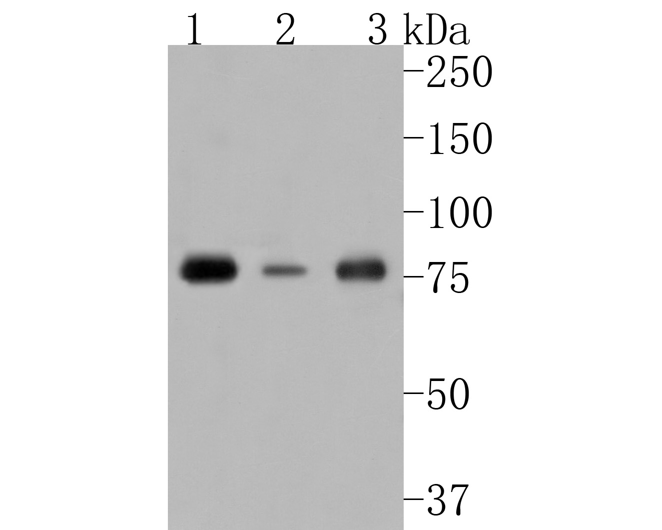 Sample:
Lane 1: 293 cell lysate
Lane 2: Hela cell lysate
Lane 3: Jurkat cell lysate
Primary: Anti-Phospho-PKC alpha (Thr638) (SLM-52187R) at 1/500 dilution
Secondary: Goat Anti-Rabbit IgG - HRP at 1:5000 dilution
Predicted band size: 77 kD
Observed band size: 77 kD
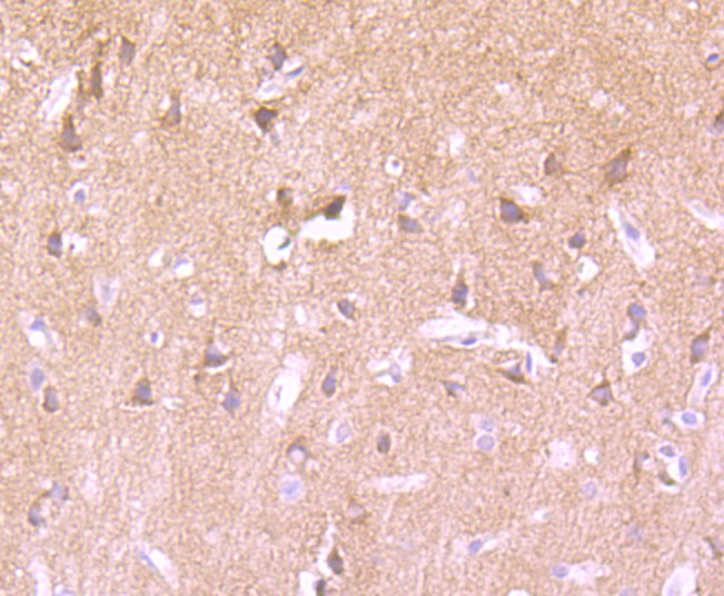 Paraformaldehyde-fixed, paraffin embedded (rat brain); Antigen retrieval by boiling in sodium citrate buffer (pH6.0) for 15min; Block endogenous peroxidase by 3% hydrogen peroxide for 20 minutes; Blocking buffer (normal goat serum) at 37°C for 30min; Antibody incubation with (Phospho-PKC alpha (Thr638)) Monoclonal Antibody, Unconjugated (SLM-52187R) at 1:50 overnight at 4°C, followed by operating according to SP Kit(Rabbit) (sp-0023) instructionsand DAB staining.
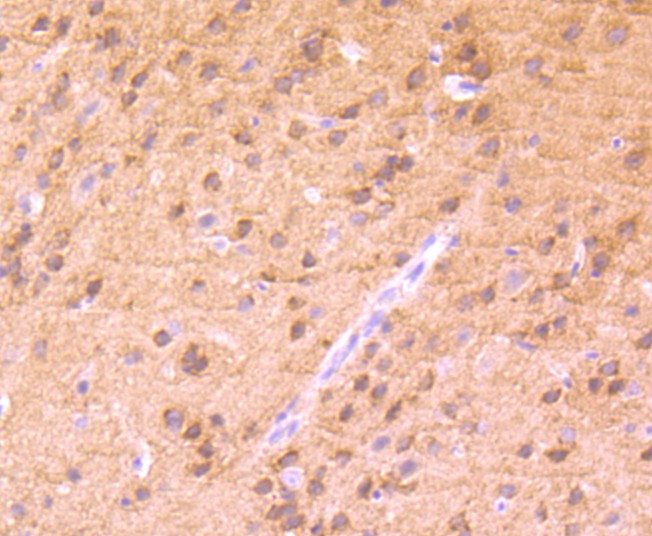 Paraformaldehyde-fixed, paraffin embedded (mouse brain); Antigen retrieval by boiling in sodium citrate buffer (pH6.0) for 15min; Block endogenous peroxidase by 3% hydrogen peroxide for 20 minutes; Blocking buffer (normal goat serum) at 37°C for 30min; Antibody incubation with (Phospho-PKC alpha (Thr638)) Monoclonal Antibody, Unconjugated (SLM-52187R) at 1:50 overnight at 4°C, followed by operating according to SP Kit(Rabbit) (sp-0023) instructionsand DAB staining.
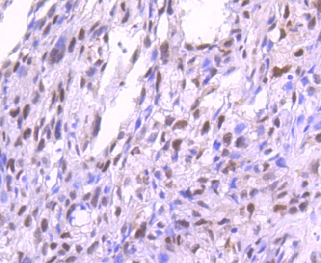 Paraformaldehyde-fixed, paraffin embedded (human breast carcinoma); Antigen retrieval by boiling in sodium citrate buffer (pH6.0) for 15min; Block endogenous peroxidase by 3% hydrogen peroxide for 20 minutes; Blocking buffer (normal goat serum) at 37°C for 30min; Antibody incubation with (Phospho-PKC alpha (Thr638)) Monoclonal Antibody, Unconjugated (SLM-52187R) at 1:50 overnight at 4°C, followed by operating according to SP Kit(Rabbit) (sp-0023) instructionsand DAB staining.
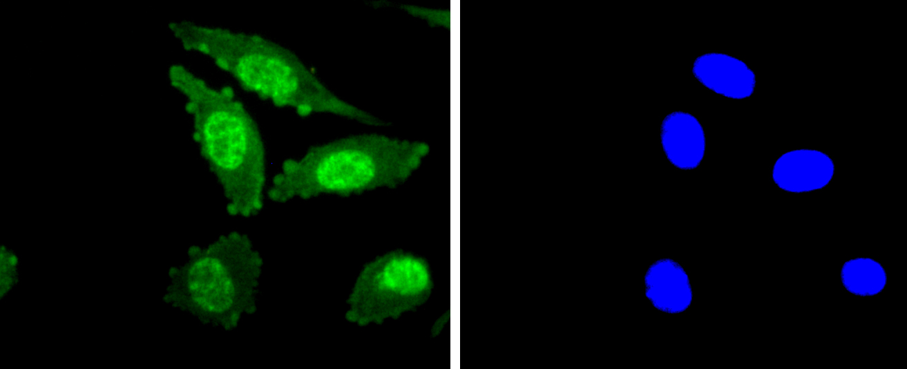 SH-SY5Y cell; 4% Paraformaldehyde-fixed; Triton X-100 at room temperature for 20 min; Blocking buffer (normal goat serum, C-0005) at 37°C for 20 min; Antibody incubation with (Phospho-PKC alpha (T638) ) monoclonal Antibody, Unconjugated (SLM-52187R) 1:50, 90 minutes at 37°C; followed by a conjugated Goat Anti-Rabbit IgG antibody at 37°C for 90 minutes, DAPI (blue, C02-04002) was used to stain the cell nuclei.
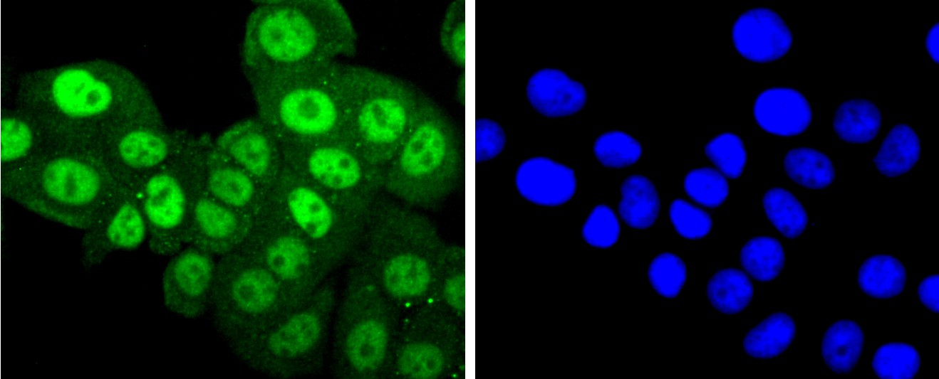 MCF-7 cell; 4% Paraformaldehyde-fixed; Triton X-100 at room temperature for 20 min; Blocking buffer (normal goat serum, C-0005) at 37°C for 20 min; Antibody incubation with (Phospho-PKC alpha (T638) in) monoclonal Antibody, Unconjugated (SLM-52187R) 1:50, 90 minutes at 37°C; followed by a conjugated Goat Anti-Rabbit IgG antibody at 37°C for 90 minutes, DAPI (blue, C02-04002) was used to stain the cell nuclei.
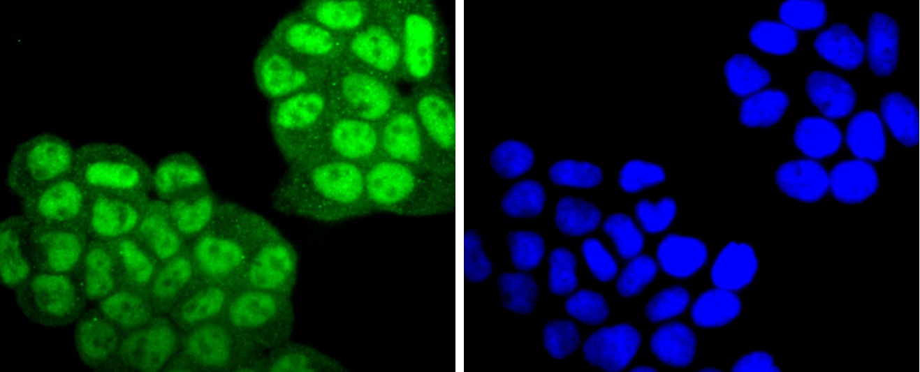 Hela cell; 4% Paraformaldehyde-fixed; Triton X-100 at room temperature for 20 min; Blocking buffer (normal goat serum, C-0005) at 37°C for 20 min; Antibody incubation with (Phospho-PKC alpha (T638) in) monoclonal Antibody, Unconjugated (SLM-52187R) 1:50, 90 minutes at 37°C; followed by a conjugated Goat Anti-Rabbit IgG antibody at 37°C for 90 minutes, DAPI (blue, C02-04002) was used to stain the cell nuclei.
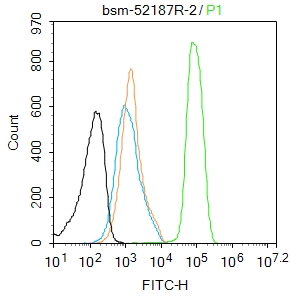 Blank control:MCF7.
Primary Antibody (green line): Rabbit Anti-Phospho-PKC alpha (Thr638) antibody (SLM-52187R)
Dilution: 2μg /10^6 cells;
Isotype Control Antibody (orange line): Rabbit IgG .
Secondary Antibody : Goat anti-rabbit IgG-AF488
Dilution: 1μg /test.
Protocol
The cells were fixed with 4% PFA (10min at room temperature)and then permeabilized with 90% ice-cold methanol for 20 min at-20℃. The cells were then incubated in 5%BSA to block non-specific protein-protein interactions for 30 min at room temperature .Cells stained with Primary Antibody for 30 min at room temperature. The secondary antibody used for 40 min at room temperature. Acquisition of 20,000 events was performed.
Cartpieces
Totalgoods,subtotals:¥Checkout
Bought notes(bought amounts latest0)
No one bought this product
User Comment(Total0User Comment Num)
- No comment


 +86 571 56623320
 +86 18668110335

