
Rabbit Anti-phospho-HSF1 (Ser326)antibody
HSF1(S326); HSF1 (phospho S326); p-HSF1 (phospho S326); Heat shock factor 1; Heat shock factor protein 1; Heat shock transcription factor 1; HSF 1; hsf1; HSTF 1; HSTF1; HSF1_HUMAN.
View History [Clear]
Details
Product Name phospho-HSF1 (Ser326) Chinese Name 磷酸化热休克因子1Recombinant rabbit monoclonal anti Alias HSF1(S326); HSF1 (phospho S326); p-HSF1 (phospho S326); Heat shock factor 1; Heat shock factor protein 1; Heat shock transcription factor 1; HSF 1; hsf1; HSTF 1; HSTF1; HSF1_HUMAN. Product Type Phosphorylated anti Recombinant rabbit monoclonal anti Research Area Tumour Signal transduction Apoptosis transcriptional regulatory factor Immunogen Species Rabbit Clonality Monoclonal Clone NO. 37E4 React Species Human, (predicted: Mouse, Rat, ) Applications WB=1:500-2000 IHC-P=1:50-200 IHC-F=1:50-200 Flow-Cyt=2ug/Test ICC=1:50-200 IF=1:50-200 (Paraffin sections need antigen repair)
not yet tested in other applications.
optimal dilutions/concentrations should be determined by the end user.Theoretical molecular weight 57kDa Cellular localization The nucleus cytoplasmic Form Liquid Concentration 1mg/ml immunogen KLH conjugated Synthesised phosphopeptide derived from human HSF1 around the phosphorylation site of Ser326: L(p-S)PT Lsotype IgG Purification affinity purified by Protein A Buffer Solution 0.01M TBS(pH7.4) with 1% BSA, 0.03% Proclin300 and 50% Glycerol. Storage Shipped at 4℃. Store at -20 °C for one year. Avoid repeated freeze/thaw cycles. Attention This product as supplied is intended for research use only, not for use in human, therapeutic or diagnostic applications. PubMed PubMed Product Detail The product of this gene is a heat-shock transcription factor. Transcription of heat-shock genes is rapidly induced after temperature stress. Hsp90, by itself and/or associated with multichaperone complexes, is a major repressor of this gene. [provided by RefSeq, Jul 2008]
Function:
DNA-binding protein that specifically binds heat shockpromoter elements (HSE) and activates transcription. In highereukaryotes, HSF is unable to bind to the HSE unless the cells areheat shocked.
Subunit:
Monomer. Under normal conditions, interacts with HSP90AA1in the HSP90 multichaperone complex; the interaction preventstrimerization and activation of HSF1. On activation by heat-stressor by other factors such as metal ions, HSF1 is released from thecomplex, homotrimerizes, is hyperphosphorylated and translocated tothe nucleus where, subsequently, it can activate transcription.Binds the complex through the regulatory domain. Interacts withSYMPK and CSTF2 in heat-stressed cells. Interacts with FKBP4 in theHSP90 multichaperone complex; the interaction is independent of thephosphorylation state of HSF1. Interacts with MAPKAPK2.
Subcellular Location:
Cytoplasm. Nucleus. Note=Cytoplasmic duringnormal growth. On activation, translocates to nuclear stressgranules. Colocalizes with SUMO1 in nuclear stress granules.
Post-translational modifications:
Phosphorylated on multiple serine residues, a subset of whichare involved in stress-related regulation of transcriptionactivation. Constitutive phosphorylation represses transcriptionalactivity at normal temperatures. Levels increase on specificresidues heat-shock and enhance HSF1 transactivation activity.Phosphorylation on Ser-307 derepresses activation on heat-stressand in combination with Ser-303 phosphorylation appears to beinvolved in recovery after heat-stress. Phosphorylated on Ser-230by CAMK2, in vitro. Cadmium also enhances phosphorylation at thissite. Phosphorylation on Ser-303 is a prerequisite for HSF1sumoylation. Phosphorylation on Ser-121 inhibits transactivationand promotes HSP90 binding. Phosphorylation on Thr-142 alsomediates transcriptional activity induced by heat.
Sumoylated with SUMO1 and SUMO2 on heat-shock. Heat-induciblesumoylation occurs after 15 min of heat-shock, after which levelsdecrease and at 4 hours, levels return to control levels.Sumoylation has no effect on HSE binding nor on transcriptionalactivity. Phosphorylation on Ser-303 is a prerequisite forsumoylation.
Similarity:
Belongs to the HSF family.
SWISS:
Q00613
Gene ID:
3297
Database links:Entrez Gene: 3297 Human
Omim: 140580 Human
SwissProt: Q00613 Human
Unigene: 530227 Human
Product Picture 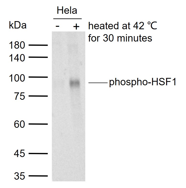 Sample:
Lane 1: Normal human HeLa cell lysates
Lane 2: Hela cells heated at 42 ℃ for 30 minutes
Primary: Anti-phospho-HSF1 (Ser326) (SLM-52166R) at 1/1000 dilution
Secondary: IRDye800CW Goat Anti-Rabbit IgG at 1/20000 dilution
Predicted band size: 57 kDa
Observed band size: 80 kDa
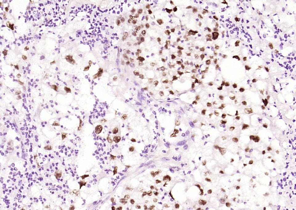 Paraformaldehyde-fixed, paraffin embedded (human endometrial carcinoma); Antigen retrieval by boiling in sodium citrate buffer (pH6.0) for 15min; Block endogenous peroxidase by 3% hydrogen peroxide for 20 minutes; Blocking buffer (normal goat serum) at 37°C for 30min; Antibody incubation with (phospho-HSF1 (Ser326)) Polyclonal Antibody, Unconjugated (SLM-52166R) at 1:200 overnight at 4°C, followed by operating according to SP Kit(Rabbit) (sp-0023) instructionsand DAB staining.
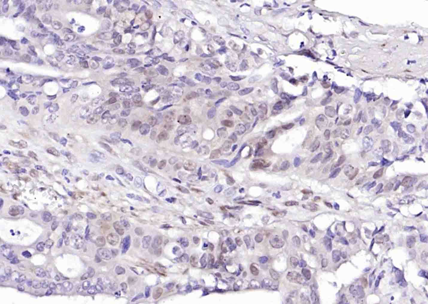 Paraformaldehyde-fixed, paraffin embedded (human colon carcinoma); Antigen retrieval by boiling in sodium citrate buffer (pH6.0) for 15min; Block endogenous peroxidase by 3% hydrogen peroxide for 20 minutes; Blocking buffer (normal goat serum) at 37°C for 30min; Antibody incubation with (phospho-HSF1 (Ser326) ) Monoclonal Antibody, Unconjugated (SLM-52166R) at 1:200 overnight at 4°C, followed by operating according to SP Kit(Rabbit) (sp-0023) instructionsand DAB staining.
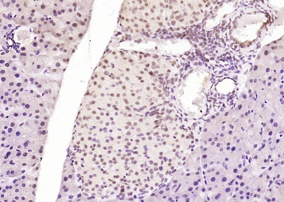 Paraformaldehyde-fixed, paraffin embedded (mouse pancreas); Antigen retrieval by boiling in sodium citrate buffer (pH6.0) for 15min; Block endogenous peroxidase by 3% hydrogen peroxide for 20 minutes; Blocking buffer (normal goat serum) at 37°C for 30min; Antibody incubation with (phospho-HSF1 (Ser326)) Polyclonal Antibody, Unconjugated (SLM-52166R) at 1:200 overnight at 4°C, followed by operating according to SP Kit(Rabbit) (sp-0023) instructionsand DAB staining.
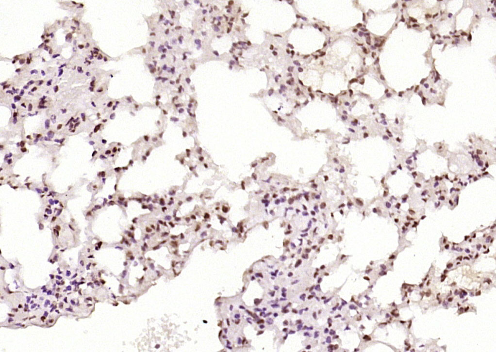 Paraformaldehyde-fixed, paraffin embedded (mouse lung); Antigen retrieval by boiling in sodium citrate buffer (pH6.0) for 15min; Block endogenous peroxidase by 3% hydrogen peroxide for 20 minutes; Blocking buffer (normal goat serum) at 37°C for 30min; Antibody incubation with (phospho-HSF1 (Ser326)) Polyclonal Antibody, Unconjugated (SLM-52166R) at 1:200 overnight at 4°C, followed by operating according to SP Kit(Rabbit) (sp-0023) instructionsand DAB staining.
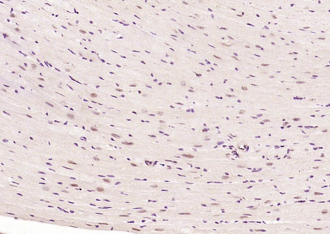 Paraformaldehyde-fixed, paraffin embedded (mouse heart); Antigen retrieval by boiling in sodium citrate buffer (pH6.0) for 15min; Block endogenous peroxidase by 3% hydrogen peroxide for 20 minutes; Blocking buffer (normal goat serum) at 37°C for 30min; Antibody incubation with (phospho-HSF1 (Ser326)) Polyclonal Antibody, Unconjugated (SLM-52166R) at 1:200 overnight at 4°C, followed by operating according to SP Kit(Rabbit) (sp-0023) instructionsand DAB staining.
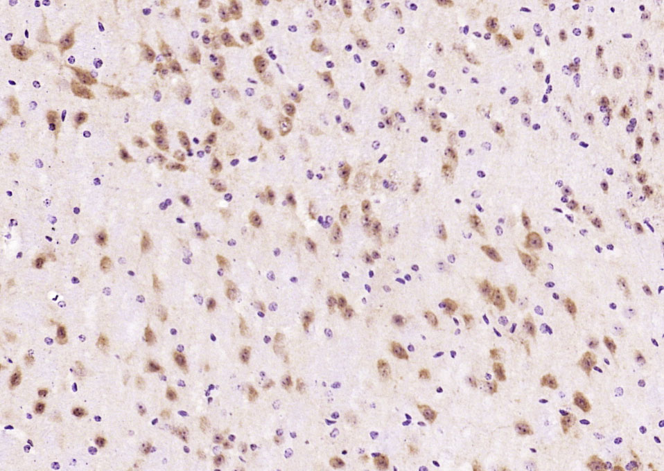 Paraformaldehyde-fixed, paraffin embedded (mouse brain); Antigen retrieval by boiling in sodium citrate buffer (pH6.0) for 15min; Block endogenous peroxidase by 3% hydrogen peroxide for 20 minutes; Blocking buffer (normal goat serum) at 37°C for 30min; Antibody incubation with (phospho-HSF1 (Ser326)) Polyclonal Antibody, Unconjugated (SLM-52166R) at 1:200 overnight at 4°C, followed by operating according to SP Kit(Rabbit) (sp-0023) instructionsand DAB staining.
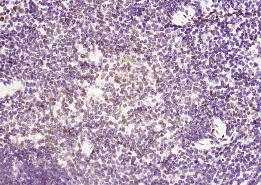 Paraformaldehyde-fixed, paraffin embedded (mouse spleen); Antigen retrieval by boiling in sodium citrate buffer (pH6.0) for 15min; Block endogenous peroxidase by 3% hydrogen peroxide for 20 minutes; Blocking buffer (normal goat serum) at 37°C for 30min; Antibody incubation with (phospho-HSF1 (Ser326)) Polyclonal Antibody, Unconjugated (SLM-52166R) at 1:200 overnight at 4°C, followed by operating according to SP Kit(Rabbit) (sp-0023) instructionsand DAB staining.
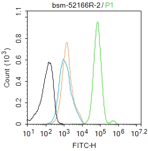 Blank control:MCF7.
Primary Antibody (green line): Rabbit Anti-phospho-HSF1 (Ser326) antibody (SLM-52166R)
Dilution: 2μg /10^6 cells;
Isotype Control Antibody (orange line): Rabbit IgG .
Secondary Antibody : Goat anti-rabbit IgG-AF488
Dilution: 1μg /test.
Protocol
The cells were fixed with 4% PFA (10min at room temperature)and then permeabilized with 90% ice-cold methanol for 20 min at-20℃. The cells were then incubated in 5%BSA to block non-specific protein-protein interactions for 30 min at room temperature .Cells stained with Primary Antibody for 30 min at room temperature. The secondary antibody used for 40 min at room temperature. Acquisition of 20,000 events was performed.
Cartpieces
Totalgoods,subtotals:¥Checkout
References (0)
No References
Bought notes(bought amounts latest0)
No one bought this product
User Comment(Total0User Comment Num)
- No comment


 +86 571 56623320
 +86 18668110335

