
Rabbit Anti-BCL2A1 antibody
ACC 1; ACC 2; B2LA1_HUMAN; Bcl 2 related protein A1; Bcl-2-like protein 5; Bcl-2-related protein A1; BCL2 related protein A1; Bcl2-L-5; BCL2L5; BFL1; GRS; HBPA1; Hematopoietic BCL2 related protein A1; Hemopoietic specific early response protein; Hemopoiet
View History [Clear]
Details
Product Name BCL2A1 Chinese Name BCL2相关蛋白A1Recombinant rabbit monoclonal anti Alias ACC 1; ACC 2; B2LA1_HUMAN; Bcl 2 related protein A1; Bcl-2-like protein 5; Bcl-2-related protein A1; BCL2 related protein A1; Bcl2-L-5; BCL2L5; BFL1; GRS; HBPA1; Hematopoietic BCL2 related protein A1; Hemopoietic specific early response protein; Hemopoietic-specific early response protein; Protein BFL 1; Protein BFL-1; Protein GRS. Research Area Cell biology Apoptosis Immunogen Species Rabbit Clonality Monoclonal Clone NO. 3E4 React Species (predicted: Human, Mouse, Rat, ) Applications WB=1:500-2000 IHC-P=1:100-500 IHC-F=1:50-200 ICC=1:50-100 IF=1:50-100 (Paraffin sections need antigen repair)
not yet tested in other applications.
optimal dilutions/concentrations should be determined by the end user.Theoretical molecular weight 29kDa Cellular localization cytoplasmic Form Liquid Concentration 1mg/ml immunogen KLH conjugated synthetic peptide derived from human BCL2A1 Lsotype IgG Purification affinity purified by Protein A Buffer Solution 0.01M TBS(pH7.4) with 1% BSA, 0.03% Proclin300 and 50% Glycerol. Storage Shipped at 4℃. Store at -20 °C for one year. Avoid repeated freeze/thaw cycles. Attention This product as supplied is intended for research use only, not for use in human, therapeutic or diagnostic applications. PubMed PubMed Product Detail This gene encodes a member of the BCL-2 protein family. The proteins of this family form hetero- or homodimers and act as anti- and pro-apoptotic regulators that are involved in a wide variety of cellular activities such as embryonic development, homeostasis and tumorigenesis. The protein encoded by this gene is able to reduce the release of pro-apoptotic cytochrome c from mitochondria and block caspase activation. This gene is a direct transcription target of NF-kappa B in response to inflammatory mediators, and is up-regulated by different extracellular signals, such as granulocyte-macrophage colony-stimulating factor (GM-CSF), CD40, phorbol ester and inflammatory cytokine TNF and IL-1, which suggests a cytoprotective function that is essential for lymphocyte activation as well as cell survival. Alternatively spliced transcript variants encoding different isoforms have been found for this gene. [provided by RefSeq, Jul 2008]
Function:
Retards apoptosis induced by IL-3 deprivation. May function in the response of hemopoietic cells to external signals and in maintaining endothelial survival during infection.
Subunit:
Interacts directly with BAK1, BID, BMF and BBC3. Interacts directly with BCL2L11/BIM. Interacts with BAX isoform Sigma.
Subcellular Location:
Cytoplasm.
Tissue Specificity:
Seems to be restricted to the hematopoietic compartment. Expressed in peripheral blood, spleen, and bone marrow, at moderate levels in lung, small intestine and testis, at a minimal levels in other tissues. Also found in vascular smooth muscle cells and hematopoietic malignancies.
Similarity:
Belongs to the Bcl-2 family.
SWISS:
Q16548
Gene ID:
597
Database links:Entrez Gene: 597 Human
Entrez Gene: 12044 Mouse
Omim: 601056 Human
SwissProt: Q16548 Human
SwissProt: Q07440 Mouse
Unigene: 227817 Human
Unigene: 425593 Mouse
Unigene: 479217 Mouse
Unigene: 19770 Rat
Product Picture 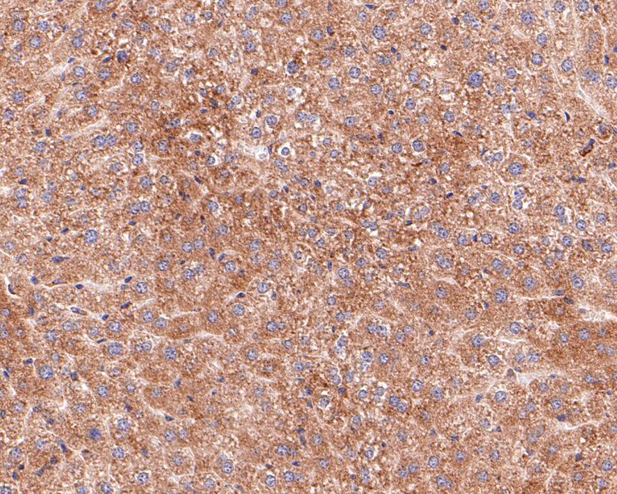 Immunohistochemical analysis of paraffin-embedded mouse liver tissue with Rabbit anti-BCL2A1 antibody (SLM-52023R) at 1/100 dilution. The section was pre-treated using heat mediated antigen retrieval with Tris-EDTA buffer (pH 9.0) for 20 minutes. The tissues were blocked in 1% BSA for 20 minutes at room temperature, washed with ddH2O and PBS, and then probed with the primary antibody ( SLM-52023R ) at 1/100 dilution for 1 hour at room temperature. The detection was performed using an HRP conjugated compact polymer system. DAB was used as the chromogen. Tissues were counterstained with hematoxylin and mounted with DPX.
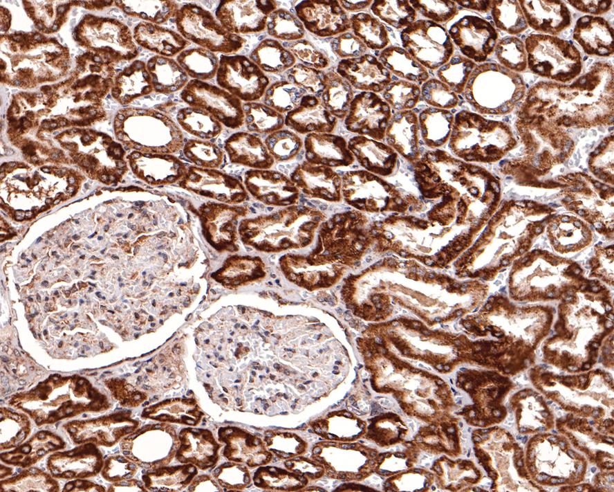 Immunohistochemical analysis of paraffin-embedded human kidney tissue with Rabbit anti-BCL2A1 antibody (SLM-52023R) at 1/400 dilution. The section was pre-treated using heat mediated antigen retrieval with Tris-EDTA buffer (pH 9.0) for 20 minutes. The tissues were blocked in 1% BSA for 20 minutes at room temperature, washed with ddH2O and PBS, and then probed with the primary antibody (SLM-52023R) at 1/400 dilution for 1 hour at room temperature. The detection was performed using an HRP conjugated compact polymer system. DAB was used as the chromogen. Tissues were counterstained with hematoxylin and mounted with DPX
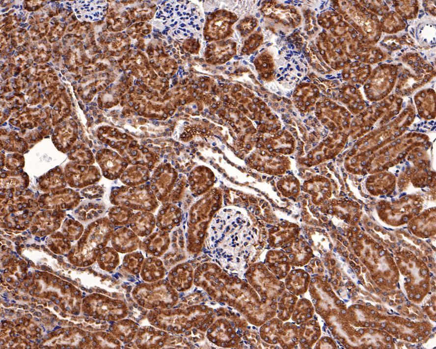 Immunohistochemical analysis of paraffin-embedded mouse kidney tissue with Rabbit anti-BCL2A1 antibody (SLM-52023R) at 1/400 dilution. The section was pre-treated using heat mediated antigen retrieval with Tris-EDTA buffer (pH 9.0) for 20 minutes. The tissues were blocked in 1% BSA for 20 minutes at room temperature, washed with ddH2O and PBS, and then probed with the primary antibody (SLM-52023R0) at 1/400 dilution for 1 hour at room temperature. The detection was performed using an HRP conjugated compact polymer system. DAB was used as the chromogen. Tissues were counterstained with hematoxylin and mounted with DPX.
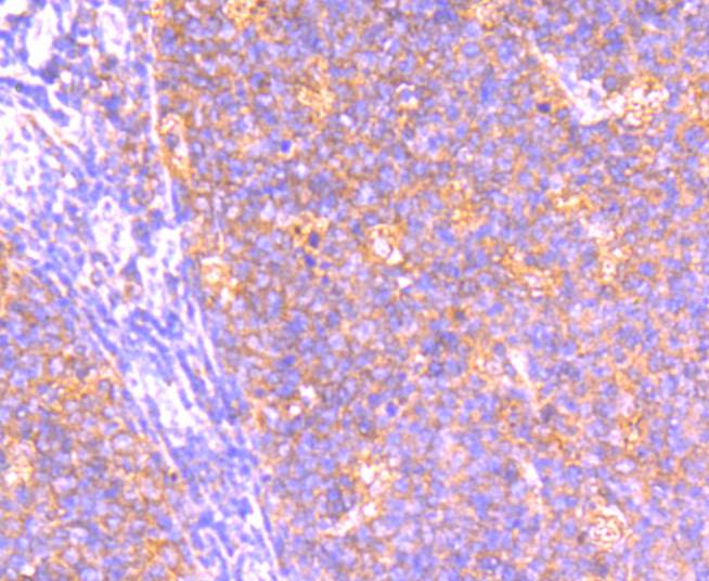 Immunohistochemical analysis of paraffin-embedded human tonsil tissue with Rabbit anti-BCL2A1 antibody (SLM-52023R) at 1/50 dilution. The section was pre-treated using heat mediated antigen retrieval with Tris-EDTA buffer (pH 9.0) for 20 minutes. The tissues were blocked in 1% BSA for 20 minutes at room temperature, washed with ddH2O and PBS, and then probed with the primary antibody (SLM-52023R) at 1/50 dilution for 1 hour at room temperature. The detection was performed using an HRP conjugated compact polymer system. DAB was used as the chromogen. Tissues were counterstained with hematoxylin and mounted with DPX.
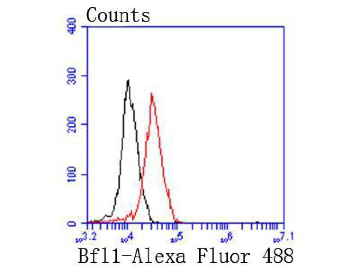 Flow cytometric analysis of BCL2A1 was done on 293 cells. The cells were fixed, permeabilized and stained with the primary antibody ( SLM-52023R , 1/50) (red). After incubation of the primary antibody at room temperature for an hour, the cells were stained with a Alexa Fluor 488-conjugated Goat anti-Rabbit IgG Secondary antibody at 1/1000 dilution for 30 minutes.Unlabelled sample was used as a control (cells without incubation with primary antibody; black).
Cartpieces
Totalgoods,subtotals:¥Checkout
References (0)
No References
Bought notes(bought amounts latest0)
No one bought this product
User Comment(Total0User Comment Num)
- No comment


 +86 571 56623320
 +86 18668110335

