
Mouse Anti-VCP antibody
valosin-containing protein; 15S Mg(2+) ATPase p97 subunit; ATPase p97; IBMPFD; MGC131997; MGC148092; MGC8560; p97; TER ATPase; TERA; transitional endoplasmic reticulum ATPase; valosin-containing protein; yeast Cdc48p homolog; Transitional endoplasmic reti
View History [Clear]
Details
Product Name VCP Chinese Name 含缬酪肽蛋白单克隆抗体 Alias valosin-containing protein; 15S Mg(2+) ATPase p97 subunit; ATPase p97; IBMPFD; MGC131997; MGC148092; MGC8560; p97; TER ATPase; TERA; transitional endoplasmic reticulum ATPase; valosin-containing protein; yeast Cdc48p homolog; Transitional endoplasmic reticulum ATPase; TER ATPase; 15S Mg(2+)-ATPase p97 subunit; p97; TERA_HUMAN. Research Area Tumour Neurobiology Signal transduction Growth factors and hormones Diabetes glycoprotein Immunogen Species Mouse Clonality Monoclonal Clone NO. N5A9 React Species Human, Mouse, Rat, Applications WB=1:500-2000 IHC-P=1:25 IHC-F=1:25 Flow-Cyt=1:25 ICC=1:25 IF=1:25 (Paraffin sections need antigen repair)
not yet tested in other applications.
optimal dilutions/concentrations should be determined by the end user.Theoretical molecular weight 97kDa Cellular localization The nucleus cytoplasmic Form Liquid Concentration 1mg/ml immunogen Recombinant human VCP. Lsotype IgG1,κ Purification affinity purified by Protein G Buffer Solution 0.01M TBS(pH7.4) with 1% BSA, 0.03% Proclin300 and 50% Glycerol. Storage Shipped at 4℃. Store at -20 °C for one year. Avoid repeated freeze/thaw cycles. Attention This product as supplied is intended for research use only, not for use in human, therapeutic or diagnostic applications. PubMed PubMed Product Detail The protein encoded by this gene is a member of a family that includes putative ATP-binding proteins involved in vesicle transport and fusion, 26S proteasome function, and assembly of peroxisomes. This protein, as a structural protein, is associated with clathrin, and heat-shock protein Hsc70, to form a complex. It has been implicated in a number of cellular events that are regulated during mitosis, including homotypic membrane fusion, spindle pole body function, and ubiquitin-dependent protein degradation. [provided by RefSeq, Jul 2008]
Function:
Necessary for the fragmentation of Golgi stacks during mitosis and for their reassembly after mitosis. Involved in the formation of the transitional endoplasmic reticulum (tER). The transfer of membranes from the endoplasmic reticulum to the Golgi apparatus occurs via 50-70 nm transition vesicles which derive from part-rough, part-smooth transitional elements of the endoplasmic reticulum (tER). Vesicle budding from the tER is an ATP-dependent process. The ternary complex containing UFD1L, VCP and NPLOC4 binds ubiquitinated proteins and is necessary for the export of misfolded proteins from the ER to the cytoplasm, where they are degraded by the proteasome. The NPLOC4-UFD1L-VCP complex regulates spindle disassembly at the end of mitosis and is necessary for the formation of a closed nuclear envelope (By similarity). Regulates E3 ubiquitin-protein ligase activity of RNF19A.
Subcellular Location:
Cytoplasm > cytosol. Nucleus. Present in the neuronal hyaline inclusion bodies specifically found in motor neurons from amyotrophic lateral sclerosis patients. Present in the Lewy bodies specifically found in neurons from Parkinson disease patients.
Post-translational modifications:
Phosphorylated by tyrosine kinases in response to T-cell antigen receptor activation.
Phosphorylated upon DNA damage, probably by ATM or ATR. ISGylated.
DISEASE:
Defects in VCP are the cause of inclusion body myopathy with early-onset Paget disease and frontotemporal dementia (IBMPFD) [MIM:167320]; also known as muscular dystrophy, limb-girdle, with Paget disease of bone or pagetoid amyotrophic lateral sclerosis or pagetoid neuroskeletal syndrome or lower motor neuron degeneration with Paget-like bone disease. IBMPFD features adult-onset proximal and distal muscle weakness (clinically resembling limb girdle muscular dystrophy), early-onset Paget disease of bone in most cases and premature frontotemporal dementia.
Similarity:
Belongs to the AAA ATPase family.
SWISS:
P55072
Gene ID:
7415
Database links:Entrez Gene: 7415 Human
Entrez Gene: 269523 Mouse
Omim: 601023 Human
SwissProt: P55072 Human
SwissProt: Q01853 Mouse
Product Picture 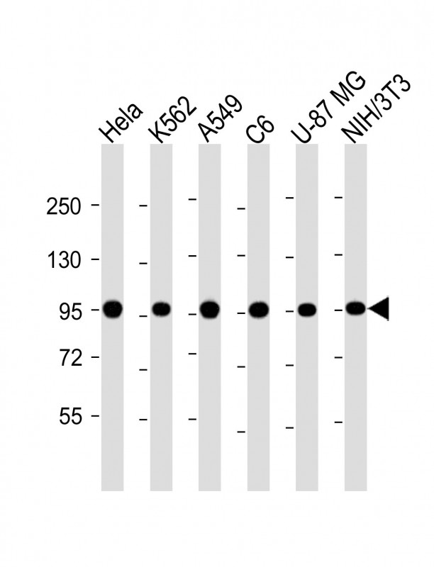 Sample:
Lane 1: Hela cell lysates
Lane 2: K562 cell lysates
Lane 3: A549 cell lysates
Lane 4: C6 cell lysates
Lane 5: U-87 MG cell lysates
Lane 6: NIH/3T3 cell lysates
Primary: Anti-VCP (SLM-51611M) at 1/4000 dilution
Secondary: IRDye800CW Goat Anti-Mouse IgG at 1/20000 dilution
Predicted band size: 97 kD
Observed band size: 97 kD
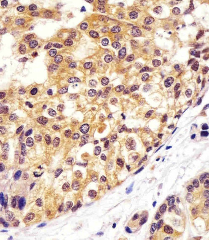 Paraformaldehyde-fixed, paraffin embedded (human breast carcinoma sections); Antigen retrieval by boiling in sodium citrate buffer (pH6.0) for 15min; Block endogenous peroxidase by 3% hydrogen peroxide for 20 minutes; Blocking buffer (normal goat serum) at 37°C for 30min; Antibody incubation with (VCP) Monoclonal Antibody, Unconjugated (SLM-51611M) at 1:200 overnight at 4°C, followed by operating according to SP Kit(Mouse)(sp-0024) instructionsand DAB staining.
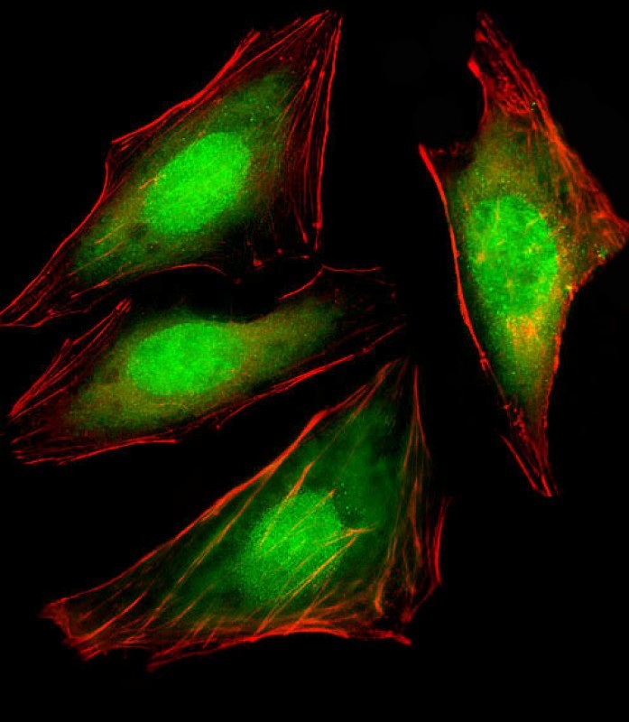 Hela cell; 4% Paraformaldehyde-fixed; Triton X-100 at room temperature for 20 min; Blocking buffer (normal goat serum) at 37°C for 20 min; Antibody incubation with (VCP) monoclonal Antibody, Unconjugated (SLM-51611M) 1:25, 90 minutes at 37°C; followed by a conjugated Goat Anti-Mouse IgG antibody at 37°C for 90 minutes,Dylight® 554 Phalloidin (red) was used to stain the cell Cytoplasmic actin.
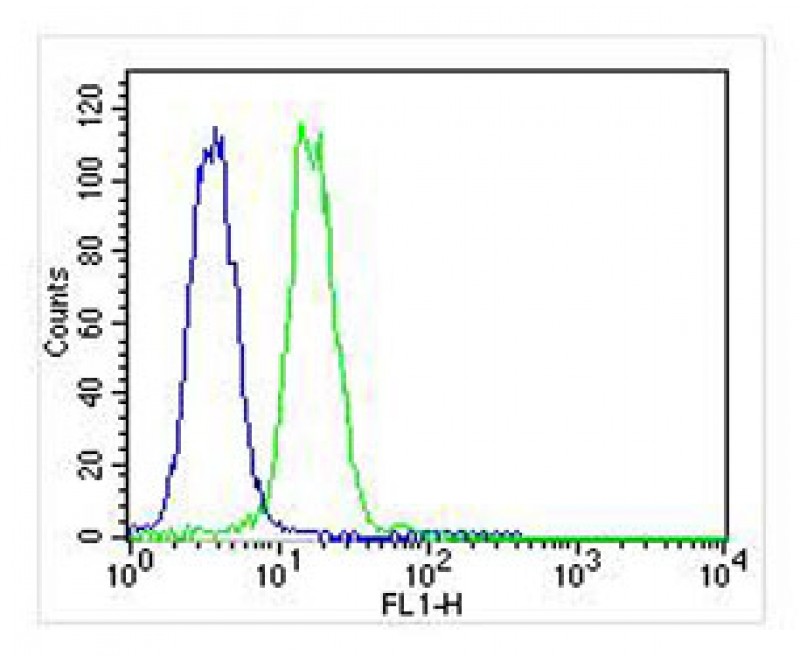 Blank control:K562.
Primary Antibody (green line): Mouse Anti-VCP antibody (SLM-51611M)
Dilution: 1:25;
Isotype Control Antibody (blue line): Mouse IgG Secondary Antibody : Goat anti-mouse IgG-AF488
Dilution: 1:400 Protocol
The cells were fixed with 4% PFA (10min at room temperature)and then permeabilized with 90% ice-cold methanol for 20 min at-20℃.The cells were then incubated in 5%BSA to block non-specific protein-protein interactions for 30 min at room temperature .Cells stained with Primary Antibody for 30 min at room temperature. The secondary antibody used for 40 min at room temperature. Acquisition of 20,000 events was performed.
Cartpieces
Totalgoods,subtotals:¥Checkout
References (0)
No References
Bought notes(bought amounts latest0)
No one bought this product
User Comment(Total0User Comment Num)
- No comment


 +86 571 56623320
 +86 18668110335

