
Mouse Anti-XIAP antibody
X-linked Inhibitor of Apoptosis Protein; RIAP-3; Baculoviral IAP repeat-containing protein 4; E3 ubiquitin-protein ligase XIAP; Inhibitor of apoptosis protein 3; X-linked IAP; IAP-like protein; HILP; BIRC4; HILP; XLP2; ILP1; Xiap; MIHA; hILP; IAP3; API3;
View History [Clear]
Details
Product Name XIAP Chinese Name X-连锁凋亡蛋白/性连锁凋亡抑制蛋白单克隆抗体 Alias X-linked Inhibitor of Apoptosis Protein; RIAP-3; Baculoviral IAP repeat-containing protein 4; E3 ubiquitin-protein ligase XIAP; Inhibitor of apoptosis protein 3; X-linked IAP; IAP-like protein; HILP; BIRC4; HILP; XLP2; ILP1; Xiap; MIHA; hILP; IAP3; API3; XIAP_HUMAN; ILP; hILP; IAP-3; hIAP-3; hIAP3; X-linked inhibitor of apoptosis protein. Research Area Tumour Cell biology Apoptosis The new supersedes the old Immunogen Species Mouse Clonality Monoclonal Clone NO. D1G6 React Species Human, Applications WB=1:500-2000 IHC-P=1:20-100 IHC-F=1:20-100 ICC=1:25 (Paraffin sections need antigen repair)
not yet tested in other applications.
optimal dilutions/concentrations should be determined by the end user.Theoretical molecular weight 55kDa Cellular localization cytoplasmic Form Liquid Concentration 1mg/ml immunogen Recombinant human XIAP Lsotype IgG1,k Purification affinity purified by Protein G Buffer Solution 0.01M TBS(pH7.4) with 1% BSA, 0.03% Proclin300 and 50% Glycerol. Storage Shipped at 4℃. Store at -20 °C for one year. Avoid repeated freeze/thaw cycles. Attention This product as supplied is intended for research use only, not for use in human, therapeutic or diagnostic applications. PubMed PubMed Product Detail This gene encodes a protein that belongs to a family of apoptotic suppressor proteins. Members of this family share a conserved motif termed, baculovirus IAP repeat, which is necessary for their anti-apoptotic function. This protein functions through binding to tumor necrosis factor receptor-associated factors TRAF1 and TRAF2 and inhibits apoptosis induced by menadione, a potent inducer of free radicals, and interleukin 1-beta converting enzyme. This protein also inhibits at least two members of the caspase family of cell-death proteases, caspase-3 and caspase-7. Mutations in this gene are the cause of X-linked lymphoproliferative syndrome. Alternate splicing results in multiple transcript variants. Pseudogenes of this gene are found on chromosomes 2 and 11.[provided by RefSeq, Feb 2011]
Function:
Apoptotic suppressor. Has E3 ubiquitin-protein ligase activity. Mediates the proteasomal degradation of target proteins, such as caspase-3, SMAC or AIFM1. Inhibitor of caspase-3, -7 and -9. Mediates activation of MAP3K7/TAK1, leading to the activation of NF-kappa-B.
Subunit:
Monomer, and homodimer. Interacts with SMAC and with PRSS25; these interactions inhibit apoptotic suppressor activity. Interacts with MAP3K7IP1 and AIFM1. Interaction with SMAC hinders binding of MAP3K7IP1 and AIFM1. Interacts with TCF25.
Subcellular Location:
Cytoplasm.
Tissue Specificity:
Ubiquitous, except peripheral blood leukocytes.
Post-translational modifications:
Ubiquitinated and degraded by the proteasome in apoptotic cells. Phosphorylation by PKB/AKT protects XIAP against ubiquitination and protects the protein against proteasomal degradation.
DISEASE:
Defects in XIAP are the cause of lymphoproliferative syndrome X-linked type 2 (XLP2) [MIM:300635]. XLP is a rare immunodeficiency characterized by extreme susceptibility to infection with Epstein-Barr virus (EBV). Symptoms include severe or fatal mononucleosis, acquired hypogammaglobulinemia, pancytopenia and malignant lymphoma.
Similarity:
Belongs to the IAP family.
Contains 3 BIR repeats.
Contains 1 RING-type zinc finger.
SWISS:
P98170
Gene ID:
331
Database links:Entrez Gene: 331 Human
SwissProt: P98170 Human
Product Picture 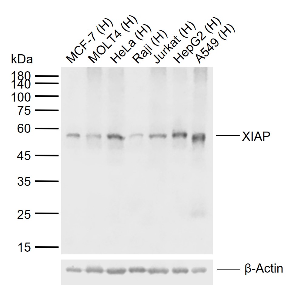 Sample:
Lane 1: Human MCF-7 cell lysates
Lane 2: Human MOLT4 cell lysates
Lane 3: Human HeLa cell lysates
Lane 4: Human Raji cell lysates
Lane 5: Human Jurkat cell lysates
Lane 6: Human HepG2 cell lysates
Lane 7: Human A549 cell lysates
Primary: Anti-XIAP (SLM-51607M) at 1/1000 dilution
Secondary: IRDye800CW Goat Anti-Mouse IgG at 1/20000 dilution
Predicted band size: 55 kDa
Observed band size: 55 kDa
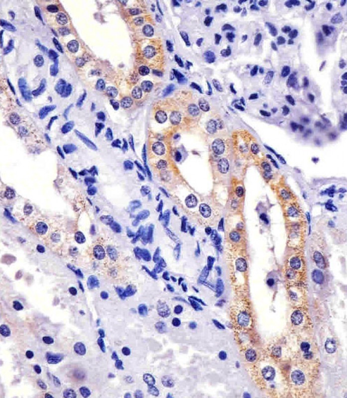 Paraformaldehyde-fixed, paraffin embedded (Human kidney section); Antigen retrieval by boiling in sodium citrate buffer (pH6.0) for 15min; Block endogenous peroxidase by 3% hydrogen peroxide for 20 minutes; Blocking buffer (normal goat serum) at 37°C for 30min; Antibody incubation with (XIAP) Monoclonal Antibody, Unconjugated (SLM-51607M) at 1:200 overnight at 4°C, followed by operating according to SP Kit(Mouse)(sp-0024) instructionsand DAB staining.
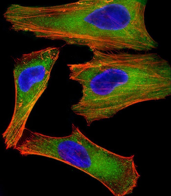 Hela cell; 4% Paraformaldehyde-fixed; Triton X-100 at room temperature for 20 min; Blocking buffer (normal goat serum) at 37°C for 20 min; Antibody incubation with (XIAP) monoclonal Antibody, Unconjugated (SLM-51607M) 1:25, 90 minutes at 37°C; followed by a conjugated Goat Anti-Mouse IgG antibody at 37°C for 90 minutes, Alexa Fluor® 555 conjugated with Phalloidin(red) was used to stain the cell Cytoplasmic actin.
Cartpieces
Totalgoods,subtotals:¥Checkout
References (0)
No References
Bought notes(bought amounts latest0)
No one bought this product
User Comment(Total0User Comment Num)
- No comment


 +86 571 56623320
 +86 18668110335

