
Mouse Anti-GATA3 antibody
GATA 3; GATA-3; GATA binding factor 3; GATA binding protein 3; HDR; MGC2346; MGC5199; MGC5445; Trans acting T cell specific transcription factor GATA 3; GATA3_HUMAN; Trans-acting T-cell-specific transcription factor GATA-3; GATA-binding factor 3.
View History [Clear]
Details
Product Name GATA3 Chinese Name GATABinding protein3单克隆抗体 Alias GATA 3; GATA-3; GATA binding factor 3; GATA binding protein 3; HDR; MGC2346; MGC5199; MGC5445; Trans acting T cell specific transcription factor GATA 3; GATA3_HUMAN; Trans-acting T-cell-specific transcription factor GATA-3; GATA-binding factor 3. Research Area immunology Developmental biology Chromatin and nuclear signals Stem cells t-lymphocyte Zinc finger protein Epigenetics Immunogen Species Mouse Clonality Monoclonal Clone NO. G3G4 React Species Human, Applications WB=1:500-1000 Flow-Cyt=1:25 ICC=1:25
not yet tested in other applications.
optimal dilutions/concentrations should be determined by the end user.Theoretical molecular weight 49kDa Cellular localization The nucleus Form Liquid Concentration 1mg/ml immunogen Recombinant human GATA3. Lsotype IgG2b,κ Purification affinity purified by Protein G Buffer Solution 0.01M TBS(pH7.4) with 1% BSA, 0.03% Proclin300 and 50% Glycerol. Storage Shipped at 4℃. Store at -20 °C for one year. Avoid repeated freeze/thaw cycles. Attention This product as supplied is intended for research use only, not for use in human, therapeutic or diagnostic applications. PubMed PubMed Product Detail Members of the GATA family share a conserved zinc finger DNA-binding domain and are capable of binding the WGATAR consensus sequence. GATA-1 is erythroid-specific and is responsible for the regulated transcription of erythroid genes. It is an essential component in the generation of the erythroid lineage. GATA-2 is expressed in embryonic brain and liver, HeLa and endothelial cells, as well as in erythroid cells. Studies with a modified GATA consensus sequence, AGATCTTA, have shown that GATA-2 and GATA-3 recognize this mutated consensus while GATA-1 has poor recognition of this sequence. This indicates broader regulatory capabilities of GATA-2 and GATA-3 than GATA-1. GATA-3 is highly expressed in T lymphocytes. GATA-4, GATA-5 and GATA-6 comprise a subfamily of transcription factors. Both GATA-4 and GATA-6 are found in heart, pancreas and ovary; lung and liver tissues exhibit GATA-6, but not GATA-4 expression. GATA-5 expression has been observed in differentiated heart and gut tissues and is present throughout the course of development in the heart. Although expression patterns of the various GATA transcription factors may overlap, it is not yet apparent how the GATA factors are able to discriminate in binding their appropriate target sites.
Function:
Transcriptional activator which binds to the enhancer of the T-cell receptor alpha and delta genes. Binds to the consensus sequence 5'-AGATAG-3'.
Subcellular Location:
Nucleus.
Tissue Specificity:
T-cells and endothelial cells.
DISEASE:
Defects in GATA3 are the cause of hypoparathyroidism with sensorineural deafness and renal dysplasia (HDR) [MIM:146255]; also known as Barakat syndrome.
Similarity:
Contains 2 GATA-type zinc fingers.
SWISS:
P23771
Gene ID:
2625
Database links:Entrez Gene: 2625 Human
SwissProt: P23771 Human
Product Picture 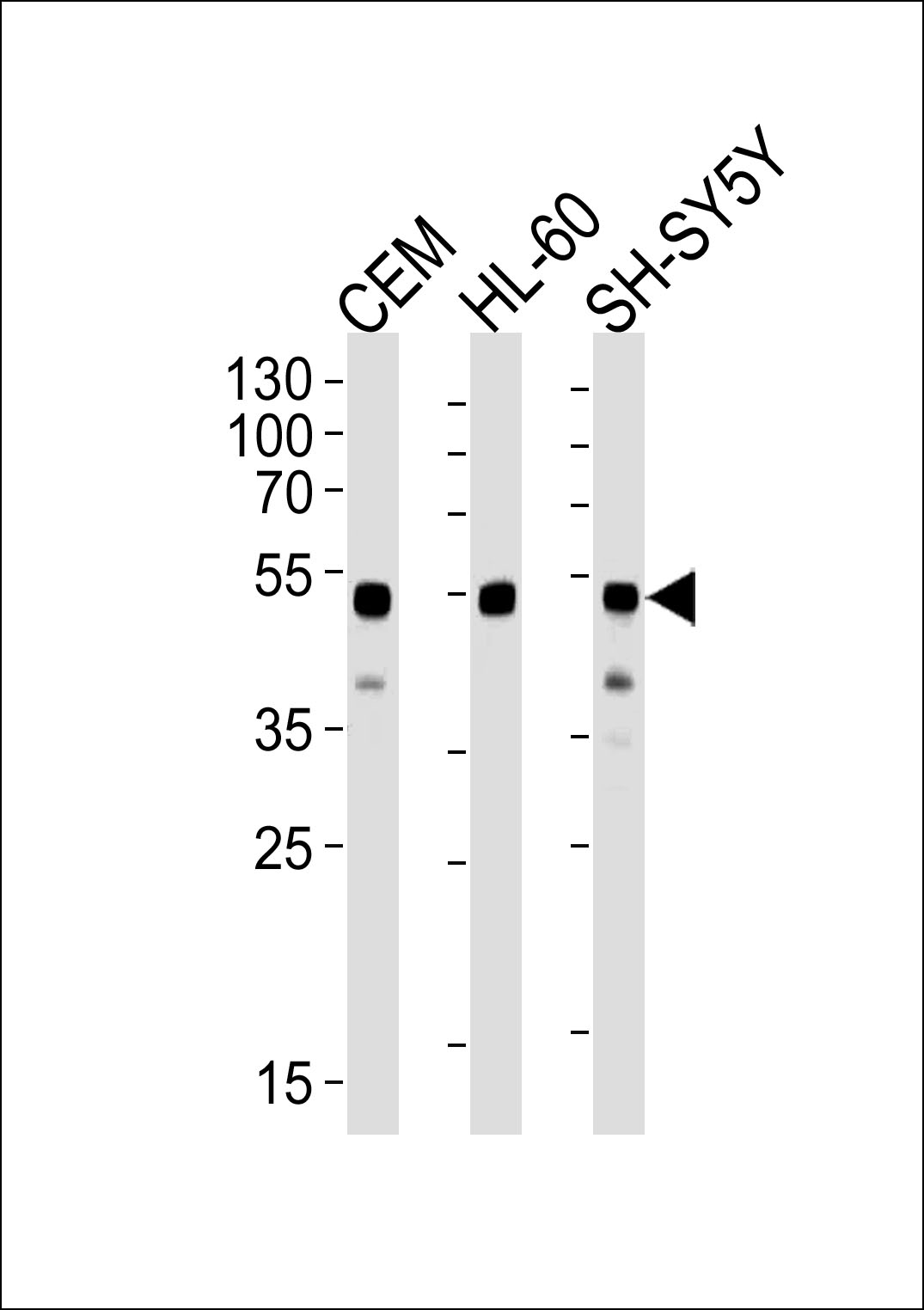 Sample:
Lane 1: CEM cell lysates
Lane 2: HL60 cell lysates
Lane 3: SH-SY5Y cell lysates
Primary: Anti-GATA3 (SLM-51575M) at 1/1000 dilution
Secondary: IRDye800CW Goat Anti-Mouse IgG at 1/20000 dilution
Predicted band size: 49 kD
Observed band size: 52 kD
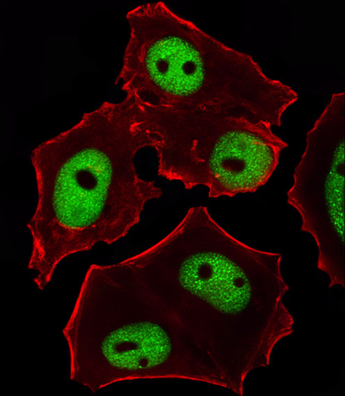 MCF7 cell; 4% Paraformaldehyde-fixed; Triton X-100 at room temperature for 20 min; Blocking buffer (normal goat serum) at 37°C for 20 min; Antibody incubation with (GATA3) monoclonal Antibody, Unconjugated (SLM-51575M) 1:25, 90 minutes at 37°C; followed by a conjugated Goat Anti-Mouse IgG antibody at 37°C for 90 minutes,Alexa Fluor® 555 conjugated with Phalloidin(red) was used to stain the cell Cytoplasmic actin.
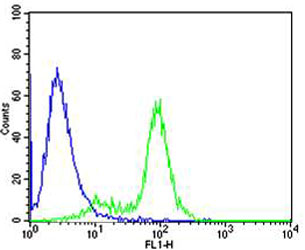 Blank control: MCF7.
Primary Antibody (green line): Mouse Anti-GATA3 antibody (SLM-51575M)
Dilution: 1:25;
Isotype Control Antibody (blue line): Mouse IgG2b Secondary Antibody : Goat anti-mouse IgG-AF488
Dilution:1:400 Protocol
The cells were fixed with 4% PFA (10min at room temperature)and then permeabilized with 90% ice-cold methanol for 20 min at-20℃.The cells were then incubated in 5%BSA to block non-specific protein-protein interactions for 30 min at room temperature .Cells stained with Primary Antibody for 30 min at room temperature. The secondary antibody used for 40 min at room temperature. Acquisition of 20,000 events was performed.
Cartpieces
Totalgoods,subtotals:¥Checkout
References (0)
No References
Bought notes(bought amounts latest0)
No one bought this product
User Comment(Total0User Comment Num)
- No comment


 +86 571 56623320
 +86 18668110335

