
Mouse Anti-LC3 antibody
MLP3A_HUMAN; Microtubule-associated proteins 1A/1B light chain 3A; MAP1LC3A; Autophagy-related protein LC3 A; Autophagy-related ubiquitin-like modifier LC3 A; MAP1 light chain 3-like protein 1; MAP1A/MAP1B light chain 3 A (MAP1A/MAP1B LC3 A); Microtubule-
View History [Clear]
Details
Product Name LC3 Chinese Name 自噬微管相关蛋白轻链3单克隆抗体 Alias MLP3A_HUMAN; Microtubule-associated proteins 1A/1B light chain 3A; MAP1LC3A; Autophagy-related protein LC3 A; Autophagy-related ubiquitin-like modifier LC3 A; MAP1 light chain 3-like protein 1; MAP1A/MAP1B light chain 3 A (MAP1A/MAP1B LC3 A); Microtubule-associated protein 1 light chain 3 alpha; MLP3B_HUMAN; Microtubule-associated proteins 1A/1B light chain 3B; MAP1LC3B; MAP1ALC3; Autophagy-related protein LC3 B; Autophagy-related ubiquitin-like modifier LC3 B; MAP1 light chain 3-like protein 2; MAP1A/MAP1B light chain 3 B (MAP1A/MAP1B LC3 B); Microtubule-associated protein 1 light chain 3 beta; literatures Research Area Tumour Signal transduction Apoptosis transcriptional regulatory factor Cell differentiation Immunogen Species Mouse Clonality Monoclonal Clone NO. L3G1 React Species Human, Mouse, Rat, Applications WB=1:500-1000
not yet tested in other applications.
optimal dilutions/concentrations should be determined by the end user.Theoretical molecular weight 13kDa Cellular localization cytoplasmic Form Liquid Concentration 1mg/ml immunogen Recombinant protein of human MAP1LC3A/B Lsotype IgG1,k Purification affinity purified by Protein G Buffer Solution 0.01M TBS(pH7.4) with 1% BSA, 0.03% Proclin300 and 50% Glycerol. Storage Shipped at 4℃. Store at -20 °C for one year. Avoid repeated freeze/thaw cycles. Attention This product as supplied is intended for research use only, not for use in human, therapeutic or diagnostic applications. PubMed PubMed Product Detail A major contributor to cellular homeostasis is the ability of the cell to strike a balance between the formation and degradation/removal of its cellular components. This process of internal cellular turn-over is called autophagy (self-eating), and is facilitated by a pathway of around 16 interacting proteins in the human. LC3, a ubiquitin-like modifier protein, is the human homolog of yeast Apg8 and is involved in the formation of autophagosomal vacuoles, called autophagosomes. LC3 is expressed as 3 splice variants (LC3A, LC3B and LC3C), which exhibit different tissue distributions and are processed into cytosolic and autophagosomal membrane-bound forms, termed LC3-I and LC3-II, respectively. A disruption to the autophagic process is now associated with the progression of several cancers, neurodegenerative disorders and cardiac pathologies, where LC3 is widely employed as a marker for autophagy.
Function:
Probably involved in formation of autophagosomal vacuoles (autophagosomes).
Subunit:
3 different light chains, LC1, LC2 and LC3, can associate with MAP1A and MAP1B proteins.
Subcellular Location:
Cytoplasm, cytoskeleton. Endomembrane system; Lipid-anchor. Cytoplasmic vesicle, autophagosome membrane; Lipid-anchor. Note=LC3-II binds to the autophagic membranes.
Tissue Specificity:
Most abundant in heart, brain, liver, skeletal muscle and testis but absent in thymus and peripheral blood leukocytes.
Post-translational modifications:
The precursor molecule is cleaved by APG4B/ATG4B to form the cytosolic form, LC3-I. This is activated by APG7L/ATG7, transferred to ATG3 and conjugated to phospholipid to form the membrane-bound form, LC3-II.
Similarity:
Belongs to the MAP1 LC3 family.
SWISS:
Q9GZQ8; Q9H492
Gene ID:
81631
Database links:Entrez Gene: 81631 Human
SwissProt: Q9GZQ8 Human
Product Picture 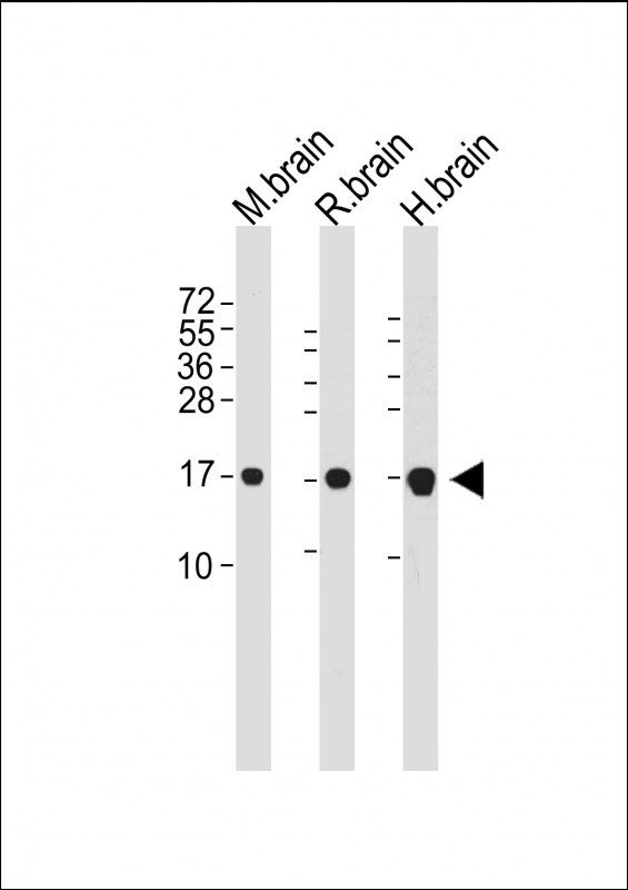 Sample:
Lane 1: Brain (Mouse) Tissue Lysate
Lane 2: Brain (Rat) Tissue Lysate
Lane 3: Brain (Human) Tissue Lysate
Primary:
Anti-LC3 (SLM-51460M) at 1/1000 dilution
Secondary: IRDye800CW Goat Anti-Mouse IgG at 1/20000 dilution
Predicted band size: 13 kD
Observed band size: 17 kD
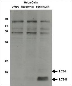 Sample:
Human Hela cell lysates, which were treated with rapamycin or bafilomycin overnight. Data courtesy of Dr. David Rubinsztein, Cambridge Institute for Medical Research.
Primary:
Anti-LC3 (SLM-51460M) at 1/1000 dilution
Secondary: IRDye800CW Goat Anti-Mouse IgG at 1/20000 dilution
Predicted band size: 13 kD
Observed band size: 13 kD
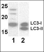 Sample:
Lane 1: Y79 (soluble fraction of cell extract) cell lysate
Lane 2: 293 transfected with human LC3 (whole cell extract) cell lysate
Primary:
Anti-LC3 (SLM-51460M) at 1/1000 dilution
Secondary: IRDye800CW Goat Anti-Mouse IgG at 1/20000 dilution
Predicted band size: 13 kD
Observed band size: 18/16 kD
Cartpieces
Totalgoods,subtotals:¥Checkout
References (0)
No References
Bought notes(bought amounts latest0)
No one bought this product
User Comment(Total0User Comment Num)
- No comment


 +86 571 56623320
 +86 18668110335

