
Mouse Anti-SARS-CoV-2 (2019-nCoV) Spike RBD antibody
SARS-CoV-2 spike protein; 2019-nCOV Spike protein; Spike glycoprotein; Spike protein S1; Surface glycoprotein; Spike protein RBD; SPIKE_SARS2.
View History [Clear]
Details
Product Name SARS-CoV-2 (2019-nCoV) Spike RBD Chinese Name SARS冠状病毒2 Spike RBD单克隆抗体 Alias SARS-CoV-2 spike protein; 2019-nCOV Spike protein; Spike glycoprotein; Spike protein S1; Surface glycoprotein; Spike protein RBD; SPIKE_SARS2. literatures Research Area Bacteria and viruses Immunogen Species Mouse Clonality Monoclonal Clone NO. 2B1 Applications WB=1:500-2000
not yet tested in other applications.
optimal dilutions/concentrations should be determined by the end user.Theoretical molecular weight 140kDa Form Liquid Concentration Lot Dependent immunogen Recombinant SARS-CoV-2 Spike S1 Protein: 14-685/1213 Lsotype Purification affinity purified by Protein A Buffer Solution 0.01M PBS(pH7.4) Storage Shipped at 4℃. Store at -20 °C for one year. Avoid repeated freeze/thaw cycles. Attention This product as supplied is intended for research use only, not for use in human, therapeutic or diagnostic applications. PubMed PubMed Product Detail The spike (S) glycoprotein of coronaviruses contains protrusions that will only bind to certain receptors on the host cell. Known receptors bind S1 are ACE2, angiotensin-converting enzyme 2; DPP4, dipeptidyl peptidase-4; APN, aminopeptidase N; CEACAM, carcinoembryonic antigen-related cell adhesion molecule 1; Sia, sialic acid; O-ac Sia, O-acetylated sialic acid. The spike is essential for both host specificity and viral infectivity. The term 'peplomer' is typically used to refer to a grouping of heterologous proteins on the virus surface that function together. The spike (S) glycoprotein of coronaviruses is known to be essential in the binding of the virus to the host cell at the advent of the infection process. It's been reported that 2019-nCoV can infect the human respiratory epithelial cells through interaction with the human ACE2 receptor. The spike protein is a large type I transmembrane protein containing two subunits, S1 and S2. S1 mainly contains a receptor binding domain (RBD), which is responsible for recognizing the cell surface receptor. S2 contains basic elements needed for the membrane fusion.The S protein plays key parts in the induction of neutralizing-antibody and T-cell responses, as well as protective immunity. The main functions for the Spike protein are summarized as: Mediate receptor binding and membrane fusion; Defines the range of the hosts and specificity of the virus; Main component to bind with the neutralizing antibody; Key target for vaccine design; Can be transmitted between different hosts through gene recombination or mutation of the receptor binding domain (RBD), leading to a higher mortality rate.
Function:
attaches the virion to the cell membrane by interacting with host receptor, initiating the infection (By similarity). Binding to human ACE2 receptor and internalization of the virus into the endosomes of the host cell induces conformational changes in the Spike glycoprotein (PubMed:32142651, PubMed:32075877, PubMed:32155444). Uses also human TMPRSS2 for priming in human lung cells which is an essential step for viral entry (PubMed:32142651). Proteolysis by cathepsin CTSL may unmask the fusion peptide of S2 and activate membranes fusion within endosomes.
Subunit:
Spike glycoprotein:
Homotrimer; each monomer consists of a S1 and a S2 subunit (PubMed:32155444, PubMed:32075877). The resulting peplomers protrude from the virus surface as spikes (By similarity).
Interacts with the accessory proteins 3a and 7a.
Spike protein S1:
Binds to human ACE2.
Subcellular Location:
Virion membrane ; Single-pass type I membrane protein ; Host endoplasmic reticulum-Golgi intermediate compartment membrane ; Single-pass type I membrane protein ; Host cell membrane ; Single-pass type I membrane protein ;
Note: Accumulates in the endoplasmic reticulum-Golgi intermediate compartment, where it participates in virus particle assembly. Colocalizes with S in the host endoplasmic reticulum-Golgi intermediate compartment. Some S oligomers are transported to the host plasma membrane, where they may mediate cell-cell fusion.
Tissue Specificity:
The cytoplasmic Cys-rich domain is palmitoylated. Spike glycoprotein is digested within host endosomes.
Specific enzymatic cleavages in vivo yield mature proteins. The precurssor is processed into S1 and S2 by host cell furin or another cellular protease to yield the mature S1 and S2 proteins (PubMed:32155444). Additionally, a second cleavage leads to the release of a fusion peptide after viral attachment to host cell receptor (By similarity). The presence of a furin polybasic cleavage site sets SARS-CoV-2 S apart from SARS-CoV S that possesses a monobasic S1/S2 cleavage site processed upon entry of target cells (PubMed:32155444).
Highly decorated by heterogeneous N-linked glycans protruding from the trimer surface.
SWISS:
P0DTC2
Gene ID:
43740568
Product Picture 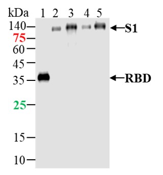 Sample:
Lane 1: SARS-CoV-2 Spike RBD Protein(WT) at 500ng
Lane 2: SARS-CoV-2 Spike S1 Protein(E484Q, L452R, D614G,P681R) at 500ng
Lane 3: SARS-CoV-2 Spike S1 Protein (D80A, D215G, del241/243, K417N, E484K, N501Y, D614G) at 500ng
Lane 4: SARS-CoV-2 Spike S1 Protein (L18F, T20N, P26S, D138Y, R190S,K417T,E484K,N501Y,D614G,H655Y) at 500ng
Lane 5: SARS-CoV-2 Spike S1 Protein (WT) at 500ng
Primary: Anti- SARS-CoV-2 Spike S1 Protein at 1/1000 dilution
Secondary: IRDye800CW Goat Anti-Mouse IgG at 1/20000 dilution
Predicted band size: 27kDa /77.2 kDa
Observed band size: 35kDa/114 kDa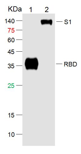 Sample:
Lane 1: SARS-CoV-2 Spike RBD Protein (His-Avi,HEK293) at 500ng
Lane 2: SARS-CoV-2 S1 Protein (His-Avi,HEK293) at 500ng
Primary: Mouse Anti-SARS-CoV-2 Spike RBD Protein Antibody at 1/1000 dilution
Secondary: IRDye800CW Goat Anti-Mouse IgG at 1/20000 dilution
Predicted band size: 27/78kD
Observed band size: kD
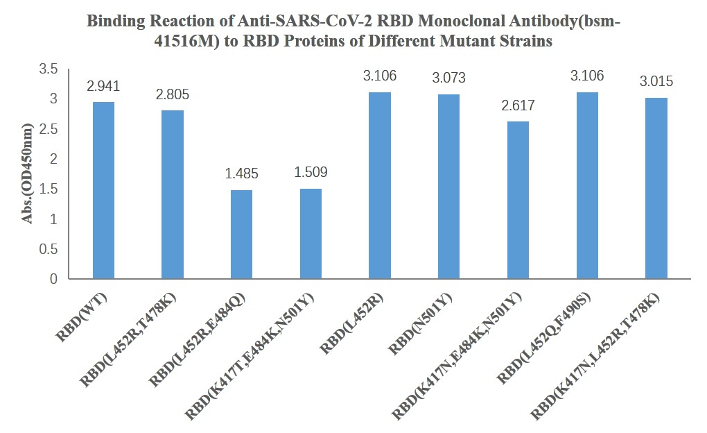 Direct ELISA was used to detect the binding ability of anti-RBD monoclonal antibody to RBD domain proteins of different SARS-CoV-2 Mutant Strains. Immobilized SARS-CoV-2 RBD proteins, at 2μg/ml (100ul/Well) can bind Anti-RBD monoclonal antibody-HRP at 10ug/ml (100ul/Well).
Cartpieces
Totalgoods,subtotals:¥Checkout
References (0)
No References
Bought notes(bought amounts latest0)
No one bought this product
User Comment(Total0User Comment Num)
- No comment


 +86 571 56623320
 +86 18668110335

