
Mouse Anti-LC3A antibody
ATG8E; Autophagy-related protein LC3 A; Autophagy-related ubiquitin-like modifier LC3 A; LC3; MAP1 light chain 3 like protein 1; MAP1 light chain 3-like protein 1; MAP1A/1B light chain 3 A; MAP1A/MAP1B LC3 A; MAP1A/MAP1B light chain 3 A; MAP1ALC3; MAP1BLC
View History [Clear]
Details
Product Name LC3A Chinese Name 自噬微管相关蛋白轻链3单克隆抗体 Alias ATG8E; Autophagy-related protein LC3 A; Autophagy-related ubiquitin-like modifier LC3 A; LC3; MAP1 light chain 3 like protein 1; MAP1 light chain 3-like protein 1; MAP1A/1B light chain 3 A; MAP1A/MAP1B LC3 A; MAP1A/MAP1B light chain 3 A; MAP1ALC3; MAP1BLC3; Map1lc3a; Microtubule associated proteins 1A/1B light chain 3; Microtubule-associated protein 1 light chain 3 alpha; Microtubule-associated proteins 1A and 1B, light chain 3; Microtubule-associated proteins 1A/1B light chain 3A; MLP3A_HUMAN. literatures Research Area Tumour Cell biology immunology Apoptosis Autophagy Immunogen Species Mouse Clonality Monoclonal Clone NO. 7G10 React Species Human, Mouse, Rat, Applications WB=1:500-1000 IHC-P=1:100-500 IHC-F=1:100-500 ICC=1:100 IF=1:200-800 (Paraffin sections need antigen repair)
not yet tested in other applications.
optimal dilutions/concentrations should be determined by the end user.Theoretical molecular weight 14/16kDa Cellular localization cytoplasmic The cell membrane Form Liquid Concentration 1mg/ml immunogen KLH conjugated synthetic peptide derived from human LC3A: 1-100/121 Lsotype IgG Purification affinity purified by Protein G Buffer Solution 0.01M TBS(pH7.4) with 1% BSA, 0.03% Proclin300 and 50% Glycerol. Storage Shipped at 4℃. Store at -20 °C for one year. Avoid repeated freeze/thaw cycles. Attention This product as supplied is intended for research use only, not for use in human, therapeutic or diagnostic applications. PubMed PubMed Product Detail Microtubule-associated MAPILC3A constitutes nearly half of the mass of all the microtubule associated proteins that copurify with brain microtubules. MAP1LC3A is one of three human orthologs of the rat Map1LC3, (named MAP1LC3A, MAP1LC3B, and MAP1LC3C). The three isoforms of human MAP1LC3 exhibit distinct expression patterns in different human tissues and also differ in their post-translation modifications. MAP1LC3A and MAP1LC3C are produced by the proteolytic cleavage after the conserved C-terminal Gly residue; MAP1LC3B does not undergo C-terminal cleavage and exists in a single modified form.
Function:
Probably involved in formation of autophagosomal vacuoles (autophagosomes).
Subunit:
3 different light chains, LC1, LC2 and LC3, can associate with MAP1A and MAP1B proteins. Interacts with SQSTM1. Interacts with TP53INP1 and TP53INP2.
Subcellular Location:
Cytoplasm, cytoskeleton. Endomembrane system; Lipid-anchor. Cytoplasmic vesicle, autophagosome membrane; Lipid-anchor. Cytoplasmic vesicle, autophagosome. Note=LC3-II binds to the autophagic membranes.
Tissue Specificity:
Most abundant in heart, brain, liver, skeletal muscle and testis but absent in thymus and peripheral blood leukocytes.
Post-translational modifications:
The precursor molecule is cleaved by APG4B/ATG4B to form the cytosolic form, LC3-I. This is activated by APG7L/ATG7, transferred to ATG3 and conjugated to phospholipid to form the membrane-bound form, LC3-II.
Similarity:
Detects a band of approximately 16 kDa (predicted molecular weight: 14 kDa).
SWISS:
Q9H492
Gene ID:
84557
Database links:Entrez Gene: 84557 Human
Entrez Gene: 66734 Mouse
Omim: 601242 Human
SwissProt: Q9H492 Human
SwissProt: Q91VR7 Mouse
Unigene: 632273 Human
Unigene: 196239 Mouse
Unigene: 3135 Rat
Product Picture 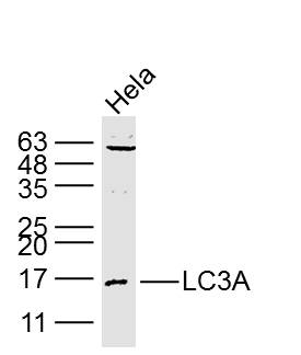 Sample:
Hela Cell (Human) Lysate at 40 ug
Primary: Anti- LC3A (SLM-33309M) at 1/1000 dilution
Secondary: IRDye800CW Goat Anti-Mouse IgG at 1/20000 dilution
Predicted band size: 14/16 kD
Observed band size: 16 kD
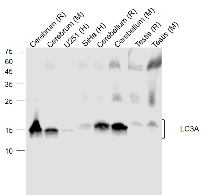 Sample:
Lane 1: Cerebrum (Rat) Lysate at 40 ug
Lane 2: Cerebrum (Mouse) Lysate at 40 ug
Lane 3: U251 (Human) Cell Lysate at 30 ug
Lane 4: SiHa (Human) Cell Lysate at 30 ug
Lane 5: Cerebellum (Rat) Lysate at 40 ug
Lane 6: Cerebellum (Mouse) Lysate at 40 ug
Lane 7: Testis (Rat) Lysate at 40 ug
Lane 8: Testis (Mouse) Lysate at 40 ug
Primary: Anti-LC3A (SLM-33309M) at 1/1000 dilution
Secondary: IRDye800CW Goat Anti-Mouse IgG at 1/20000 dilution
Predicted band size: 17 kD
Observed band size: 15 kD
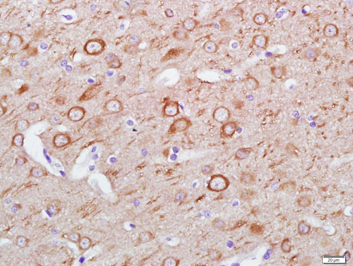 Paraformaldehyde-fixed, paraffin embedded (Rat brain); Antigen retrieval by boiling in sodium citrate buffer (pH6.0) for 15min; Block endogenous peroxidase by 3% hydrogen peroxide for 20 minutes; Blocking buffer (normal goat serum) at 37°C for 30min; Antibody incubation with (LC3A) Monoclonal Antibody, Unconjugated (SLM-33309M) at 1:400 overnight at 4°C, followed by a conjugated secondary (sp-0023) for 20 minutes and DAB staining.
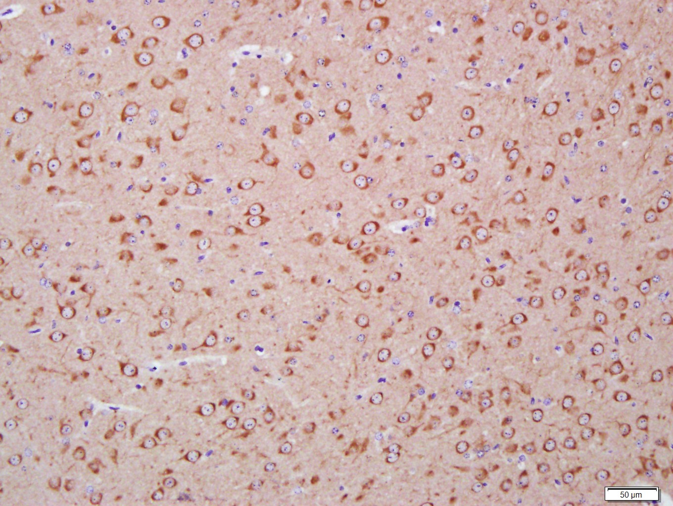 Paraformaldehyde-fixed, paraffin embedded (Mouse brain); Antigen retrieval by boiling in sodium citrate buffer (pH6.0) for 15min; Block endogenous peroxidase by 3% hydrogen peroxide for 20 minutes; Blocking buffer (normal goat serum) at 37°C for 30min; Antibody incubation with (LC3A) Monoclonal Antibody, Unconjugated (SLM-33309M) at 1:400 overnight at 4°C, followed by a conjugated secondary (sp-0023) for 20 minutes and DAB staining.
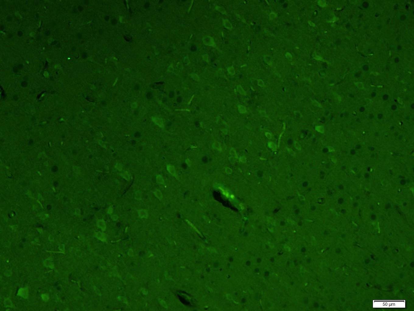 Paraformaldehyde-fixed, paraffin embedded (Rat brain); Antigen retrieval by boiling in sodium citrate buffer (pH6.0) for 15min; Block endogenous peroxidase by 3% hydrogen peroxide for 20 minutes; Blocking buffer (normal goat serum) at 37°C for 30min; Antibody incubation with (LC3A) Monoclonal Antibody, Unconjugated (SLM-33309M) at 1:400 overnight at 4°C, followed by a conjugated Goat Anti-Mouse IgG antibody (SL0296G-FITC) for 90 minutes, and DAPI for nuclei staining.
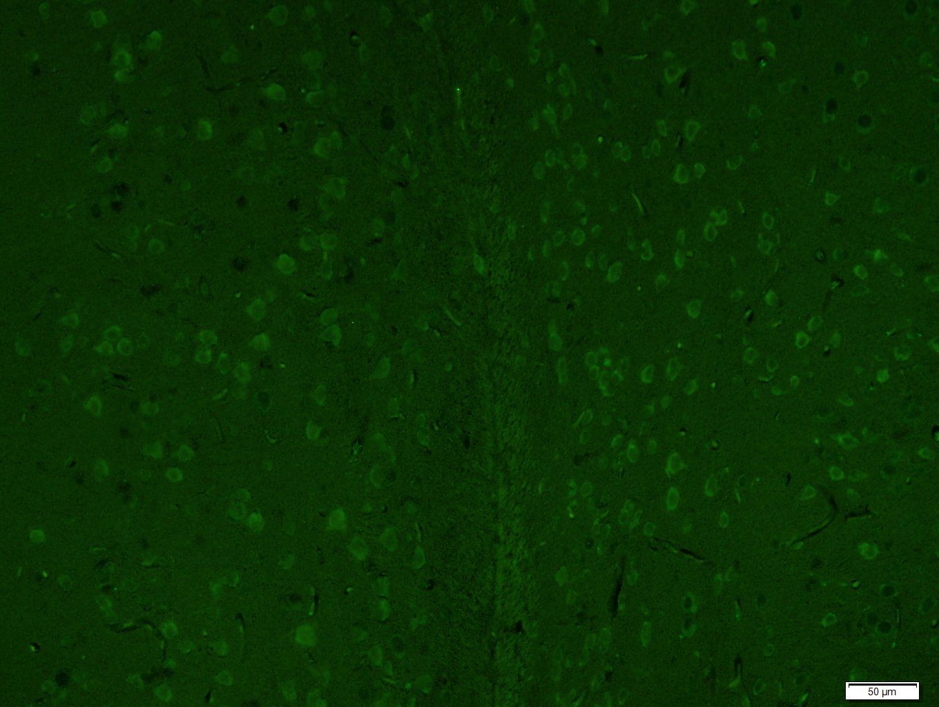 Paraformaldehyde-fixed, paraffin embedded (Mouse brain); Antigen retrieval by boiling in sodium citrate buffer (pH6.0) for 15min; Block endogenous peroxidase by 3% hydrogen peroxide for 20 minutes; Blocking buffer (normal goat serum) at 37°C for 30min; Antibody incubation with (LC3A) Monoclonal Antibody, Unconjugated (SLM-33309M) at 1:400 overnight at 4°C, followed by a conjugated Goat Anti-Mouse IgG antibody (SL0296G-FITC) for 90 minutes, and DAPI for nuclei staining.
Cartpieces
Totalgoods,subtotals:¥Checkout
References (0)
No References
Bought notes(bought amounts latest0)
No one bought this product
User Comment(Total0User Comment Num)
- No comment


 +86 571 56623320
 +86 18668110335

