
Mouse Anti-STAT3 antibody
1110034C02Rik; Acute Phase Response Factor; APRF; AW109958; DNA binding protein APRF; FLJ20882; HIES; MGC128731; MGC16063; MGC93551; Signal transducer and activator of transcription 3 (acute-phase response factor); Signal transducer and activator of trans
View History [Clear]
Details
Product Name [KO validated anti] STAT3 Chinese Name Signal transduction和转录激活因子3单克隆抗体 Alias 1110034C02Rik; Acute Phase Response Factor; APRF; AW109958; DNA binding protein APRF; FLJ20882; HIES; MGC128731; MGC16063; MGC93551; Signal transducer and activator of transcription 3 (acute-phase response factor); Signal transducer and activator of transcription 3; Signal Transductor and Activator of Transcription 3; STAT 3; Transcription factor; STAT3_HUMAN. literatures Research Area Tumour Cell biology Signal transduction Apoptosis transcriptional regulatory factor Immunogen Species Mouse Clonality Monoclonal Clone NO. 3F5 React Species Human, Mouse, Rat, Applications WB=1:500-2000 IHC-P=1:100-500 IHC-F=1:100-500 ICC=1:100-500 IF=1:100-500 (Paraffin sections need antigen repair)
not yet tested in other applications.
optimal dilutions/concentrations should be determined by the end user.Theoretical molecular weight 88kDa Cellular localization The nucleus cytoplasmic Form Liquid Concentration 1mg/ml immunogen Recombinant human STAT3 Protein: 9-265/770 Lsotype IgG Purification affinity purified by Protein G Buffer Solution 0.01M TBS(pH7.4) with 1% BSA, 0.03% Proclin300 and 50% Glycerol. Storage Shipped at 4℃. Store at -20 °C for one year. Avoid repeated freeze/thaw cycles. Attention This product as supplied is intended for research use only, not for use in human, therapeutic or diagnostic applications. PubMed PubMed Product Detail The protein encoded by this gene is a member of the STAT protein family. In response to cytokines and growth factors, STAT family members are phosphorylated by the receptor associated kinases, and then form homo- or heterodimers that translocate to the cell nucleus where they act as transcription activators. This protein is activated through phosphorylation in response to various cytokines and growth factors including IFNs, EGF, IL5, IL6, HGF, LIF and BMP2. This protein mediates the expression of a variety of genes in response to cell stimuli, and thus plays a key role in many cellular processes such as cell growth and apoptosis. The small GTPase Rac1 has been shown to bind and regulate the activity of this protein. PIAS3 protein is a specific inhibitor of this protein. Three alternatively spliced transcript variants encoding distinct isoforms have been described. Gene Symbol: APRF; FLJ20882; HIES; MGC16063.
Subunit:
Forms a homodimer or a heterodimer with a related family member (at least STAT1). Interacts with IL31RA, NCOA1, PELP1, SIPAR, SOCS7, STATIP1 and TMF1. Interacts with HCV core protein. Interacts with IL23R in presence of IL23. Interacts (via SH2 domain) with NLK. Interacts with ARL2BP; the interaction is enhanced by LIF and JAK1 expression (By similarity). Interacts with KPNA4 and KPNA5; KPNA4 may be the primary mediator of nuclear import (By similarity). Interacts with CAV2; the interaction is increased on insulin-induced tyrosine phosphorylation of CAV2 and leads to STAT3 activation (By similarity). Interacts with ARL2BP; interaction is enhanced with ARL2. Interacts with NEK6 (By similarity). Binds to CDK9 when activated and nuclear. Interacts with BMX. Interacts with ZIPK/DAPK3. Interacts with PIAS3; the interaction occurs on stimulation by IL6, CNTF or OSM and inhibits the DNA binding activity of STAT3. In prostate cancer cells, interacts with STAT3 and promotes DNA binding activity of STAT3. Interacts with STMN3, antagonizing its microtubule-destabilizing activity.
Subcellular Location:
Cytoplasm. Nucleus. Note=Shuttles between the nucleus and the cytoplasm. Translocated into the nucleus upon tyrosine phosphorylation and dimerization, in response to signaling by activated FGFR1, FGFR2, FGFR3 or FGFR4. Constitutive nuclear presence is independent of tyrosine phosphorylation. Predominantly present in the cytoplasm without stimuli. Upon leukemia inhibitory factor (LIF) stimulation, accumulates in the nucleus. The complex composed of BART and ARL2 plays an important role in the nuclear translocation and retention of STAT3. Identified in a complex with LYN and PAG1.
Tissue Specificity:
Heart, brain, placenta, lung, liver, skeletal muscle, kidney and pancreas.
Similarity:
Belongs to the transcription factor STAT family.
Contains 1 SH2 domain.
SWISS:
P40763
Gene ID:
6774
Database links:Entrez Gene: 6774 Human
Entrez Gene: 20848 Mouse
Omim: 102582 Human
SwissProt: P40763 Human
SwissProt: P42227 Mouse
Unigene: 463059 Human
Stat3在Cell differentiation、增殖、凋亡等方面有着重要生理作用。有学者认为:STAT-3对Tumour有一定的抑制作用,由于其与Tumour的关系较为密切,越来越受到人们的重视。Product Picture 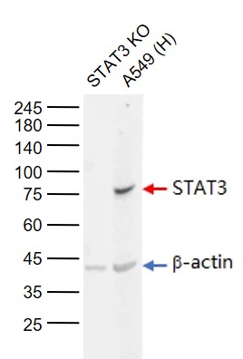 Sample:
Lane 1: STAT3 knockout (KO) A549 Cell Lysate
Lane 2: Human A549 Cell (Control) Lysate
Primary: Anti-STAT3 (SLM-33218M) at 1/1000 dilution
Secondary: IRDye800CW Goat Anti-Mouse IgG at 1/20000 dilution
Predicted band size: 88 kD
Observed band size: 88 kD
 Sample:
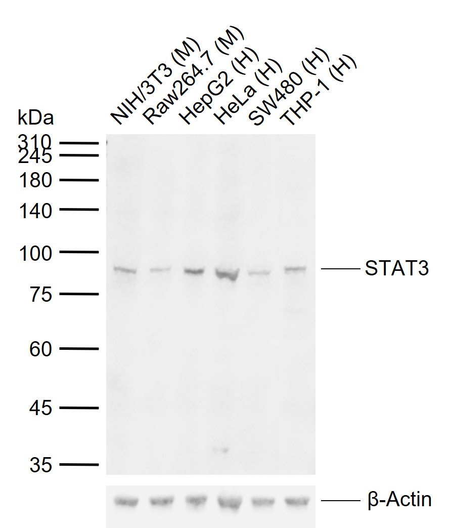
Lane 1: Mouse NIH/3T3 cell lysates
Lane 2: Mouse Raw264.7 cell lysates
Lane 3: Human HepG2 cell lysates
Lane 4: Human HeLa cell lysates
Lane 5: Human SW480 cell lysates
Lane 6: Human THP-1 cell lysates
Primary: Anti-STAT3 (SLM-33218M) at 1/500 dilution
Secondary: IRDye800CW Goat Anti-Mouse IgG at 1/20000 dilution
Predicted band size: 88 kDa
Observed band size: 88 kDa
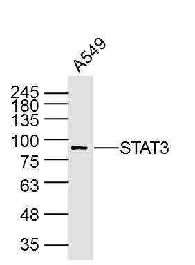 Sample: A549 Cell (Human) Lysate at 40 ug
Primary: Anti-STAT3 (SLM-33218M) at 1/1000 dilution
Secondary: IRDye800CW Goat Anti-Mouse IgG at 1/20000 dilution
Predicted band size: 88 kD
Observed band size: 88 kD
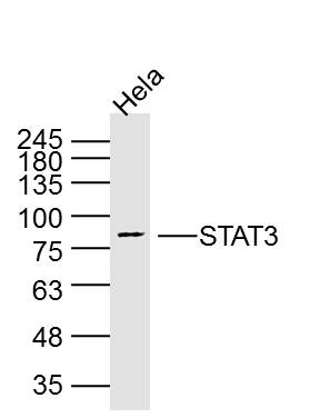 Sample: Hela Cell (Human) Lysate at 40 ug
Primary: Anti-STAT3 (SLM-33218M) at 1/1000 dilution
Secondary: IRDye800CW Goat Anti-Mouse IgG at 1/20000 dilution
Predicted band size: 88 kD
Observed band size: 88 kD
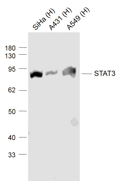 Sample:
Lane 1: SiHa (Human) Cell Lysate at 30 ug
Lane 2: A431 (Human) Cell Lysate at 30 ug
Lane 3: A549 (Human) Cell Lysate at 30 ug
Primary: Anti-STAT3 (SLM-33218M) at 1/1000 dilution
Secondary: IRDye800CW Goat Anti-Mouse IgG at 1/20000 dilution
Predicted band size: 88 kD
Observed band size: 88 kD
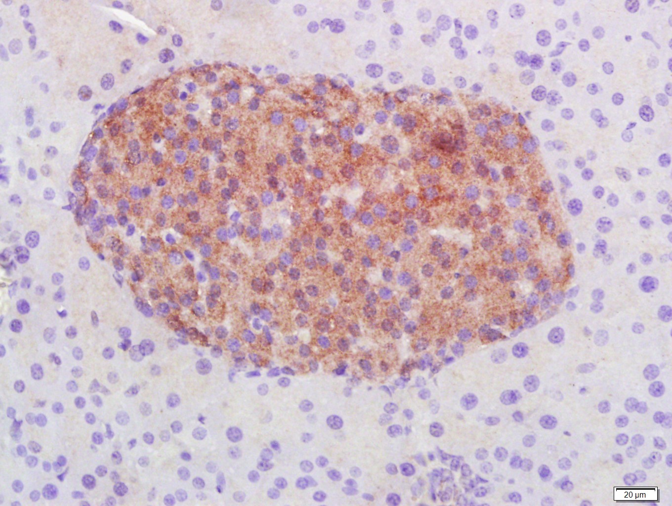 Paraformaldehyde-fixed, paraffin embedded (Mouse pancreas); Antigen retrieval by boiling in sodium citrate buffer (pH6.0) for 15min; Block endogenous peroxidase by 3% hydrogen peroxide for 20 minutes; Blocking buffer (normal goat serum) at 37°C for 30min; Antibody incubation with (STAT3) Monoclonal Antibody, Unconjugated (SLM-33218M) at 1:400 overnight at 4°C, followed by a conjugated secondary (sp-0023) for 20 minutes and DAB staining.
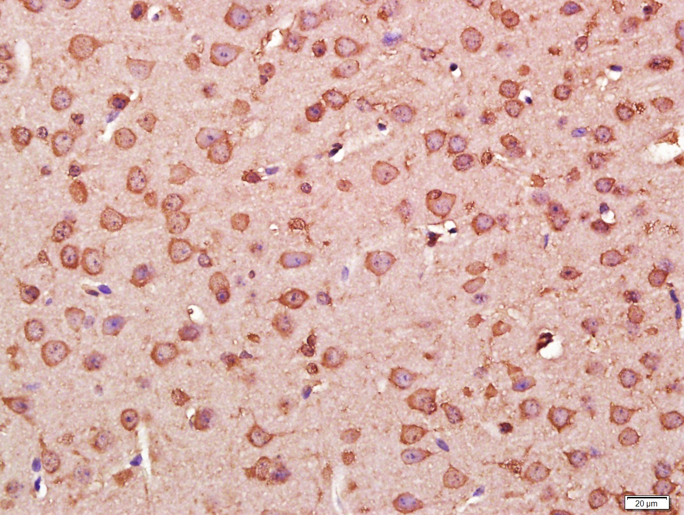 Paraformaldehyde-fixed, paraffin embedded (Mouse brain); Antigen retrieval by boiling in sodium citrate buffer (pH6.0) for 15min; Block endogenous peroxidase by 3% hydrogen peroxide for 20 minutes; Blocking buffer (normal goat serum) at 37°C for 30min; Antibody incubation with (STAT3) Monoclonal Antibody, Unconjugated (SLM-33218M) at 1:400 overnight at 4°C, followed by a conjugated secondary (sp-0023) for 20 minutes and DAB staining.
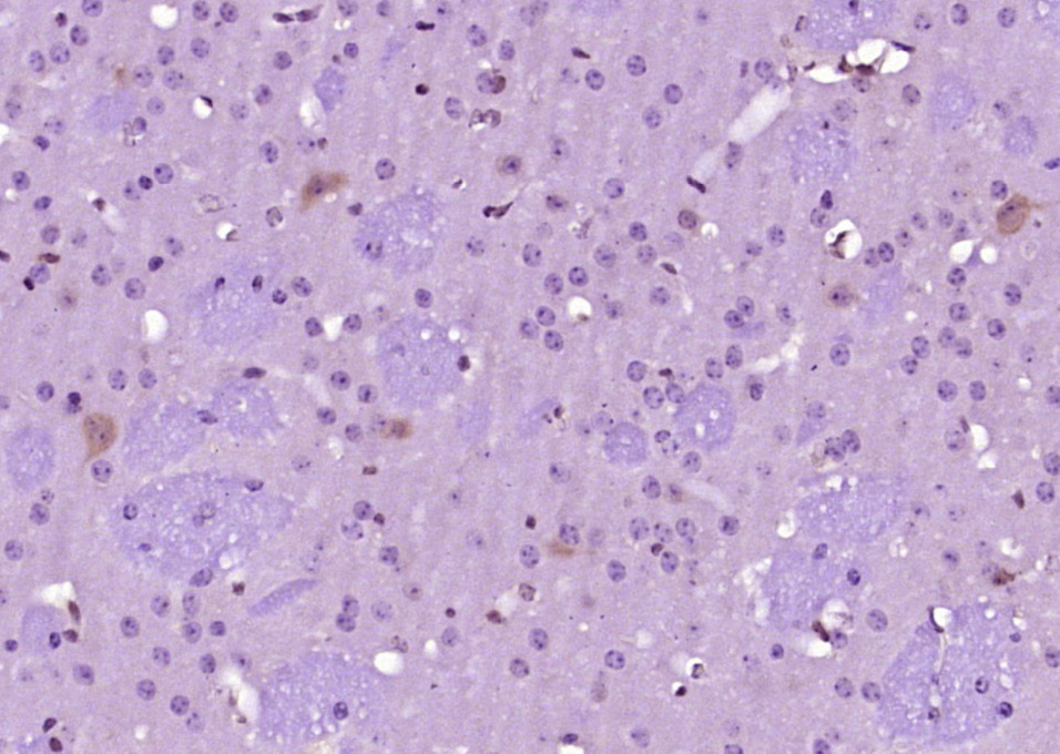 Paraformaldehyde-fixed, paraffin embedded (mouse brain tissue); Antigen retrieval by boiling in sodium citrate buffer (pH6.0) for 15min; Block endogenous peroxidase by 3% hydrogen peroxide for 20 minutes; Blocking buffer (normal goat serum) at 37°C for 30min; Antibody incubation with (STAT3) Monoclonal Antibody, Unconjugated (ascites of SLM-33218M) at 1:2000 overnight at 4°C, followed by a conjugated secondary (sp-0024) for 20 minutes and DAB staining.
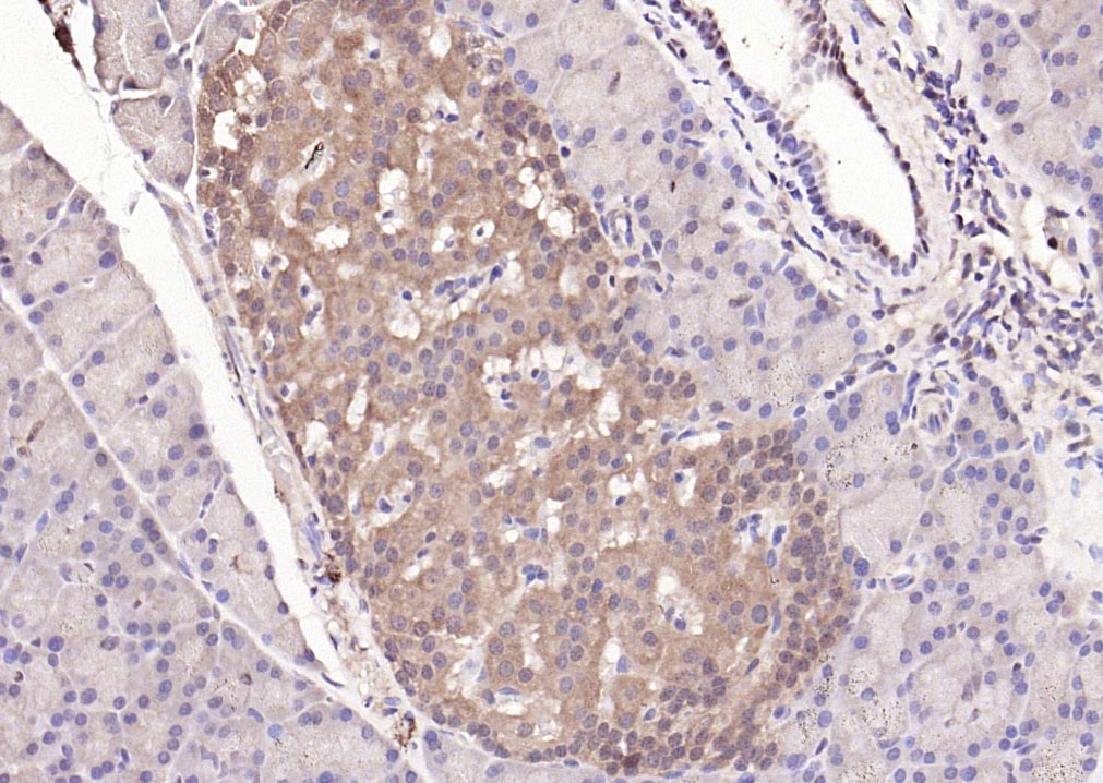 Paraformaldehyde-fixed, paraffin embedded (rat pancreas); Antigen retrieval by boiling in sodium citrate buffer (pH6.0) for 15min; Block endogenous peroxidase by 3% hydrogen peroxide for 20 minutes; Blocking buffer (normal goat serum) at 37°C for 30min; Antibody incubation with (STAT3) Polyclonal Antibody, Unconjugated (SLM-33218M) at 1:200 overnight at 4°C, followed by operating according to SP Kit(Mouse)(sp-0024) instructionsand DAB staining.
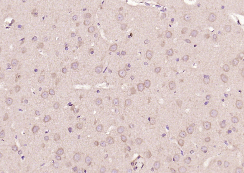 Paraformaldehyde-fixed, paraffin embedded (rat brain); Antigen retrieval by boiling in sodium citrate buffer (pH6.0) for 15min; Block endogenous peroxidase by 3% hydrogen peroxide for 20 minutes; Blocking buffer (normal goat serum) at 37°C for 30min; Antibody incubation with (STAT3) Polyclonal Antibody, Unconjugated (SLM-33218M) at 1:200 overnight at 4°C, followed by operating according to SP Kit(Mouse)(sp-0024) instructionsand DAB staining.
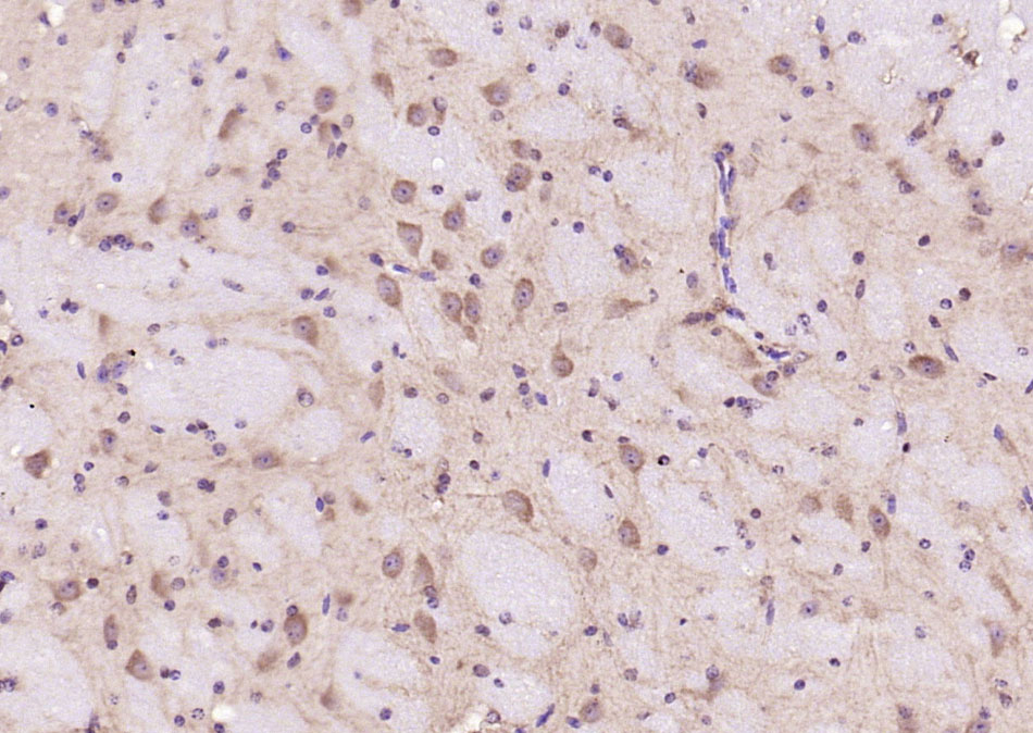 Paraformaldehyde-fixed, paraffin embedded (mouse brain); Antigen retrieval by boiling in sodium citrate buffer (pH6.0) for 15min; Block endogenous peroxidase by 3% hydrogen peroxide for 20 minutes; Blocking buffer (normal goat serum) at 37°C for 30min; Antibody incubation with (STAT3) Polyclonal Antibody, Unconjugated (SLM-33218M) at 1:200 overnight at 4°C, followed by operating according to SP Kit(Mouse)(sp-0024) instructionsand DAB staining.
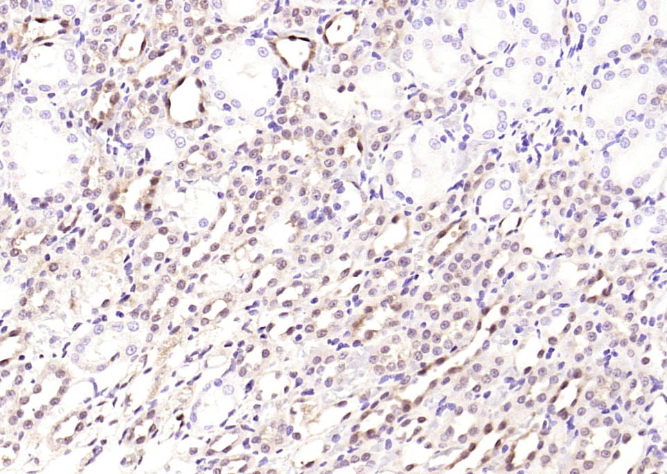 Paraformaldehyde-fixed, paraffin embedded (rat kidney); Antigen retrieval by boiling in sodium citrate buffer (pH6.0) for 15min; Block endogenous peroxidase by 3% hydrogen peroxide for 20 minutes; Blocking buffer (normal goat serum) at 37°C for 30min; Antibody incubation with (STAT3) Monoclonal Antibody, Unconjugated (ascites of SLM-33218M 3F5) at 1:2000 overnight at 4°C, followed by operating according to SP Kit(Mouse) (sp-0024) instructions and DAB staining.
Cartpieces
Totalgoods,subtotals:¥Checkout
References (0)
No References
Bought notes(bought amounts latest0)
No one bought this product
User Comment(Total0User Comment Num)
- No comment


 +86 571 56623320
 +86 18668110335

