
Mouse Anti-CK18 antibody
K1C18_HUMAN; Keratin, type I cytoskeletal 18; KRT18; CYK18; Cell proliferation-inducing gene 46 protein; Cytokeratin-18 (CK-18); Keratin-18 (K18); PIG46; Keratin 18;
View History [Clear]
Details
Product Name CK18 Chinese Name 细胞角蛋白18单克隆抗体 Alias K1C18_HUMAN; Keratin, type I cytoskeletal 18; KRT18; CYK18; Cell proliferation-inducing gene 46 protein; Cytokeratin-18 (CK-18); Keratin-18 (K18); PIG46; Keratin 18; Research Area Tumour Cell biology immunology Signal transduction Immunogen Species Mouse Clonality Monoclonal Clone NO. 4H7 React Species Human, (predicted: Mouse, ) Applications WB=1:500-1000 ICC=1:100
not yet tested in other applications.
optimal dilutions/concentrations should be determined by the end user.Theoretical molecular weight 48kDa Cellular localization cytoplasmic The cell membrane Form Liquid Concentration 1mg/ml immunogen KLH conjugated synthetic peptide derived from human CK18 Lsotype IgG Purification affinity purified by Protein G Buffer Solution 0.01M TBS(pH7.4) with 1% BSA, 0.03% Proclin300 and 50% Glycerol. Storage Shipped at 4℃. Store at -20 °C for one year. Avoid repeated freeze/thaw cycles. Attention This product as supplied is intended for research use only, not for use in human, therapeutic or diagnostic applications. PubMed PubMed Product Detail KRT18 encodes the type I intermediate filament chain keratin 18. Keratin 18, together with its filament partner keratin 8, are perhaps the most commonly found members of the intermediate filament gene family. They are expressed in single layer epithelial tissues of the body. Mutations in this gene have been linked to cryptogenic cirrhosis. Two transcript variants encoding the same protein have been found for this gene. [provided by RefSeq, Jul 2008]
Function:
When phosphorylated, plays a role in filament reorganization. Involved in the delivery of mutated CFTR to the plasma membrane. Involved in the uptake of thrombin-antithrombin complexes by hepatic cells (By similarity). Together with KRT8, is involved in interleukin-6 (IL-6)-mediated barrier protection.
Post-translational modifications:
Phosphorylation increases by IL-6.
Proteolytically cleaved by caspases during epithelial cell apoptosis. Cleavage occurs at Asp-231 by either caspase-3, caspas-6 or caspase-7.
O-glycosylated at multiple sites; glycans consist of single N-acetylglucosamine residue.
Similarity:
Belongs to the intermediate filament family.
SWISS:
P05783
Gene ID:
3875
Database links:
Entrez Gene: 3875 Human
Entrez Gene: 16668 Mouse
SwissProt: P05783 Human
SwissProt: P05784 Mouse
结构蛋白(Structural Proteins)
常用于Tumour细胞的分化、增殖及转移方面的研究。Product Picture 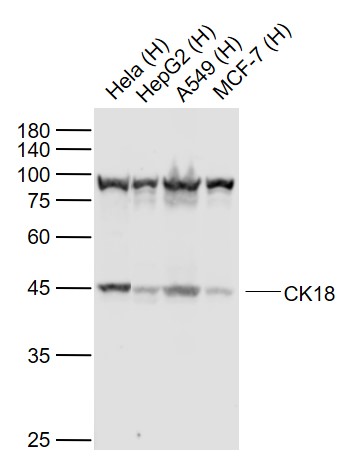 Sample:
Lane 1: Hela (Human) cell Lysate at 30 ug
Lane 2: HepG2 (Human) cell Lysate at 30 ug
Lane 3: A549 (Human) cell Lysate at 30 ug
Lane 4: MCF-7 (Human) cell Lysate at 30 ug
Primary: Anti-CK18 (SLM-33102M ) at 1/1000 dilution
Secondary: IRDye800CW Goat Anti-Mouse IgG at 1/20000 dilution
Predicted band size: 48 kD
Observed band size: 45 kD
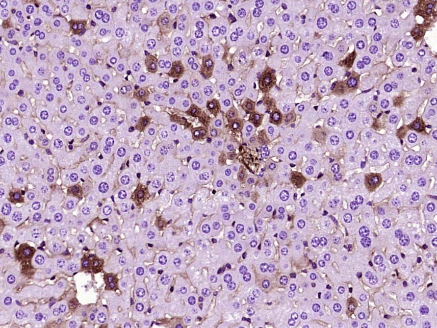 Paraformaldehyde-fixed, paraffin embedded (Mouse liver); Antigen retrieval by boiling in sodium citrate buffer (pH6.0) for 15min; Block endogenous peroxidase by 3% hydrogen peroxide for 20 minutes; Blocking buffer (normal goat serum) at 37°C for 30min; Antibody incubation with (CK18) Monoclonal Antibody, Unconjugated (ascites of SLM-33102M 4H7) at 1:2000 overnight at 4°C, followed by a conjugated secondary (sp-0024) for 20 minutes and DAB staining.
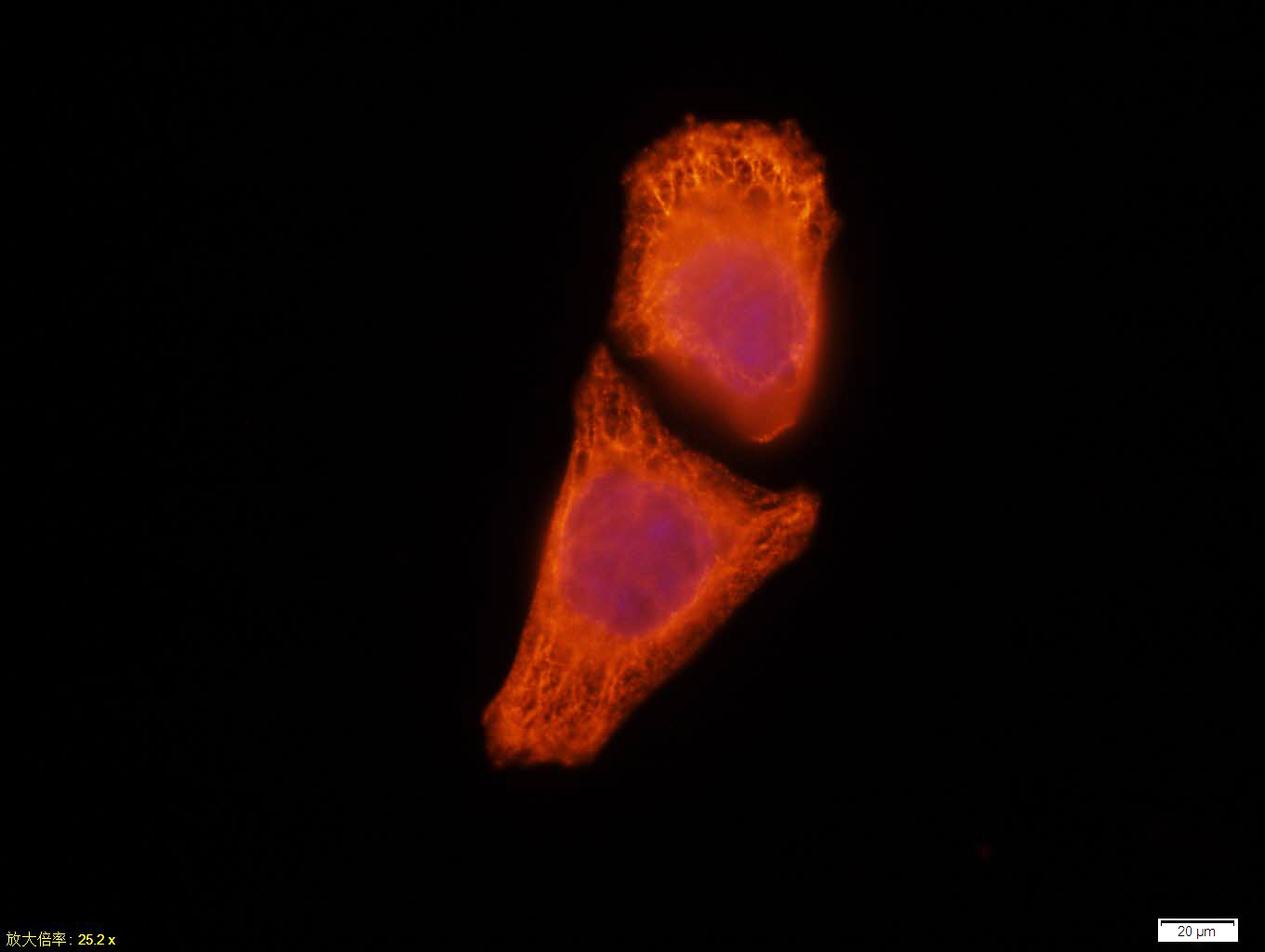 Tissue/cell:Hela cell; 4% Paraformaldehyde-fixed; Triton X-100 at room temperature for 20 min; Blocking buffer (normal goat serum, C-0005) at 37°C for 20 min; Antibody incubation with (CK18) monoclonal Antibody, Unconjugated (SLM-33102M) 1:100, 90 minutes at 37°C; followed by a CY3 conjugated Goat Anti-Mouse IgG antibody at 37°C for 90 minutes, DAPI (blue, C02-04002) was used to stain the cell nuclei.
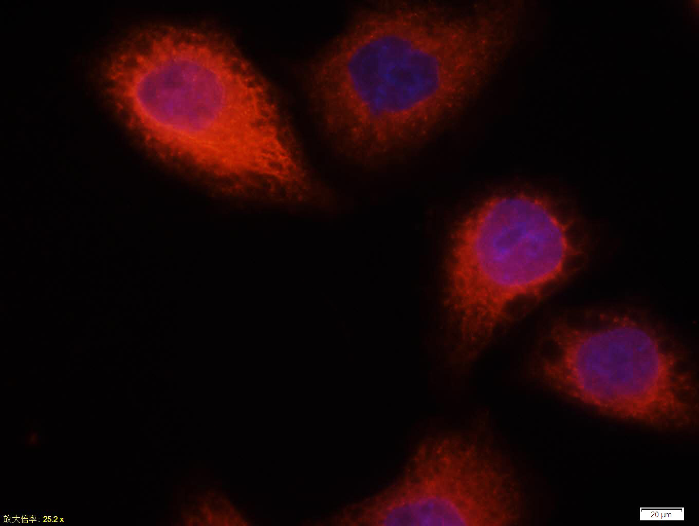 Tissue/cell:Hela cell; 4% Paraformaldehyde-fixed; Triton X-100 at room temperature for 20 min; Blocking buffer (normal goat serum, C-0005) at 37°C for 20 min; Antibody incubation with (CK18) monoclonal Antibody, Unconjugated (SLM-33102M) 1:100, 90 minutes at 37°C; followed by a CY3 conjugated Goat Anti-Mouse IgG antibody at 37°C for 90 minutes, DAPI (blue, C02-04002) was used to stain the cell nuclei.
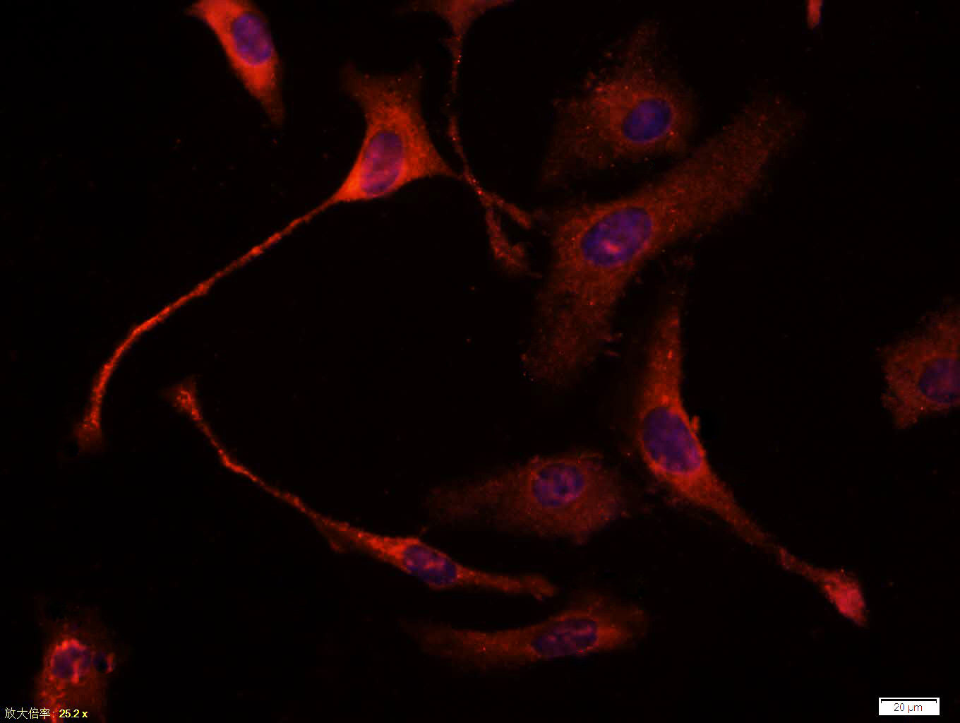 Tissue/cell:Hela cell; 4% Paraformaldehyde-fixed; Triton X-100 at room temperature for 20 min; Blocking buffer (normal goat serum, C-0005) at 37°C for 20 min; Antibody incubation with (CK18) monoclonal Antibody, Unconjugated (SLM-33102M) 1:100, 90 minutes at 37°C; followed by a CY3 conjugated Goat Anti-Mouse IgG antibody at 37°C for 90 minutes, DAPI (blue, C02-04002) was used to stain the cell nuclei.
Cartpieces
Totalgoods,subtotals:¥Checkout
Bought notes(bought amounts latest0)
No one bought this product
User Comment(Total0User Comment Num)
- No comment


 +86 571 56623320
 +86 18668110335

