
Mouse Anti-Ki-67 antibody
Antigen identified by monoclonal antibody Ki 67; Antigen KI67; KIA; Ki67; MKI67; Proliferation related Ki 67 antigen; Antigen KI-67; KI67_HUMAN.
View History [Clear]
Details
Product Name Ki-67 Chinese Name Ki67蛋白单克隆抗体 Alias Antigen identified by monoclonal antibody Ki 67; Antigen KI67; KIA; Ki67; MKI67; Proliferation related Ki 67 antigen; Antigen KI-67; KI67_HUMAN. literatures Research Area Tumour Cell biology Neurobiology Cyclin Cell differentiation Cell type markers Epigenetics Immunogen Species Mouse Clonality Monoclonal Clone NO. 6B9 React Species Human, Rat, (predicted: Mouse, ) Applications IHC-P=1:100-500 IHC-F=1:100-500 Flow-Cyt=1ug/Test IF=1:100-500 (Paraffin sections need antigen repair)
not yet tested in other applications.
optimal dilutions/concentrations should be determined by the end user.Theoretical molecular weight 358kDa Cellular localization The nucleus Form Liquid Concentration 1mg/ml immunogen KLH conjugated synthetic peptide derived from human Ki-67 Lsotype IgG Purification affinity purified by Protein G Buffer Solution 0.01M TBS(pH7.4) with 1% BSA, 0.03% Proclin300 and 50% Glycerol. Storage Shipped at 4℃. Store at -20 °C for one year. Avoid repeated freeze/thaw cycles. Attention This product as supplied is intended for research use only, not for use in human, therapeutic or diagnostic applications. PubMed PubMed Product Detail Ki67 antigen is the prototypic cell cycle related nuclear protein, expressed by proliferating cells in all phases of the active cell cycle (G1, S, G2 and M phase). It is absent in resting (G0) cells. Ki67 antibodies are useful in establishing the cell growing fraction in neoplasms (immunohistochemically quantified by determining the number of Ki67 positive cells among the total number of resting cells = Ki67 index). In neoplastic tissues the prognostic value is comparable to the tritiated thymidine labelling index. The correlation between low Ki67 index and histologically low grade tumours is strong. Ki67 is routinely used as a neuronal marker of cell cycling and proliferation.
Function:
Thought to be required for maintaining cell proliferation.
Subcellular Location:
Nucleus. Chromosome. Predominantly localized in the G1 phase in the perinucleolar region, in the later phases it is also detected throughout the nuclear interior, being predominantly localized in the nuclear matrix. In mitosis, it is present on all chromosomes.
Similarity:
Contains 1 FHA domain.
SWISS:
P46013
Gene ID:
4288
Database links:
Entrez Gene: 4288 HumanEntrez Gene: 17345 Mouse
Omim: 176741 Human
SwissProt: P46013 Human
SwissProt: Q91VE6 Mouse
Unigene: 689823 Human
Unigene: 80976 Human
Unigene: 4078 Mouse
Unigene: 233802 Rat
细胞增殖Maker(Proliferation Marker)
Ki67与PCNA一样,为细胞增殖的一种标记,在Apoptosis中S、G2 、M期均有表达,G0期缺如。 Ki-67增殖指数高低与许多Tumour的分化程度、浸润、转移以及预后密切相关,因此被广泛作为各种恶性Tumour的必检项目之一。Product Picture 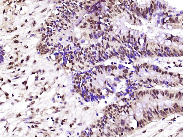 Paraformaldehyde-fixed, paraffin embedded (Human colon cancer); Antigen retrieval by boiling in sodium citrate buffer (pH6.0) for 15min; Block endogenous peroxidase by 3% hydrogen peroxide for 20 minutes; Blocking buffer (normal goat serum) at 37°C for 30min; Antibody incubation with (Ki-67) Monoclonal Antibody, Unconjugated (SLM-33070M) at 1:400 overnight at 4°C, followed by operating according to SP Kit(Mouse) (sp-0024) instructions and DAB staining.
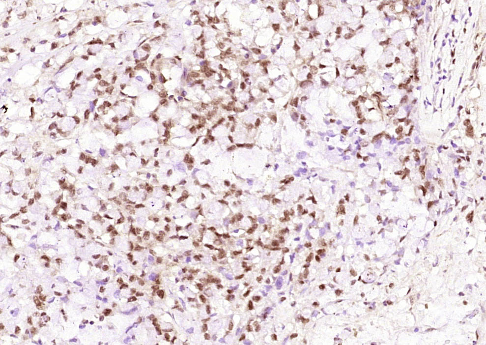 Paraformaldehyde-fixed, paraffin embedded (Human caecum cancer); Antigen retrieval by boiling in sodium citrate buffer (pH6.0) for 15min; Block endogenous peroxidase by 3% hydrogen peroxide for 20 minutes; Blocking buffer (normal goat serum) at 37°C for 30min; Antibody incubation with (Ki-67) Monoclonal Antibody, Unconjugated (SLM-33070M) at 1:200 overnight at 4°C, followed by operating according to SP Kit(Mouse)(sp-0024) instructionsand DAB staining.
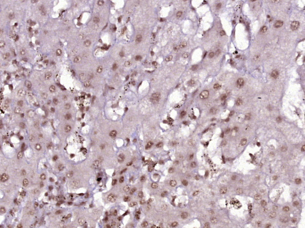 Paraformaldehyde-fixed, paraffin embedded (Human liver cancer); Antigen retrieval by boiling in sodium citrate buffer (pH6.0) for 15min; Block endogenous peroxidase by 3% hydrogen peroxide for 20 minutes; Blocking buffer (normal goat serum) at 37°C for 30min; Antibody incubation with (Ki-67) Monoclonal Antibody, Unconjugated (SLM-33070M) at 1:400 overnight at 4°C, followed by operating according to SP Kit(Mouse) (sp-0024) instructions and DAB staining.
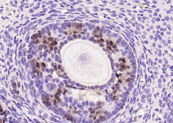 Paraformaldehyde-fixed, paraffin embedded (rat ovary); Antigen retrieval by boiling in sodium citrate buffer (pH6.0) for 15min; Block endogenous peroxidase by 3% hydrogen peroxide for 20 minutes; Blocking buffer (normal goat serum) at 37°C for 30min; Antibody incubation with (Ki-67) Monoclonal Antibody, Unconjugated (SLM-33070M) at 1:200 overnight at 4°C, followed by operating according to SP Kit(Mouse)(sp-0024) instructionsand DAB staining.
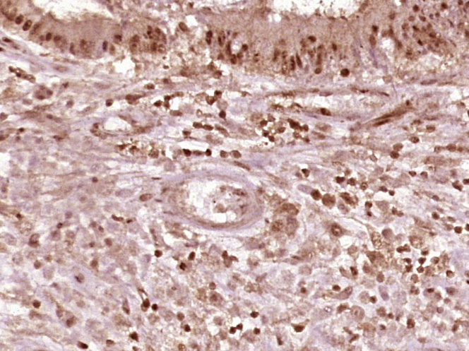 Paraformaldehyde-fixed, paraffin embedded (Human stomach cancer); Antigen retrieval by boiling in sodium citrate buffer (pH6.0) for 15min; Block endogenous peroxidase by 3% hydrogen peroxide for 20 minutes; Blocking buffer (normal goat serum) at 37°C for 30min; Antibody incubation with (Ki-67) Monoclonal Antibody, Unconjugated (SLM-33070M) at 1:400 overnight at 4°C, followed by operating according to SP Kit(Mouse) (sp-0024) instructions and DAB staining.
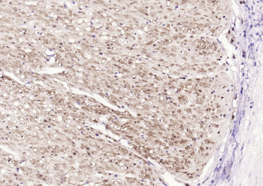 Paraformaldehyde-fixed, paraffin embedded (human rectal carcinoma); Antigen retrieval by boiling in sodium citrate buffer (pH6.0) for 15min; Block endogenous peroxidase by 3% hydrogen peroxide for 20 minutes; Blocking buffer (normal goat serum) at 37°C for 30min; Antibody incubation with (Ki-67) Monoclonal Antibody, Unconjugated (SLM-33070M) at 1:200 overnight at 4°C, followed by operating according to SP Kit(Mouse)(sp-0024) instructionsand DAB staining.
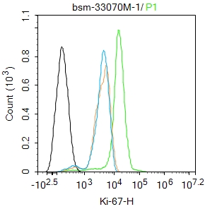 Blank control:Hela.
Primary Antibody (green line): Mouse Anti-Ki-67 antibody (SLM-33070M)
Dilution: 1ug/Test;
Secondary Antibody : Goat anti-mouse IgG-FITC
Dilution: 0.5ug/Test.
Protocol
The cells were fixed with 4% PFA (10min at room temperature)and then permeabilized with 90% ice-cold methanol for 20 min at -20℃.The cells were then incubated in 5%BSA to block non-specific protein-protein interactions for 30 min at room temperature .Cells stained with Primary Antibody for 30 min at room temperature. The secondary antibody used for 40 min at room temperature. Acquisition of 20,000 events was performed.
Cartpieces
Totalgoods,subtotals:¥Checkout
References (0)
No References
Bought notes(bought amounts latest0)
No one bought this product
User Comment(Total0User Comment Num)
- No comment


 +86 571 56623320
 +86 18668110335

