
Mouse Anti-beta I Tubulin (Loading Control)antibody
2810484G07Rik; Beta tubulin 1, class VI; Class VI beta tubulin; dJ543J19.4; M(beta)1; TBB1_HUMAN; TUBB1; Tubulin beta 1 class VI; Tubulin beta-1 chain; Tubulin, beta 1; tubulin, beta1;
View History [Clear]
Details
Product Name beta I Tubulin (Loading Control) Chinese Name 微管蛋白β1 tubulin(内参)单克隆抗体 Alias 2810484G07Rik; Beta tubulin 1, class VI; Class VI beta tubulin; dJ543J19.4; M(beta)1; TBB1_HUMAN; TUBB1; Tubulin beta 1 class VI; Tubulin beta-1 chain; Tubulin, beta 1; tubulin, beta1; literatures Product Type Internal reference anti Research Area Cell biology immunology Signal transduction Cytoskeleton Immunogen Species Mouse Clonality Monoclonal Clone NO. 5F7 React Species Human, Mouse, Rat, Applications WB=1:5000-20000 IHC-P=1:100-500 IHC-F=1:100-500 Flow-Cyt=1:100-500 ICC=1:100-500 IF=1:100-500 (Paraffin sections need antigen repair)
not yet tested in other applications.
optimal dilutions/concentrations should be determined by the end user.Theoretical molecular weight 50kDa Cellular localization cytoplasmic Form Liquid Concentration 1mg/ml immunogen KLH conjugated synthetic peptide derived from human beta I Tubulin Lsotype IgG2a Purification affinity purified by Protein G Buffer Solution 0.01M TBS(pH7.4) with 1% BSA, 0.03% Proclin300 and 50% Glycerol. Storage Shipped at 4℃. Store at -20 °C for one year. Avoid repeated freeze/thaw cycles. Attention This product as supplied is intended for research use only, not for use in human, therapeutic or diagnostic applications. PubMed PubMed Product Detail This gene encodes a member of the beta tubulin protein family. Beta tubulins are one of two core protein families (alpha and beta tubulins) that heterodimerize and assemble to form microtubules. This protein is specifically expressed in platelets and megakaryocytes and may be involved in proplatelet production and platelet release. A mutations in this gene is associated with autosomal dominant macrothrombocytopenia. Two pseudogenes of this gene are found on chromosome Y.[provided by RefSeq, Jul 2010]
Function:
Tubulin is the major constituent of microtubules. It binds two moles of GTP, one at an exchangeable site on the beta chain and one at a non-exchangeable site on the alpha chain (By similarity).
Subunit:
Dimer of alpha and beta chains. A typical microtubule is a hollow water-filled tube with an outer diameter of 25 nm and an inner diameter of 15 nM. Alpha-beta heterodimers associate head-to-tail to form protofilaments running lengthwise along the microtubule wall with the beta-tubulin subunit facing the microtubule plus end conferring a structural polarity. Microtubules usually have 13 protofilaments but different protofilament numbers can be found in some organisms and specialized cells. Interacts with RANBP10.
Subcellular Location:
Cytoplasm, cytoskeleton
Tissue Specificity:
Hematopoietic cell-specific. Major isotype in leukocytes, where it represents 50% of all beta-tubulins.
Post-translational modifications:
Some glutamate residues at the C-terminus are polyglutamylated, resulting in polyglutamate chains on the gamma-carboxyl group (PubMed:26875866). Polyglutamylation plays a key role in microtubule severing by spastin (SPAST). SPAST preferentially recognizes and acts on microtubules decorated with short polyglutamate tails: severing activity by SPAST increases as the number of glutamates per tubulin rises from one to eight, but decreases beyond this glutamylation threshold (PubMed:26875866).
Some glutamate residues at the C-terminus are monoglycylated but not polyglycylated due to the absence of functional TTLL10 in human. Monoglycylation is mainly limited to tubulin incorporated into axonemes (cilia and flagella). Both polyglutamylation and monoglycylation can coexist on the same protein on adjacent residues, and lowering glycylation levels increases polyglutamylation, and reciprocally. The precise function of monoglycylation is still unclear (Probable).
Phosphorylated on Ser-172 by CDK1 during the cell cycle, from metaphase to telophase, but not in interphase. This phosphorylation inhibits tubulin incorporation into microtubules.
DISEASE:
Macrothrombocytopenia, autosomal dominant, TUBB1-related (MAD-TUBB1). The disease is caused by mutations affecting the gene represented in this entry. A congenital blood disorder characterized by increased platelet size and decreased number of circulating platelets.
Similarity:
Belongs to the tubulin family.
SWISS:
Q9H4B7
Gene ID:
81027
Database links:Entrez Gene: 396427 Chicken
Entrez Gene: 101836899 Hamster
Entrez Gene: 81027 Human
Entrez Gene: 545486 Mouse
Omim: 612901 Human
SwissProt: P09203 Chicken
SwissProt: Q9H4B7 Human
SwissProt: A2AQ07 Mouse
Unigene: 303023 Human
Unigene: 45285 Mouse
Product Picture 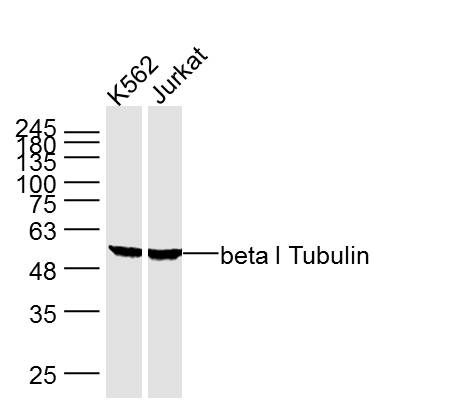 Sample:
K562 Cell (Human) Lysate at 40 ug
Jurkat Cell (Human) Lysate at 40 ug
Primary: Anti- beta I Tubulin (SLM-33041M) at 1/1000 dilution
Secondary: IRDye800CW Goat Anti-Mouse IgG at 1/20000 dilution
Predicted band size: 50 kD
Observed band size: 50 kD
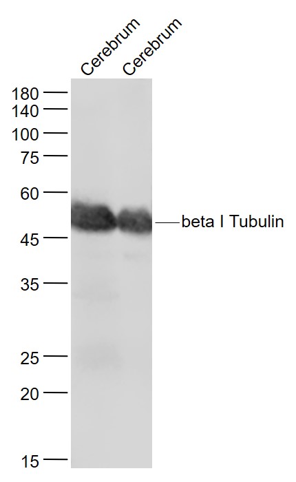 Sample:
Cerebrum (Mouse) Lysate at 40 ug
Cerebrum (Rat) Lysate at 40 ug
Primary: Anti- beta I Tubulin (SLM-33041M) at 1/1000 dilution
Secondary: IRDye800CW Goat Anti-Mouse IgG at 1/20000 dilution
Predicted band size: 50 kD
Observed band size: 50 kD
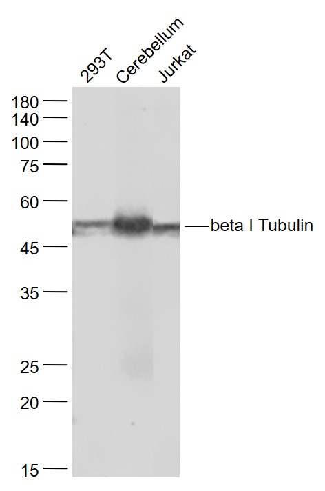 Sample:
293T(Human) Cell Lysate at 30 ug
Cerebellum (Mouse) Lysate at 40 ug
Jurkat(Human) Cell Lysate at 30 ug
Primary: Anti- beta I Tubulin (SLM-33041M) at 1/1000 dilution
Secondary: IRDye800CW Goat Anti-Mouse IgG at 1/20000 dilution
Predicted band size: 50 kD
Observed band size: 50 kD
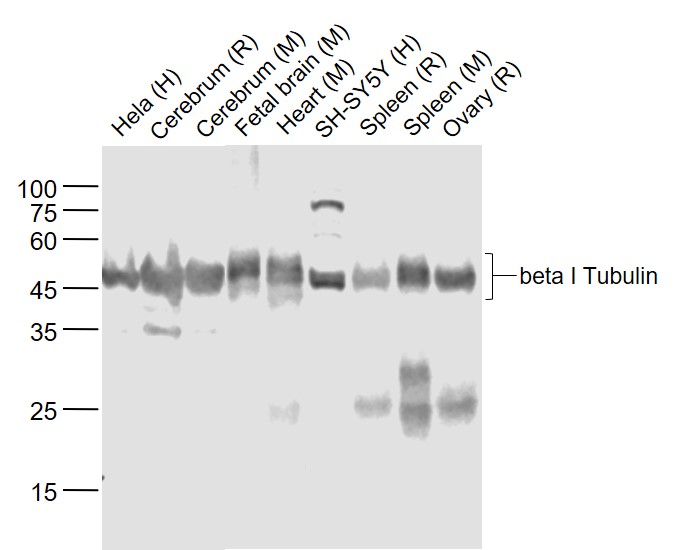 Sample:
Lane 1: Hela (Human) Cell Lysate at 30 ug
Lane 2: Cerebrum (Rat) Lysate at 40 ug
Lane 3: Cerebrum (Mouse) Lysate at 40 ug
Lane 4: Fetal brain (Mouse) Lysate at 40 ug
Lane 5: Heart (Mouse) Lysate at 40 ug
Lane 6: SH-SY5Y (Human) Cell Lysate at 30 ug
Lane 7: Spleen (Rat) Lysate at 40 ug
Lane 8: Spleen (Mouse) Lysate at 40 ug
Lane 9: Ovary (Rat) Lysate at 40 ug
Primary: Anti-beta I Tubulin (SLM-33041M) at 1/1000 dilution
Secondary: IRDye800CW Goat Anti-Mouse IgG at 1/20000 dilution
Predicted band size: 50 kD
Observed band size: 50 kD
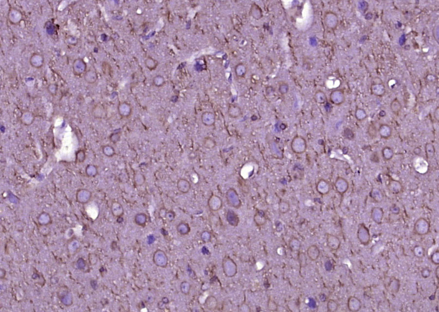 Paraformaldehyde-fixed, paraffin embedded (mouse brain tissue); Antigen retrieval by boiling in sodium citrate buffer (pH6.0) for 15min; Block endogenous peroxidase by 3% hydrogen peroxide for 20 minutes; Blocking buffer (normal goat serum) at 37°C for 30min; Antibody incubation with (beta I Tubulin) Monoclonal Antibody, Unconjugated (ascites of SLM-33041M) at 1:2000 overnight at 4°C, followed by a conjugated secondary (sp-0024) for 20 minutes and DAB staining.
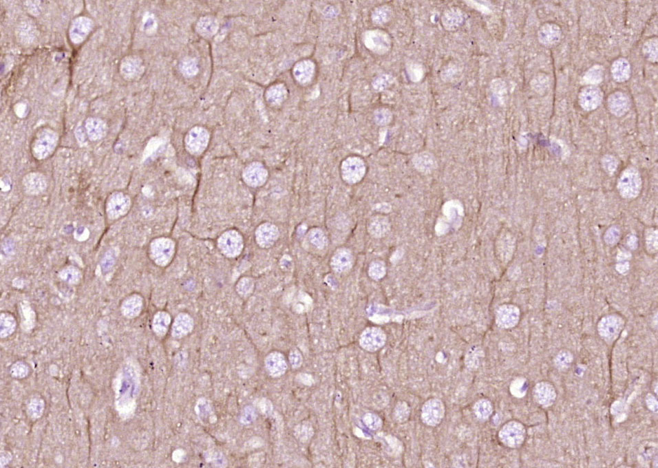 Paraformaldehyde-fixed, paraffin embedded (rat brain tissue); Antigen retrieval by boiling in sodium citrate buffer (pH6.0) for 15min; Block endogenous peroxidase by 3% hydrogen peroxide for 20 minutes; Blocking buffer (normal goat serum) at 37°C for 30min; Antibody incubation with (beta I Tubulin) Monoclonal Antibody, Unconjugated (ascites of SLM-33041M) at 1:2000 overnight at 4°C, followed by a conjugated secondary (sp-0024) for 20 minutes and DAB staining.
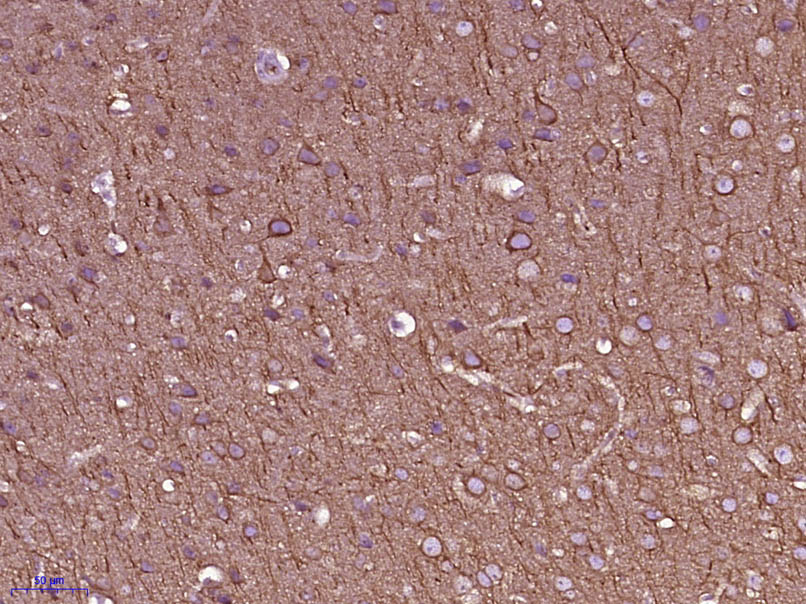 Paraformaldehyde-fixed, paraffin embedded (Mouse brain); Antigen retrieval by boiling in sodium citrate buffer (pH6.0) for 15min; Block endogenous peroxidase by 3% hydrogen peroxide for 20 minutes; Blocking buffer (normal goat serum) at 37°C for 30min; Antibody incubation with (beta I Tubulin) Monoclonal Antibody, Unconjugated (SLM-33041M 5F7) at 1:400 overnight at 4°C, followed by operating according to SP Kit(Mouse) (sp-0024) instructionsand DAB staining.
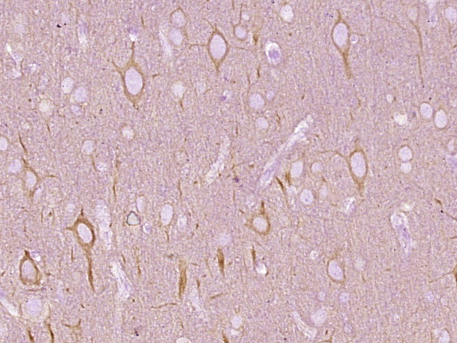 Paraformaldehyde-fixed, paraffin embedded (Rat brain); Antigen retrieval by boiling in sodium citrate buffer (pH6.0) for 15min; Block endogenous peroxidase by 3% hydrogen peroxide for 20 minutes; Blocking buffer (normal goat serum) at 37°C for 30min; Antibody incubation with (beta I Tubulin) Monoclonal Antibody, Unconjugated (SLM-33041M) at 1:400 overnight at 4°C, followed by operating according to SP Kit(Mouse) (sp-0024) instructions and DAB staining.
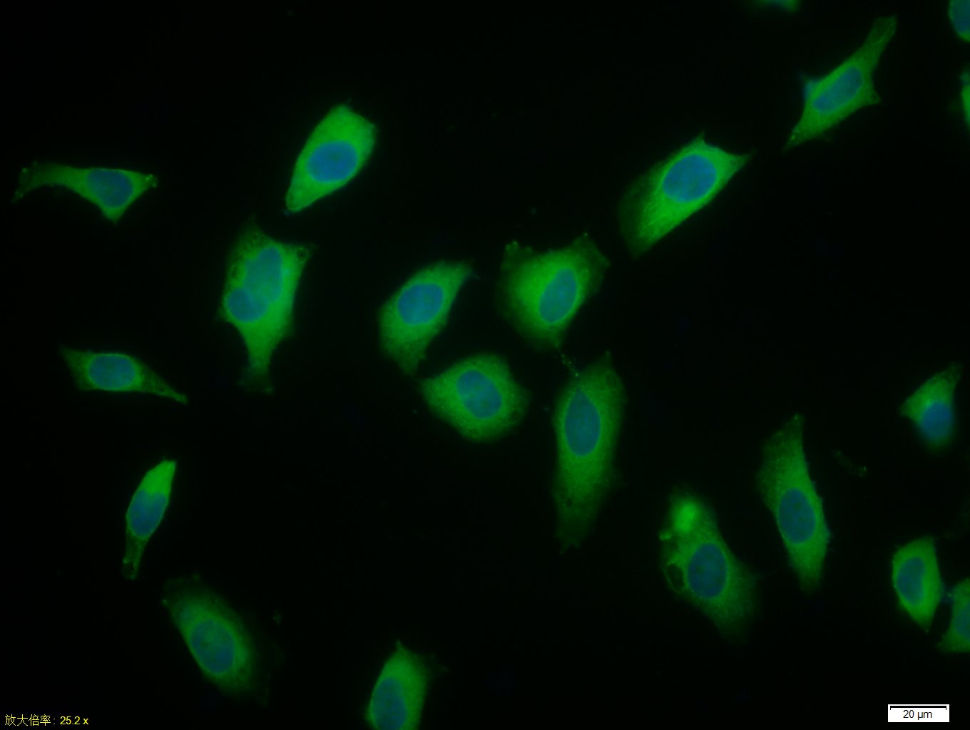 Tissue/cell: HeLa cell; 4% Paraformaldehyde-fixed; Triton X-100 at room temperature for 20 min; Blocking buffer (normal goat serum, C-0005) at 37°C for 20 min; Antibody incubation with (beta I Tubulin) Monoclonal Antibody, Unconjugated (SLM-33041M) 1:50, 90 minutes at 37°C; followed by a conjugated Goat Anti-Rabbit IgG antibody (SL0296G-FITC) at 37°C for 90 minutes, DAPI (5ug/ml, blue, C-0033) was used to stain the cell nuclei.
Cartpieces
Totalgoods,subtotals:¥Checkout
Bought notes(bought amounts latest0)
No one bought this product
User Comment(Total0User Comment Num)
- No comment


 +86 571 56623320
 +86 18668110335

