
Rabbit Anti-MUL1 antibody
E3 ubiquitin-protein ligase MUL1; C1orf166; E3 ubiquitin ligase; E3 ubiquitin protein ligase MUL1; GIDE; Growth inhibition and death E3 ligase; MAPL; Mitochondrial anchored protein ligase; Mitochondrial ubiquitin ligase activator of NFKB 1; MUL1; MULAN; P
View History [Clear]
Details
Product Name MUL1 Chinese Name E3Ubiquitin连接酶MUL1抗体 Alias E3 ubiquitin-protein ligase MUL1; C1orf166; E3 ubiquitin ligase; E3 ubiquitin protein ligase MUL1; GIDE; Growth inhibition and death E3 ligase; MAPL; Mitochondrial anchored protein ligase; Mitochondrial ubiquitin ligase activator of NFKB 1; MUL1; MULAN; Putative NF kappa B activating protein 266; RING finger protein 218; RNF218; RP23-25C1.10-002; MUL1_HUMAN. Research Area Cell biology immunology Immunogen Species Rabbit Clonality Polyclonal React Species Human, Mouse, Rat, (predicted: Dog, Pig, Cow, Rabbit, ) Applications WB=1:500-2000 ELISA=1:5000-10000 IHC-P=1:100-500 IHC-F=1:100-500 Flow-Cyt=1ug/test IF=1:50-200 (Paraffin sections need antigen repair)
not yet tested in other applications.
optimal dilutions/concentrations should be determined by the end user.Theoretical molecular weight 40kDa Cellular localization cytoplasmic The cell membrane Form Liquid Concentration 1mg/ml immunogen KLH conjugated synthetic peptide derived from human MUL1/RNF218: 1-100/352 Lsotype IgG Purification affinity purified by Protein A Buffer Solution 0.01M TBS(pH7.4) with 1% BSA, 0.03% Proclin300 and 50% Glycerol. Storage Shipped at 4℃. Store at -20 °C for one year. Avoid repeated freeze/thaw cycles. Attention This product as supplied is intended for research use only, not for use in human, therapeutic or diagnostic applications. PubMed PubMed Product Detail E3 ubiquitin-protein ligase that plays a role in the control of mitochondrial morphology. Promotes mitochondrial fragmentation and influences mitochondrial localization. Inhibits cell growth. When overexpressed, activates JNK through MAP3K7/TAK1 and induces caspase-dependent apoptosis. E3 ubiquitin ligases accept ubiquitin from an E2 ubiquitin-conjugatin.
Function:
Exhibits weak E3 ubiquitin-protein ligase activity, but preferentially acts as a SUMO E3 ligase at physiological concentrations. Plays a role in the control of mitochondrial morphology. Promotes mitochondrial fragmentation and influences mitochondrial localization. Inhibits cell growth. When overexpressed, activates JNK through MAP3K7/TAK1 and induces caspase-dependent apoptosis. E3 ubiquitin ligases accept ubiquitin from an E2 ubiquitin-conjugating enzyme in the form of a thioester and then directly transfer the ubiquitin to targeted substrates.
Subunit:
Homooligomer. Interacts with MAP3K7/TAK1. Interacts with UBC9. Interacts with and sumoylates DNM1L.
Subcellular Location:
Mitochondrion outer membrane; Multi-pass membrane protein. Peroxisome. Note: Transported in mitochondrion-derived vesicles from the mitochondrion to the peroxisome.
Tissue Specificity:
Widely expressed with highest levels in the heart, skeletal muscle, placenta, kidney and liver. Barely detectable in colon and thymus.
Similarity:
Contains 1 RING-type zinc finger.
SWISS:
Q969V5
Gene ID:
79594
Database links:Entrez Gene: 79594 Human
Entrez Gene: 68350 Mouse
Omim: 612037 Human
SwissProt: Q969V5 Human
SwissProt: Q8VCM5 Mouse
Unigene: 10101 Human
Unigene: 103413 Mouse
Product Picture 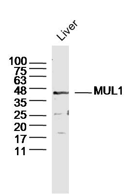 Sample: Liver (Mouse) Lysate at 40 ug
Primary: Anti-MUL1 (SL9291R)at 1/300 dilution
Secondary: IRDye800CW Goat Anti-Rabbit IgG at 1/20000 dilution
Predicted band size: 40kD
Observed band size: 40kD
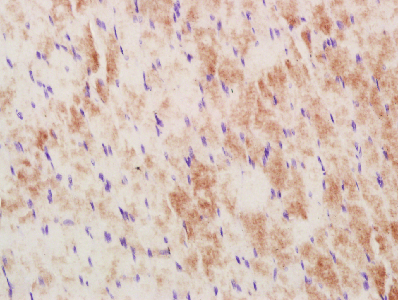 Paraformaldehyde-fixed, paraffin embedded (Rat skeletal muscle); Antigen retrieval by boiling in sodium citrate buffer (pH6.0) for 15min; Block endogenous peroxidase by 3% hydrogen peroxide for 20 minutes; Blocking buffer (normal goat serum) at 37°C for 30min; Antibody incubation with (MUL1) Polyclonal Antibody, Unconjugated (SL9291R) at 1:400 overnight at 4°C, followed by a conjugated secondary antibody (sp-0023) for 20 minutes and DAB staining.
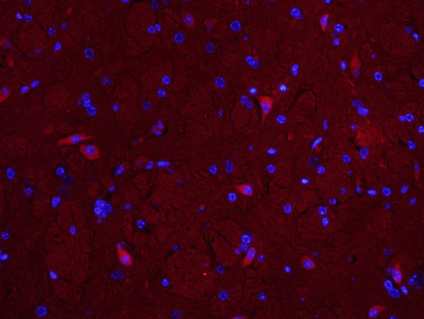 Paraformaldehyde-fixed, paraffin embedded (Mouse brain); Antigen retrieval by boiling in sodium citrate buffer (pH6.0) for 15min; Block endogenous peroxidase by 3% hydrogen peroxide for 20 minutes; Blocking buffer (normal goat serum) at 37°C for 30min; Antibody incubation with (MUL1) Polyclonal Antibody, Unconjugated (SL9291R) at 1:400 overnight at 4°C, followed by a conjugated secondary antibody (SL0295g-cy3) for 90 minutes, and DAPI for nuclei staining.
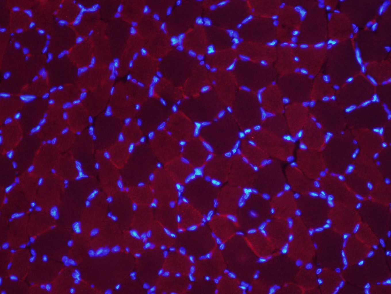 Paraformaldehyde-fixed, paraffin embedded (Rat skeletal muscle); Antigen retrieval by boiling in sodium citrate buffer (pH6.0) for 15min; Block endogenous peroxidase by 3% hydrogen peroxide for 20 minutes; Blocking buffer (normal goat serum) at 37°C for 30min; Antibody incubation with (MUL1) Polyclonal Antibody, Unconjugated (SL9291R) at 1:400 overnight at 4°C, followed by a conjugated secondary antibody (SL0295g-cy3) for 90 minutes, and DAPI for nuclei staining.
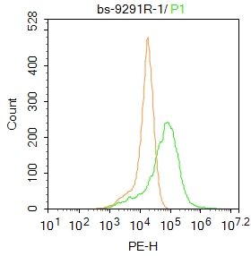 Blank control: A549.
Primary Antibody (green line): Rabbit Anti-MUL1 antibody (SL9291R)
Dilution: 1μg /10^6 cells;
Isotype Control Antibody (orange line): Rabbit IgG .
Secondary Antibody : Goat anti-rabbit IgG-PE
Dilution: 1μg /test.
Protocol
The cells were fixed with 4% PFA (10min at room temperature)and then permeabilized with PBST for 20 min at room temperature. The cells were then incubated in 5%BSA to block non-specific protein-protein interactions for 30 min at room temperature .Cells stained with Primary Antibody for 30 min at room temperature. The secondary antibody used for 40 min at room temperature. Acquisition of 20,000 events was performed.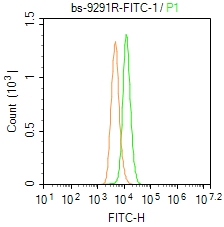 Blank control: A549.
Primary Antibody (green line): Rabbit Anti-MUL1/FITC Conjugated antibody (SL9291R-FITC)
Dilution: 1μg /10^6 cells;
Isotype Control Antibody (orange line): Rabbit IgG-FITC .
Protocol
The cells were fixed with 4% PFA (10min at room temperature)and then permeabilized with 0.1% PBST for 20 min at-20℃. The cells were then incubated in 5% BSA to block non-specific protein-protein interactions for 30 min at room temperature. The cells were stained with Primary Antibody for 30 min at room temperature. Acquisition of 20,000 events was performed.
Cartpieces
Totalgoods,subtotals:¥Checkout
References (0)
No References
Bought notes(bought amounts latest0)
No one bought this product
User Comment(Total0User Comment Num)
- No comment


 +86 571 56623320
 +86 18668110335

