
Rabbit Anti-TRPM4 antibody
Long transient receptor potential channel 4; LTrpC-4; LTrpC4; Melastatin 4; Melastatin like 2 protein; Melastatin-4; Melastatin-like 2; Mls2s; PFHB1B; Transient receptor potential cation channel subfamily M member 4; Transient receptor potential cation ch
View History [Clear]
Details
Product Name TRPM4 Chinese Name 瞬时受体电位离子Channel protein4抗体(M亚家族) Alias Long transient receptor potential channel 4; LTrpC-4; LTrpC4; Melastatin 4; Melastatin like 2 protein; Melastatin-4; Melastatin-like 2; Mls2s; PFHB1B; Transient receptor potential cation channel subfamily M member 4; Transient receptor potential cation channel, subfamily M, member 4; Trpm4; TRPM4_HUMAN; TRPM4B; 1110030C19Rik; AW047689; Calcium-activated non-selective cation channel 1; FLJ20041; hTRPM4. Research Area Cell biology immunology Neurobiology Signal transduction Channel protein Immunogen Species Rabbit Clonality Polyclonal React Species Mouse, (predicted: Human, Rat, ) Applications WB=1:500-2000 ELISA=1:5000-10000 IHC-P=1:100-500 IHC-F=1:100-500 Flow-Cyt=0.2ug/test IF=1:50-200 (Paraffin sections need antigen repair)
not yet tested in other applications.
optimal dilutions/concentrations should be determined by the end user.Theoretical molecular weight 134kDa Cellular localization cytoplasmic The cell membrane Form Liquid Concentration 1mg/ml immunogen KLH conjugated synthetic peptide derived from human TRPM4: 751-850/1214 <Extracellular> Lsotype IgG Purification affinity purified by Protein A Buffer Solution 0.01M TBS(pH7.4) with 1% BSA, 0.03% Proclin300 and 50% Glycerol. Storage Shipped at 4℃. Store at -20 °C for one year. Avoid repeated freeze/thaw cycles. Attention This product as supplied is intended for research use only, not for use in human, therapeutic or diagnostic applications. PubMed PubMed Product Detail Calcium-activated non selective (CAN) cation channel that mediates membrane depolarization. While it is activated by increase in intracellular Ca(2+), it is impermeable to it. Mediates transport of monovalent cations (Na(+) > K(+) > Cs(+) > Li(+)), leading to depolarize the membrane. It thereby plays a central role in cadiomyocytes, neurons from entorhinal cortex, dorsal root and vomeronasal neurons, endocrine pancreas cells, kidney epithelial cells, cochlea hair cells etc. Participates in T-cell activation by modulating Ca(2+) oscillations after T lymphocyte activation, which is required for NFAT-dependent IL2 production. Involved in myogenic constriction of cerebral arteries. Controls insulin secretion in pancreatic beta-cells. May also be involved in pacemaking or could cause irregular electrical activity under conditions of Ca(2+) overload. Affects T-helper 1 (Th1) and T-helper 2 (Th2) cell motility and cytokine production through differential regulation of calcium signaling and NFATC1 localization. Enhances cell proliferation through up-regulation of the beta-catenin signaling pathway.
Involvement in disease:
Defects in TRPM4 are the cause of progressive familial heart block type 1B (PFHB1B) [MIM:604559]. It is a cardiac bundle branch disorder characterized by progressive alteration of cardiac conduction through the His-Purkinje system, with a pattern of a right bundle-branch block and/or left anterior hemiblock occurring individually or together. It leads to complete atrio-ventricular block causing syncope and sudden death.
Function:
Calcium-activated non selective (CAN) cation channel that mediates membrane depolarization. While it is activated by increase in intracellular Ca(2+), it is impermeable to it. Mediates transport of monovalent cations (Na(+) > K(+) > Cs(+) > Li(+)), leading to depolarize the membrane. It thereby plays a central role in cadiomyocytes, neurons from entorhinal cortex, dorsal root and vomeronasal neurons, endocrine pancreas cells, kidney epithelial cells, cochlea hair cells etc. Participates in T-cell activation by modulating Ca(2+) oscillations after T lymphocyte activation, which is required for NFAT-dependent IL2 production. Involved in myogenic constriction of cerebral arteries. Controls insulin secretion in pancreatic beta-cells. May also be involved in pacemaking or could cause irregular electrical activity under conditions of Ca(2+) overload. Affects T-helper 1 (Th1) and T-helper 2 (Th2) cell motility and cytokine production through differential regulation of calcium signaling and NFATC1 localization. Enhances cell proliferation through up-regulation of the beta-catenin signaling pathway.
Subunit:
Homomultimer.
Subcellular Location:
Endoplasmic reticulum. Golgi apparatus and Cell membrane. Endoplasmic reticulum. Golgi apparatus
Tissue Specificity:
Widely expressed with a high expression in intestine and prostate. In brain, it is both expressed in whole cerebral arteries and isolated vascular smooth muscle cells. Prominently expressed in Purkinje fibers. Expressed at higher levels in T-helper 2 (Th2) cells as compared to T-helper 1 (Th1) cells.
Post-translational modifications:
Phosphorylation by PKC leads to increase the sensitivity to Ca(2+).
Sumoylated. Desumoylated by SENP1.
DISEASE:
Defects in TRPM4 are the cause of progressive familial heart block type 1B (PFHB1B) [MIM:604559]. It is a cardiac bundle branch disorder characterized by progressive alteration of cardiac conduction through the His-Purkinje system, with a pattern of a right bundle-branch block and/or left anterior hemiblock occurring individually or together. It leads to complete atrioventricular block causing syncope and sudden death.
Similarity:
Belongs to the transient receptor (TC 1.A.4) family. LTrpC subfamily. TRPM4 sub-subfamily.
SWISS:
Q8TD43
Gene ID:
54795
Database links:Entrez Gene: 54795 Human
Entrez Gene: 68667 Mouse
Omim: 606936 Human
SwissProt: Q8TD43 Human
SwissProt: Q7TN37 Mouse
Unigene: 467101 Human
Unigene: 439890 Mouse
Unigene: 205004 Rat
Product Picture 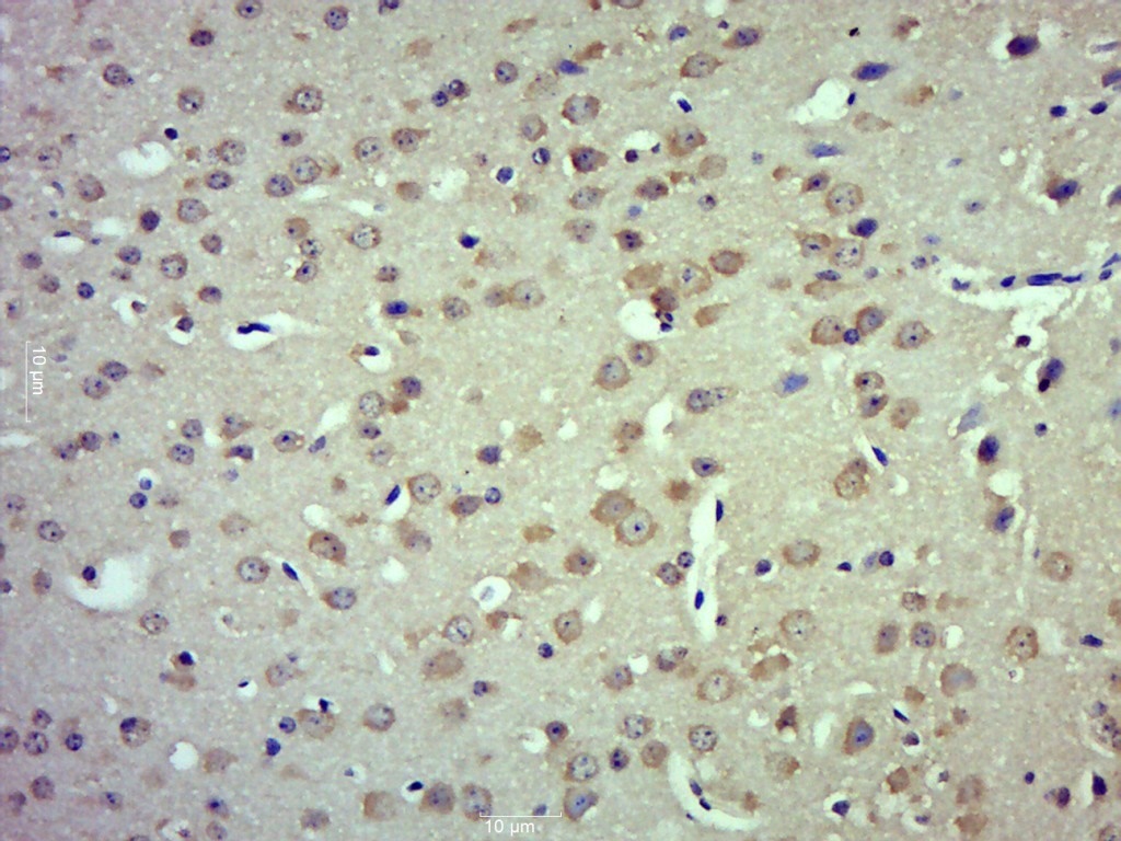 Paraformaldehyde-fixed, paraffin embedded (Mouse brain); Antigen retrieval by boiling in sodium citrate buffer (pH6.0) for 15min; Block endogenous peroxidase by 3% hydrogen peroxide for 20 minutes; Blocking buffer (normal goat serum) at 37°C for 30min; Antibody incubation with (TRPM4) Polyclonal Antibody, Unconjugated (SL9051R) at 1:500 overnight at 4°C, followed by a conjugated secondary (sp-0023) for 20 minutes and DAB staining.
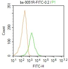 Blank control: Mouse spleen.
Primary Antibody (green line): Rabbit Anti-TRPM4/FITC Conjugated antibody (SL9051R-FITC)
Dilution: 0.2μg /10^6 cells;
Isotype Control Antibody (orange line): Rabbit IgG-FITC .
Protocol
The cells were fixed with 4% PFA (10min at room temperature)and then permeabilized with 0.1% PBST for 20 min at-20℃. The cells were then incubated in 5% BSA to block non-specific protein-protein interactions for 30 min at room temperature. The cells were stained with Primary Antibody for 30 min at room temperature. Acquisition of 20,000 events was performed.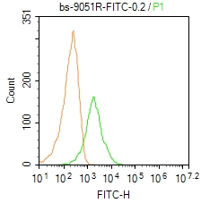 Blank control: Mouse spleen.
Primary Antibody (green line): Rabbit Anti-TRPM4/FITC Conjugated antibody (SL9051R-FITC)
Dilution: 0.2μg /10^6 cells;
Isotype Control Antibody (orange line): Rabbit IgG-FITC .
Protocol
The cells were fixed with 4% PFA (10min at room temperature)and then permeabilized with 0.1% PBST for 20 min at-20℃. The cells were then incubated in 5% BSA to block non-specific protein-protein interactions for 30 min at room temperature. The cells were stained with Primary Antibody for 30 min at room temperature. Acquisition of 20,000 events was performed.
Cartpieces
Totalgoods,subtotals:¥Checkout
References (0)
No References
Bought notes(bought amounts latest0)
No one bought this product
User Comment(Total0User Comment Num)
- No comment


 +86 571 56623320
 +86 18668110335

