
Rabbit Anti-ADA2 antibody
ADA2_HUMAN; Adenosine deaminase 2; Cat eye syndrome critical region protein 1; ADGF; CECR1; IDGFL;
View History [Clear]
Details
Product Name ADA2 Chinese Name 猫眼综合征染色体候选基因1抗体 Alias ADA2_HUMAN; Adenosine deaminase 2; Cat eye syndrome critical region protein 1; ADGF; CECR1; IDGFL; Research Area Cell biology immunology Epigenetics Immunogen Species Rabbit Clonality Polyclonal React Species Human, (predicted: Pig, ) Applications ELISA=1:5000-10000 IHC-P=1:100-500 IHC-F=1:100-500 Flow-Cyt=1ug/test IF=1:50-200 (Paraffin sections need antigen repair)
not yet tested in other applications.
optimal dilutions/concentrations should be determined by the end user.Theoretical molecular weight 56kDa Cellular localization Secretory protein Form Liquid Concentration 1mg/ml immunogen KLH conjugated synthetic peptide derived from human ADA2: 421-511/511 Lsotype IgG Purification affinity purified by Protein A Buffer Solution 0.01M TBS(pH7.4) with 1% BSA, 0.03% Proclin300 and 50% Glycerol. Storage Shipped at 4℃. Store at -20 °C for one year. Avoid repeated freeze/thaw cycles. Attention This product as supplied is intended for research use only, not for use in human, therapeutic or diagnostic applications. PubMed PubMed Product Detail CECR1 is a member of the adenosine and AMP deaminases family. It may act as a growth factor and have adenosine deaminase activity. It is a candidate gene for cat eye syndrome. Two transcript variants encoding distinct isoforms have been identified for this gene.
Adenosine deaminase is an enzyme that is present in most tissues and exists predominantly as a monomer, although in some tissues it is associated with adenosine deaminase-binding protein. Adenosine deaminase degrades extracellular adenosine, which is toxic for lymphocytes. A novel family of growth factors that share sequence similarity to adenosine deaminase has been identified. The cat eye syndrome critical region protein (CECR) family includes CECR1, CECR2, CECR3, CECR4, CECR5, CECR6, CECR7, CECR8 and CECR9. The genes encoding CECR proteins are candidates for Cat Eye Syndrome (CES), a developmental disorder associated with the duplication of a 2 Mb region of 22q11.2. CES is characterized by the combination of coloboma of the iris and anal atresia with fistula, downslanting palpebral fissures, preauricular tags and/or pits, frequent occurrence of heart and renal malformations, and normal or near-normal mental development. CECR family members are widely expressed. Specifically, CECR1 has the highest expression in adult heart, lung, lymphoblasts and placenta. CECR2 is also involved in neurulation and chromatin remodeling. Mutations in the CECR2 gene result in neural tube defects.
Function:
Adenosine deaminase that may contribute to the degradation of extracellular adenosine, a signaling molecule that controls a variety of cellular responses. Requires elevated adenosine levels for optimal enzyme activity. Binds to cell surfaces via proteoglycans and may play a role in the regulation of cell proliferation and differentiation, independently of its enzyme activity.
Subunit:
Homodimer. Interacts with adenosine receptors. Binds heparin.
Subcellular Location:
Secreted
Tissue Specificity:
Detected in blood plasma (at protein level). Widely expressed, with most abundant expression in human adult heart, lung, lymphoblasts, and placenta as well as fetal lung, liver, and kidney. In embryo, expressed in the outflow tract and atrium of the developing heart, the VII/VIII cranial nerve ganglion, and the notochord.
Similarity:
Belongs to the adenosine and AMP deaminases family. ADGF subfamily.
SWISS:
Q9NZK5
Gene ID:
51816
Database links:Entrez Gene: 51816 Human
Omim: 607575 Human
SwissProt: Q9NZK5 Human
Unigene: 170310 Human
Product Picture 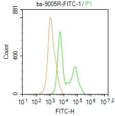 Blank control: Molt4.
Primary Antibody (green line): Rabbit Anti-CECR1/FITC Conjugated antibody (SL9005R-FITC)
Dilution: 1μg /10^6 cells;
Isotype Control Antibody (orange line): Rabbit IgG-FITC .
Protocol
The cells were fixed with 4% PFA (10min at room temperature)and then permeabilized with 0.1% PBST for 20 min at-20℃. The cells were then incubated in 5% BSA to block non-specific protein-protein interactions for 30 min at room temperature. The cells were stained with Primary Antibody for 30 min at room temperature. Acquisition of 20,000 events was performed.
Cartpieces
Totalgoods,subtotals:¥Checkout
References (0)
No References
Bought notes(bought amounts latest0)
No one bought this product
User Comment(Total0User Comment Num)
- No comment


 +86 571 56623320
 +86 18668110335

