
Rabbit Anti-LC3 antibody
Autophagy related protein LC3 A; Autophagy related protein LC3 B; Autophagy related ubiquitin like modifier LC3 A; Autophagy related ubiquitin like modifier LC3 B; MAP1 light chain 3 like protein 1; MAP1 light chain 3 like protein 2; MAP1A/1B light chain
View History [Clear]
Details
Product Name LC3 Chinese Name 自噬微管相关蛋白轻链3抗体 Alias Autophagy related protein LC3 A; Autophagy related protein LC3 B; Autophagy related ubiquitin like modifier LC3 A; Autophagy related ubiquitin like modifier LC3 B; MAP1 light chain 3 like protein 1; MAP1 light chain 3 like protein 2; MAP1A/1B light chain 3 A; MAP1A/1B light chain 3 B; MAP1A/1BLC3; MAP1A/MAP1B LC3 A; MAP1A/MAP1B LC3 B; MAP1ALC3; MAP1BLC3; MAP1LC3A; MAP1LC3B; Microtubule associated protein 1 light chain 3 alpha; Microtubule associated protein 1 light chain 3 beta; Microtubule associated proteins 1A/1B light chain 3; Microtubule associated proteins 1A/1B light chain 3A; Microtubule associated proteins 1A/1B light chain 3B; MLP3A_HUMAN. literatures Research Area Tumour Cell biology Neurobiology Signal transduction Cytoskeleton Extracellular matrix Autophagy Immunogen Species Rabbit Clonality Polyclonal React Species Human, Rat, (predicted: Mouse, Chicken, Dog, Pig, Cow, Horse, Sheep, Goat, ) Applications WB=1:500-2000 ELISA=1:5000-10000 IHC-P=1:100-500 IHC-F=1:100-500 Flow-Cyt=1μg/Test IF=1:100-500 (Paraffin sections need antigen repair)
not yet tested in other applications.
optimal dilutions/concentrations should be determined by the end user.Theoretical molecular weight 14/16kDa Cellular localization cytoplasmic Form Liquid Concentration 1mg/ml immunogen KLH conjugated synthetic peptide derived from human LC3: 31-121/121 Lsotype IgG Purification affinity purified by Protein A Buffer Solution 0.01M TBS(pH7.4) with 1% BSA, 0.03% Proclin300 and 50% Glycerol. Storage Shipped at 4℃. Store at -20 °C for one year. Avoid repeated freeze/thaw cycles. Attention This product as supplied is intended for research use only, not for use in human, therapeutic or diagnostic applications. PubMed PubMed Product Detail A major contributor to cellular homeostasis is the ability of the cell to strike a balance between the formation and degradation/removal of its cellular components. This process of internal cellular turn-over is called autophagy (self-eating), and is facilitated by a pathway of around 16 interacting proteins in the human. LC3, a ubiquitin-like modifier protein, is the human homolog of yeast Apg8 and is involved in the formation of autophagosomal vacuoles, called autophagosomes. LC3 is expressed as 3 splice variants (LC3A, LC3B and LC3C), which exhibit different tissue distributions and are processed into cytosolic and autophagosomal membrane-bound forms, termed LC3-I and LC3-II, respectively. A disruption to the autophagic process is now associated with the progression of several cancers, neurodegenerative disorders and cardiac pathologies, where LC3 is widely employed as a marker for autophagy.
Function:
Probably involved in formation of autophagosomal vacuoles (autophagosomes).
Subunit:
3 different light chains, LC1, LC2 and LC3, can associate with MAP1A and MAP1B proteins. Interacts with SQSTM1. Interacts with TP53INP1 and TP53INP2.
Subcellular Location:
Cytoplasmic. Endomembrane system; Lipid-anchor. Cytoplasmic vesicle, autophagosome membrane; Lipid-anchor. Note: LC3B binds to the autophagic membranes.
Tissue Specificity:
Most abundant in heart, brain, liver, skeletal muscle and testis but absent in thymus and peripheral blood leukocytes.
Post-translational modifications:
The precursor molecule is cleaved by APG4B/ATG4B to form the cytosolic form, LC3-I. This is activated by APG7L/ATG7, transferred to ATG3 and conjugated to phospholipid to form the membrane-bound form, LC3-II.
Similarity:
Belongs to the MAP1 LC3 family.
SWISS:
Q9H492
Gene ID:
84557
Database links:Entrez Gene: 84557 Human
Entrez Gene: 66734 Mouse
Omim: 601242 Human
SwissProt: Q9H492 Human
SwissProt: Q91VR7 Mouse
Unigene: 632273 Human
Unigene: 196239 Mouse
Unigene: 3135 Rat
Product Picture 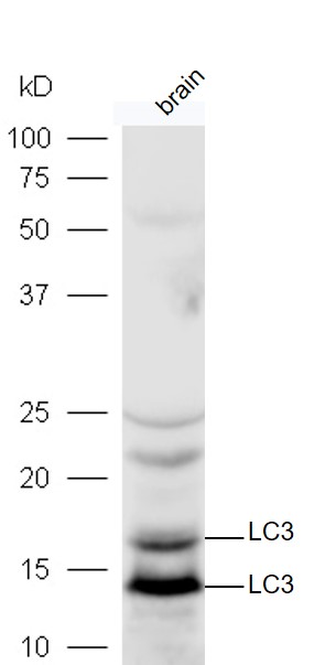 Protein: mouse brain lysates at 40ug;
Primary: rabbit Anti-LC3 (SL8878R) at 1:300;
Secondary: HRP conjugated Goat-Anti-rabbit IgG(SL0295G-HRP) at 1: 5000;
Predicted band size:14 kD Observed band size:14,16 kD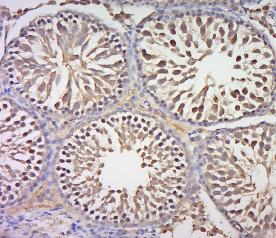 Paraformaldehyde-fixed, paraffin embedded (rat testis tissue); Antigen retrieval by boiling in sodium citrate buffer (pH6.0) for 15min; Block endogenous peroxidase by 3% hydrogen peroxide for 20 minutes; Blocking buffer (normal goat serum) at 37°C for 30min; Antibody incubation with (LC3) Polyclonal Antibody, Unconjugated (SL8878R) at 1:400 overnight at 4°C, followed by a conjugated secondary (sp-0023) for 20 minutes and DAB staining.
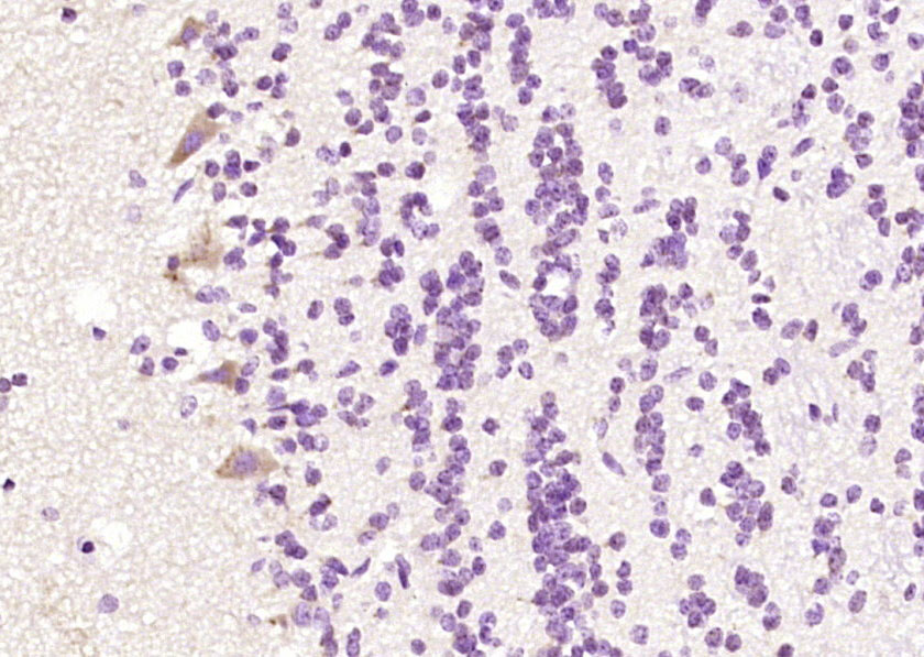 Paraformaldehyde-fixed, paraffin embedded (rat brain); Antigen retrieval by boiling in sodium citrate buffer (pH6.0) for 15min; Block endogenous peroxidase by 3% hydrogen peroxide for 20 minutes; Blocking buffer (normal goat serum) at 37°C for 30min; Antibody incubation with (LC3) Polyclonal Antibody, Unconjugated (SL8878R) at 1:200 overnight at 4°C, followed by operating according to SP Kit(Rabbit) (sp-0023) instructionsand DAB staining.
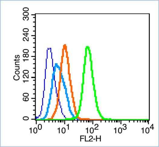 Blank control (blue line): Hela (fixed with 70% ethanol (Overmight at 4℃) and then permeabilized with 90% ice-cold methanol for 30 min at -20℃).
Primary Antibody (green line): Rabbit Anti-LC3 antibody (SL8878R),Dilution: 1μg /10^6 cells;
Isotype Control Antibody (orange line): Rabbit IgG .
Secondary Antibody (white blue line): Goat anti-rabbit IgG-PE,Dilution: 1μg /test.
Cartpieces
Totalgoods,subtotals:¥Checkout
References (0)
No References
Bought notes(bought amounts latest0)
No one bought this product
User Comment(Total0User Comment Num)
- No comment


 +86 571 56623320
 +86 18668110335

