
Rabbit Anti-IP3 receptor antibody
Type 1 inositol 1 4 5 trisphosphate receptor; Type 1 inositol 1; Type 1 InsP3 receptor.; 5-trisphosphate receptor; 5-trisphosphate receptor type 1; DKFZp313E1334; DKFZp313N1434; inositol 1 4 5 triphosphate receptor type 1; Inositol 1 4 5 trisphosphate Rec
View History [Clear]
Details
Product Name IP3 receptor Chinese Name 5-三磷酸肌醇受体1抗体 Alias Type 1 inositol 1 4 5 trisphosphate receptor; Type 1 inositol 1; Type 1 InsP3 receptor.; 5-trisphosphate receptor; 5-trisphosphate receptor type 1; DKFZp313E1334; DKFZp313N1434; inositol 1 4 5 triphosphate receptor type 1; Inositol 1 4 5 trisphosphate Receptor Type 1; Inositol 1; Inositol 145-trisphosphate Receptor Type 1; InsP3R1; IP3; IP3 receptor; IP3 receptor isoform 1; IP3R 1; IP3R; IP3R1; ITPR 1; Itpr1; ITPR1_HUMAN; SCA15; SCA16; SCA29; ACV; CLA4; INSP3R1. Research Area Tumour Cell biology Signal transduction Apoptosis The new supersedes the old Immunogen Species Rabbit Clonality Polyclonal React Species Human, (predicted: Mouse, Rat, Chicken, Dog, Pig, Cow, Horse, Rabbit, Sheep, ) Applications ELISA=1:5000-10000 IHC-F=1:100-500 Flow-Cyt=2ug/Test ICC=1:100-500 IF=1:100-500 (Paraffin sections need antigen repair)
not yet tested in other applications.
optimal dilutions/concentrations should be determined by the end user.Theoretical molecular weight 314kDa Cellular localization cytoplasmic The cell membrane Form Liquid Concentration 1mg/ml immunogen KLH conjugated synthetic peptide derived from human IP3R 1: 101-200/2758 Lsotype IgG Purification affinity purified by Protein A Buffer Solution 0.01M TBS(pH7.4) with 1% BSA, 0.03% Proclin300 and 50% Glycerol. Storage Shipped at 4℃. Store at -20 °C for one year. Avoid repeated freeze/thaw cycles. Attention This product as supplied is intended for research use only, not for use in human, therapeutic or diagnostic applications. PubMed PubMed Product Detail This gene encodes an intracellular receptor for inositol 1,4,5-trisphosphate. Upon stimulation by inositol 1,4,5-trisphosphate, this receptor mediates calcium release from the endoplasmic reticulum. Mutations in this gene cause spinocerebellar ataxia type 15, a disease associated with an heterogeneous group of cerebellar disorders. Multiple transcript variants have been identified for this gene. [provided by RefSeq, Nov 2009].
Function:
Intracellular channel that mediates calcium release from the endoplasmic reticulum following stimulation by inositol 1,4,5-trisphosphate.
Subunit:
Homotetramer. Interacts with TRPC4. The PPXXF motif binds HOM1, HOM2 and HOM3. Interacts with RYR1, RYR2, ITPR1, SHANK1 and SHANK3. Interacts with ERP44 in a pH-, redox state- and calcium-dependent manner which results in the inhibition the calcium channel activity. The strength of this interaction inversely correlates with calcium concentration. Part of cGMP kinase signaling complex at least composed of ACTA2/alpha-actin, CNN1/calponin H1, PLN/phospholamban, PRKG1 and ITPR1. Interacts with AHCYL1 (By similarity). Interacts with MRVI1 and CABP1 (via N-terminus).
Subcellular Location:
Endoplasmic reticulum membrane.
Tissue Specificity:
Widely expressed.
Post-translational modifications:
Phosphorylated by cAMP kinase.
Phosphorylation prevents the ligand-induced opening of the calcium channels.
Phosphorylated on tyrosine residues.
DISEASE:
Defects in ITPR1 are the cause of spinocerebellar ataxia type 15 (SCA15) (SCA15) [MIM:606658]. Spinocerebellar ataxia is a clinically and genetically heterogeneous group of cerebellar disorders. Patients show progressive incoordination of gait and often poor coordination of hands, speech and eye movements, due to degeneration of the cerebellum with variable involvement of the brainstem and spinal cord. SCA15 is an autosomal dominant cerebellar ataxia (ADCA). It is very slow progressing form with a wide range of onset, ranging from childhood to adult. Most patients remain ambulatory.
Similarity:
Belongs to the InsP3 receptor family.
Contains 5 MIR domains.
SWISS:
Q14643
Gene ID:
3708
Database links:Entrez Gene: 3708 Human
Entrez Gene: 16438 Mouse
Omim: 147265 Human
SwissProt: Q14643 Human
SwissProt: P11881 Mouse
Unigene: 567295 Human
Unigene: 715765 Human
Unigene: 227912 Mouse
Unigene: 2135 Rat
Product Picture 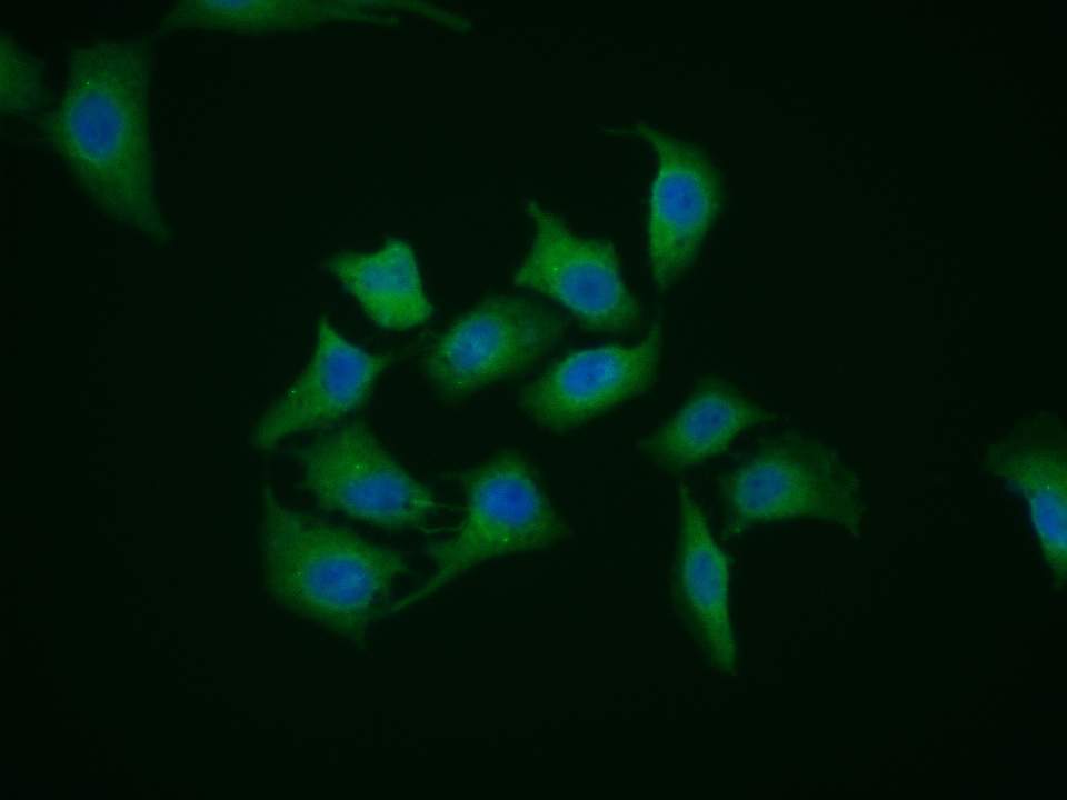 Hela cell; 4% Paraformaldehyde-fixed; Triton X-100 at room temperature for 20 min; Blocking buffer (normal goat serum, C-0005) at 37°C for 20 min; Antibody incubation with (IP3 receptor) polyclonal Antibody, Unconjugated (SL8869R) 1:100, 90 minutes at 37°C; followed by a conjugated Goat Anti-Rabbit IgG antibody at 37°C for 90 minutes, DAPI (blue, C02-04002) was used to stain the cell nuclei.
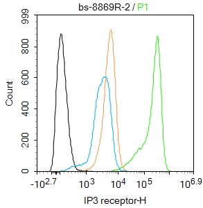 Blank control(black line):SH-SY5Y.
Primary Antibody (green line): Rabbit Anti-IP3 receptor antibody (SL8869R)
Dilution:2ug/Test;
Secondary Antibody(white blue line): Goat anti-rabbit IgG-AF488
Dilution: 0.5ug/Test.
Isotype control(orange line): Normal Rabbit IgG
Protocol
The cells were fixed with 4% PFA (10min at room temperature)and then permeabilized with 90% ice-cold methanol for 20 min at -20℃, The cells were then incubated in 5%BSA to block non-specific protein-protein interactions for 30 min at room temperature .Cells stained with Primary Antibody for 30 min at room temperature. The secondary antibody used for 40 min at room temperature. Acquisition of 20,000 events was performed.
Cartpieces
Totalgoods,subtotals:¥Checkout
References (0)
No References
Bought notes(bought amounts latest0)
No one bought this product
User Comment(Total0User Comment Num)
- No comment


 +86 571 56623320
 +86 18668110335

